


Introduction
Musculoskeletal tumors and tumor-like lesions in the foot and ankle region can present with a wide range of clinical and radiographic features. Common lesions in this area include giant cell tumors of the tendon sheath, which are among the most frequent soft-tissue tumors of the foot and ankle. These typically manifest as slowly growing, painless masses closely associated with the tendon sheath [1]. Soft-tissue sarcomas, which are malignant neoplasms, can arise from various soft-tissue structures in the foot and ankle and often present as rapidly enlarging, painful masses [2]. Benign soft-tissue lesions such as fibromas, lipomas, and ganglion cysts can appear as palpable masses of varying sizes and growth rates, depending on the specific pathology [3]. Rare benign entities, such as pseudo-malignant osseous soft-tissue tumors, can mimic neoplastic processes radiographically by appearing as bony protrusions from the cortical surface [4]. Bizarre parosteal osteochondromatous proliferation, also known as Nora’s lesion, is a benign, tumor-like lesion that arises from the periosteum and presents as a bony outgrowth on imaging [4]. Another uncommon lesion to consider is the fibro-osseous pseudotumor of the digit (FOPD), which typically originates from the cortical surface of the affected phalanx or metatarsal bone [5].
FOPD is a rare benign lesion characterized by fibro-osseous proliferation in the soft tissues of the digits. It most commonly affects the hands and wrists of young adult females, with the index finger being the predominant site [6, 7]. FOPD involving the feet is exceedingly uncommon, with only a few cases reported in the literature [8, 9, 10].
We present an unusual case of a 15-year-old male patient who developed FOPD in the hallux (great toe) of his right foot without antecedent trauma. This case does not involve the nailbed. The patient presented with a gradually enlarging mass over the proximal phalanx, causing local irritation and limiting joint mobility. Radiologic findings were concerning for a giant cell tumor, prompting surgical intervention [10].
Case Report
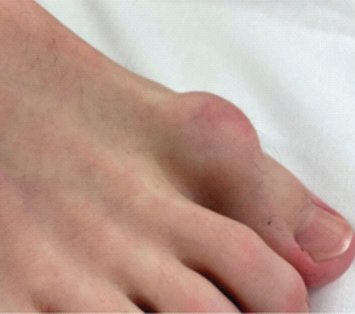
This case involves a 15-year-old male who presented to the pediatric orthopedic outpatient clinic with a rapidly growing lesion on the proximal phalanx of his right hallux, without nail bed involvement, as shown in Figure 1. The lesion had gradually increased in size over the past year. The patient reported that the swelling was aggravated by the rubbing of shoe wear and experienced pain only when the toe was flexed or extended. He denied any history of trauma to the toe. A neurovascular assessment revealed normal results, with a palpable dorsalis pedis pulse, preserved digital nerve function, and no changes to the nail bed.
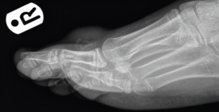
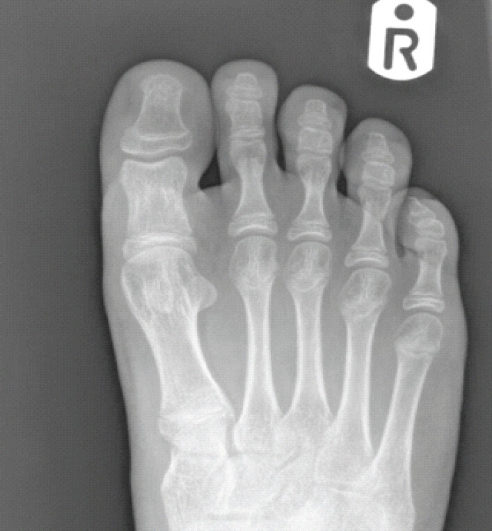
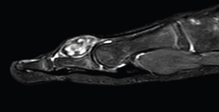
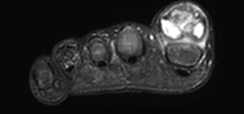
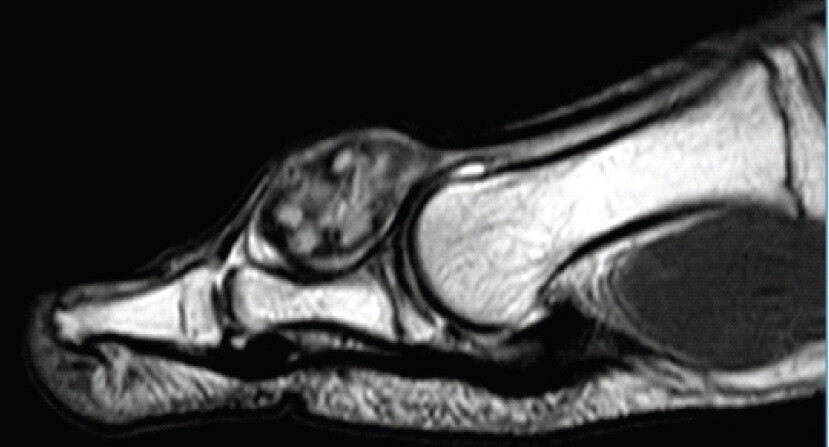
Radiological examinations included X-rays and magnetic resonance imaging (MRI); the radiograph revealed soft-tissue shadowing and slight scalloping of the bone dorsally, as shown in Figure 2a and b. MRI findings suggested a giant cell tumor of the extensor hallucis longus (EHL) tendon sheath, as shown in Figures 3a, b, c. Radiologically, FOPD lesions appear as ill-defined, calcified soft-tissue masses, lacking the distinct zoning pattern seen in myositis ossificans, though histologically, FOPD resembles this condition.
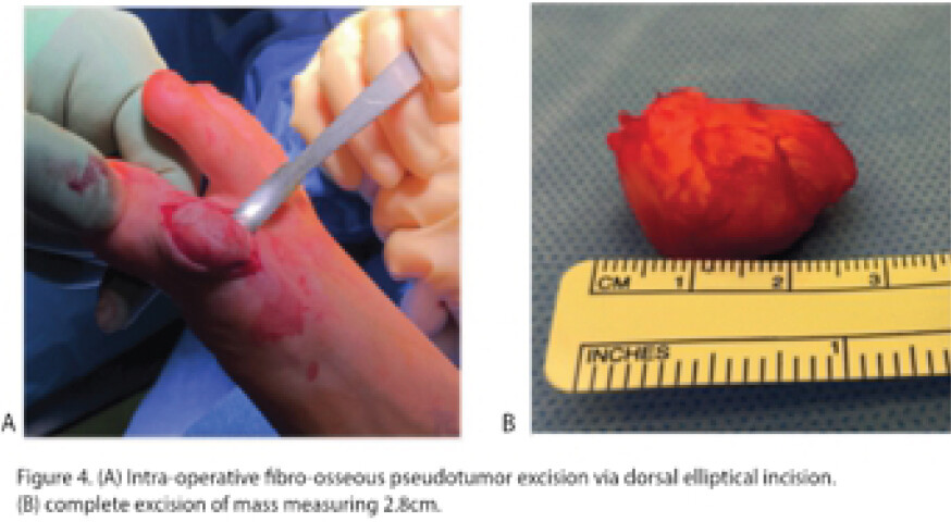
The patient underwent surgery in which the mass was carefully dissected from the EHL tendon, to which it was attached through the tendon sheath, through a dorsal elliptical incision, as shown in Figure 4a. There was noted scalloping of the dorsal proximal phalanx due to compression, but no significant changes to the periosteum or physis were observed. The circumscribed tumor weighed 4.5 g and measured 28 × 22 × 15 mm, as show in in Figure 4b. Histological analysis revealed bony trabeculae with osteoclastic and osteoblastic proliferation, as well as a cartilaginous cap. These findings were pathologically consistent with FOPD.
Discussion
Benign tumors and tumor-like lesions affecting the foot and ankle are relatively common, with up to 80% of musculoskeletal tumors in this region being benign [11]. In contrast, malignant tumors, particularly malignant bone tumors of the foot, are rare. Tumors in the foot represent approximately 3–6% of all pediatric bone tumors, highlighting their lower incidence in younger populations [12, 13].
One uncommon benign lesion that can occur in the foot and digits is the FOPD. This entity is characterized by the proliferation of fibroblast cells with areas of bone formation and predominantly affects the hands and wrists of young adult females, with the index finger being the most frequently reported site [6, 9]. The index finger is the most frequently reported site, while only 1 out of 21 cases in the study by Dupree and Enzinger (1986) involved the feet [7]. A recent case report from 2021 described a 26-year-old female diagnosed with FOPD on the dorsal aspect of her left hallux [8], unlike our case, where there was no involvement of the nailbed. Another similar case in 2020 involved a 40-year-old man who was initially treated for pyogenic granuloma, but further investigations revealed FOPD [10].
Although several cases of FOPD have been reported since first described by Dupree and Enzinger in 1986 [7] our case is unique due to the extremely rare location of FOPD in the hallux without a history of trauma. We experienced FOPD formation in the hallux with no history of trauma to the great toe in a 15-year-old male. While the etiology of FOPD is unclear, trauma has been observed in approximately 40% of cases [7].
Among young patients, bone tumors like osteosarcoma or Ewing’s sarcoma are common, yet rarely manifest in the foot region [2]. Instead, synovial sarcoma is the most common histological diagnosis for soft-tissue tumors in this age group and anatomical location. In the context of foot tumors, it is important to note that most lesions encountered are benign or tumor-like conditions. Malignant soft tissue and skeletal tumors of the foot are exceedingly rare, and their diagnosis is often delayed, leading to referrals to specialized teams after initial inappropriate procedures or unplanned excisions. Misdiagnosis of foot tumors can have adverse consequences, including an increased risk of amputation or local recurrence. Therefore, thorough imaging studies and histopathological examinations are essential when undertaking any local treatment for foot lumps. Early recognition and careful evaluation are crucial.
Despite the rarity of FOPD occurring in the foot, it should be considered a potential diagnosis in this location. Our patient underwent complete surgical excision, which is the gold standard treatment approach for FOPD. This procedure typically yields an excellent prognosis with a low risk of recurrence when the lesion is entirely removed. Extensive resection beyond the lesion margins is generally unnecessary, as FOPD is a benign condition, and malignant transformation is exceedingly rare. However, it is crucial to rule out malignancy and ensure clear margins during excision to prevent inappropriate amputation and preserve the function of the digit [5]. Previous reports have documented instances of unnecessary amputations resulting from misdiagnosis [5,14]. Therefore, we emphasize the importance of not ruling out the hallux for FOPD immediately; excisional biopsy and comprehensive histopathological examination to accurately diagnose FOPD, guide timely and correct management.
Conclusion
We report a rare case of FOPD in the hallux of a 15-year-old male with no history of trauma or identifiable cause. Complete surgical excision with clear margins, the recommended treatment for FOPD, was successfully performed, resulting in an excellent prognosis with a low risk of recurrence. Early recognition and accurate diagnosis through excisional biopsy and comprehensive histopathological evaluation are essential for appropriate management.
Clinical Message
This case underscores the importance of considering FOPD in the differential diagnosis for digital masses, even in atypical locations like the foot. It highlights the necessity of thorough clinical, radiologic, and histopathologic assessments to establish an accurate diagnosis and guide treatment, emphasizing that early recognition and complete surgical excision are crucial.
Conflict of Interest:
Nil
Source of Support:
Nil
Consent
The authors confirm that informed consent was obtained from the patient for publication of this article
How to Cite this Article
Buddhdev P, Gosatkar S, Ahmed M. Hallux Mystery: A Rare Case of Fibro-Osseous Pseudotumor. J Orthop Case Rep. 2026 November;16(11): 74-78. doi: 10.13107/jocr.2026.v16.i11.8392.
References
- Scheele C, Toepfer A, Beischl S, Dammerer D, Harrasser N, Von Eisenhart-Rothe R. Insights into the distribution patterns of foot and ankle tumours: Update on the perspective of a university tumour institute. J Clin Med 2024;13:350. [Google Scholar] | [PubMed]
- Mascard E, Gaspar N, Brugières L, Glorion C, Pannier S, Gomez-Brouchet A. Malignant tumours of the foot and ankle. EFORT Open Rev 2017;2:261-71. [Google Scholar] | [PubMed]
- Van Hul E, Vanhoenacker F, Van Dyck P, De Schepper A, Parizel PM. Pseudotumoural soft tissue lesions of the foot and ankle: A pictorial review. Insights Imaging 2011;2:439-52. [Google Scholar] | [PubMed]
- Edoardo I, Elisa F, Damiano RA, Silvia F, Rodolfo C, Lorenzo A. Bizarre parosteal osteochondromatous proliferation (Nora lesion): A narrative review. Acta Med Litu 2022;29:176-93. [Google Scholar] | [PubMed]
- Moosavi CA, Al-Nahar LA, Murphey MD, Fanburg-Smith JC. Fibrosseous pseudotumor of the digit: A clinicopathologic study of 43 new cases. Ann Diagn Pathol 2008;12:21-8. [Google Scholar] | [PubMed]
- Chaudhry IH, Kazakov DV, Michal M, Mentzel T, Luzar B, Calonje E. Fibro-osseous pseudotumor of the digit: A clinicopathological study of 17 cases. J Cutan Pathol 2010;37:323-9. [Google Scholar] | [PubMed]
- Dupree WB, Enzinger FM. Fibro-osseous pseudotumor of the digits. Cancer 1986;58:2103-9. [Google Scholar] | [PubMed]
- Olvi LG, Da W, Santini-Araujo E, Kalil RK. Fibro-osseous pseudotumor of digits. Berlin:Springer eBooks;2020 821-4. [Google Scholar] | [PubMed]
- Sakuda T, Kubo T, Shinomiya R, Furuta T, Adachi N. Rapidly growing Fibro-osseous pseudotumor of the digit: A case report. Medicine (Baltimore) 2020;99:e21116. [Google Scholar] | [PubMed]
- Atomura D, Makiguchi T, Yamaguchi T, Nakamura H, Yamatsu Y, Shirabe K. Fibro-osseous pseudotumor of the toe. Eplasty 2020;20:ic2. [Google Scholar] | [PubMed]
- Fritzsche H, Weidlich A, Schaser KD, Mehnert E, Winkler D, Rammelt S. Benign tumours of foot and ankle. EFORT Open Rev 2023;8:397-408. [Google Scholar] | [PubMed]
- Rammelt S, Fritzsche H, Hofbauer C, Schaser KD. Malignant tumours of the foot and ankle. Foot Ankle Surg 2020;26:363-70. [Google Scholar] | [PubMed]
- Ozdemir HM, Yildiz Y, Yilmaz C, Saglik Y. Tumors of the foot and ankle: Analysis of 196 cases. J Foot Ankle Surg 1997;36:403-8. [Google Scholar] | [PubMed]
- Coleman RA. Fibro-osseous pseudotumour of the digit--amputation for a benign but aggressive lesion. J Hand Surg Br 2005;30:504-6. [Google Scholar] | [PubMed]
© 2026 Journal of Orthopaedic Case Reports - Published by Indian Orthopaedic Research Group
About the Authors
-

Pranai Buddhdev
-

Shraddha Gosatkar
-

Mohammad Ahmed
Related Articles in Journal of Orthopaedic Case Reports
 November 1, 2026 Incidentally Detected Osseous Hemangioma of the Femoral Head: Conservative Management of a Rare Case with Review of the Literature
 October 1, 2026 Posterior Fibular Entrapment in a Bosworth Fracture-Dislocation: A Case Report and Review of Diagnostic and Operative Considerations
 October 1, 2026 Recurrent Ventral Scapular Osteochondroma Presenting with Pseudo-winging in an Adolescent: A Case Report and Follow-up of a Previously Published Case
 October 1, 2026 Development of a Non-ossifying Fibroma in the Same Femur 6 Years after Limb-Salvage Surgery for Osteosarcoma: A Case Report