


Introduction
Lumbar disc herniations are the most common degenerative abnormality of the lumbar spine, affecting two to three percent of the population and often resulting in spinal surgery [1]. The vacuum phenomenon is a radiological finding on CT characterized by the presence of gas, predominantly nitrogen, in the intervertebral disk space and is associated with advanced disc degeneration [2,3]. While the vacuum phenomenon is relatively common, a gas-containing lumbar disc herniation resulting in compressive radiculopathy is rare [4,5].
Diagnosing this pathology can be challenging as it is typically not identifiable on magnetic resonance imaging (MRI) or during open decompression surgery [1,4]. In a standard surgical environment, the gas typically escapes into the atmosphere or is obscured upon entering the disc space [4]. To confirm the presence of gas in traditional open procedures, surgeons have previously had to fill the surgical field with saline and precisely manipulate the disc fragment with a probe to observe the release of air bubbles [4]. Consequently, without the specific maneuvers endoscopic technique allows, the vacuum content is frequently missed or dismissed during standard open surgery.
This case demonstrates direct endoscopic visualization of a gas-filled diverticulum compressing a nerve root, with visual confirmation of gas release during decompression, something not achievable with traditional open approaches. The observations in this case contribute meaningful clinical insight into a rare variant of disc herniation and reinforce the diagnostic value of CT. This report adds to the growing but still limited body of literature on gas-filled lumbar disc herniation and highlights the diagnostic and therapeutic advantages of minimally invasive endoscopic spine surgery for this pathology.
Case Report
A 62-year-old male with a medical history of hypertension, migraines, anxiety, and prior right shoulder surgery presented with a 3-year history of progressive right-sided low back and leg pain. Symptoms were sharp and electrical with radiation to the posterior thigh, calf, and foot. He reported sensory loss, weakness, and loss of coordination. Conservative management, including non-steroidal anti-inflammatory drugs, gabapentin, physical therapy, chiropractic care, and exercise, provided only temporary relief. On examination, the patient demonstrated diminished sensation in the S1 distribution. Key findings include a reduced right Achilles reflex, diminished sensation at the lateral heel to both light touch and pinprick, and decreased strength of the gastrocnemius muscle.
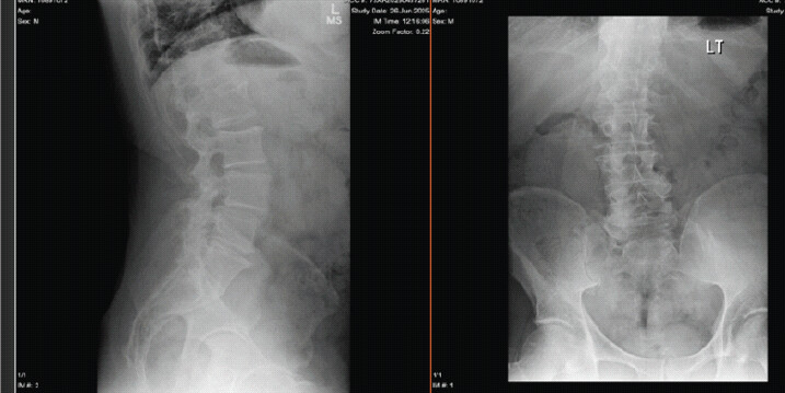
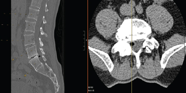
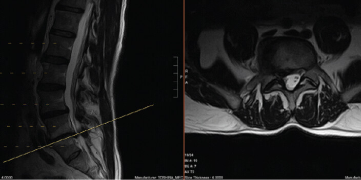
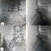
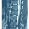
Plain radiographs demonstrated transitional lumbosacral anatomy with minor degenerative scoliosis and severe disc degeneration between the L4 and L5 vertebrae, which in this case corresponded to L5-S1 due to transitional anatomy (Fig. 1). MRI initially showed a disc herniation on the right at L5-S1 (Fig. 2). While the gas phenomenon was not clearly defined on MRI, it was easily identified on computed tomography (CT), which demonstrated a paracentral extension containing intradiscal gas within the right lateral recess, consistent with a vacuum phenomenon.
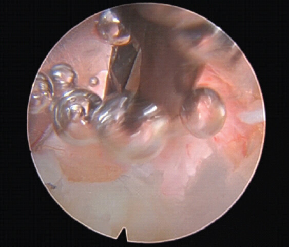
The patient underwent a right-sided endoscopic interlaminar microdiscectomy decompression at L5-S1 (Fig. 3). After gentle retraction of the nerve root, a balloon-like diverticulum of the disc was visualized (Fig. 4). Unlike a typical herniation that extrudes disc material, the lesion contained gas consistent with a gas-filled disc outpouching [A2]. The presence of gas within the lesion was confirmed during operation when the diverticulum was punctured under endoscopic visualization. This resulted in the immediate egress of gas bubbles and the subsequent decompression of the nerve root (Fig.4). The remaining redundant annulus was resected endoscopically.
In the immediate post-operative period, the patient reported marked relief of right-sided radicular pain. At a 2-week post-operative follow-up, he continued to report substantial improvement in his pain without any new neurological deficits. Since the patient remained completely asymptomatic, post-operative imaging was not performed. However, long-term clinical success was documented using the Katsuura Mini Outcomes (KMO) questionnaire. Preoperatively, the patient’s KMO score was 46 out of 100, reflecting severe impairment with his overall symptoms and mobility disability at a level of nine out of ten, with right leg pain at eight out of ten. At an 8-month follow-up on February 3rd, 2026, the patient’s KMO score improved to 0/100. This score indicated a total resolution of all axial and lower extremity symptoms, as well as the complete disappearance of numbness and functional disability.
Discussion
Micro decompressions may be performed through various surgical approaches. Techniques range from traditional open methods to endoscopic surgery. Vacuum discs are relatively common, and [6] (i.e., the radiolucent visualization of gas within the intervertebral disc space) is observed in 1–3% of lumbar spinal radiographs and most frequently in older adults (20%) [7]. This case enabled direct visualization of the decompression of the gas-filled lesion.
A traditional open microdiscectomy may be effective at treating this pathology; however, because the surgery is open to the air, it cannot visually capture the egress of gas. The advancement of endoscopic spine surgery enables easy access to the spine through small incisions, reducing complications associated with the traditional open technique [8]. In addition, as in this case, it allows for an interesting and satisfying visual confirmation of the egress of the gas-filled pathology, which is not possible in open surgery.
Several cases of gas-containing disc herniation have been reported (Table 1) [4,9,10,11,12], and other reports describe successful treatment with a minimally invasive approach using a small incision [9,10] or the occurrence of intradiscal gas bubbles during endoscopic disc removal [11]. Similarly, our case was performed in an aqueous environment that provided a video to document the gas that was venting from the disc space.
Summary of cases reporting gas-containing disc herniation
| Reference | Study design | Spinal level | Pathology | Surgical approach |
|---|---|---|---|---|
| [9] | 1 case | L5–S1 | Gas-containing herniated nucleus pulposus. | Endoscopic |
| [11] | 1 case | L5–S1 | Intraoperative findings of gas leakage from the intervertebral disc. | Endoscopic |
| [4] | 18 cases | L5–S1 | Spondylolisthesis, scoliosis, instability, or spinal stenosis. | Open |
| [10] | 2 cases | L4–L5, L5–S1 | Gas-containing lumbar disc herniation causing nerve root compression. | Open |
| [12] | 12 cases | L4, L5, or S1 nerve root | Gas in the epidural space compressing nerve roots. | Conservative treatment |
Since the disc herniation is not filled with disc material, but rather gas, it bears resemblance to a gastric diverticulum. Commonly, the removal of extruded disc material is required for a typical discectomy [1.1][9]. In this case, the gas-filled diverticulum was compressing the nerve root, pinched against bone. After the surgery, the patient expressed relief from his radicular pain. This case raises the idea that for a gas-filled compression, simple deflation may be a practical approach to relieve symptoms.
This case describes a variant of lumbar disc herniation: A gas-filled diverticulum phenomenon. It highlights a crucial diagnostic lesson that a gas-containing herniation should be considered in the differential diagnosis and may be easily identified on a pre-operative CT scan. In this case, we successfully performed an endoscopic microdiscectomy at L5–S1 for a lateral gas-containing disc herniation confirmed by MRI and CT, consistent with the vacuum phenomenon. This case highlights the unique visualization properties of endoscopic spine surgery and presents an interesting visual representation of a gas-filled diverticulum rarely caught on film.
Conclusion
Gas-filled lumbar disc herniation is a rare but important cause of radiculopathy that may be missed on MRI. This case demonstrates that CT plays a key diagnostic role in identifying intradiscal gas associated with the vacuum phenomenon and reinforces the need to consider this condition in patients with degenerative disc disease. Endoscopic decompression provides direct visualization of the gas-filled diverticulum, allows controlled deflation and removal of residual pathology, and results in effective symptom relief with minimal morbidity. This case adds to the limited literature by showing that minimally invasive endoscopic microdiscectomy can serve as both a diagnostic and definitive therapeutic approach, advancing our understanding of the clinical presentation and management of gas-containing disc herniation within the field of spine surgery.
Clinical Message
In patients with lumbar radiculopathy, the presence of intradiscal gas on computed tomography should raise suspicion for a gas-filled disc herniation, and endoscopic decompression offers a minimally invasive approach that allows direct visualization, definitive diagnosis, and effective symptom relief.
Conflict of Interest:
Nil
Source of Support:
Nil
Consent:
The authors confirm that informed consent was obtained from the patient for publication of this article
References
- Vialle LR, Vialle EN, Suárez Henao JE, Giraldo G. Lumbar disc herniation. Rev Bras Ortop 2015;45:17-22. [Google Scholar] | [PubMed]
- Pierpaolo L, Luciano M, Fabrizio P, Paolo M. Gas-containing lumbar disc herniation. A case report and review of the literature. Spine (Phila Pa 1976) 1993;18:2533-6. [Google Scholar] | [PubMed]
- Cawley DT, Simpkin A, Abrahim E, Doyle T, Elsheikh N, Fallon J. Natural history of intradiscal vacuum phenomenon and its role in advanced disc degeneration. Spine (Phila Pa 1976) 2024;49:1130-6. [Google Scholar] | [PubMed]
- Choi KC, Kim JS, Lee SH. Surgical experience of gas-containing disk herniation. Neurol Med Chir (Tokyo) 2010;50:905-9. [Google Scholar] | [PubMed]
- Tamburrelli F, Leone A, Pitta L. A rare cause of lumbar radiculopathy: Spinal gas collection. J Spinal Disord 2000;13:451-4. [Google Scholar] | [PubMed]
- Lewandrowski KU, Zhang X, Ramírez León JF, De Carvalho PS, Hellinger S, Yeung A. Lumbar vacuum disc, vertical instability, standalone endoscopic interbody fusion, and other treatments: An opinion based survey among minimally invasive spinal surgeons. J Spine Surg. 2020 Jan;6(Suppl 1):S165-S178. [Google Scholar] | [PubMed]
- Resnick D, Niwayama G, Guerra J Jr, Vint V, Usselman J. Spinal vacuum phenomena: Anatomical study and review. Radiology 1981;139:341-8. [Google Scholar] | [PubMed]
- Hagan MJ, Remacle T, Leary OP, Feler J, Shaaya E, Ali R. Navigation techniques in endoscopic spine surgery. Biomed Res Int 2022;2022:8419739. [Google Scholar] | [PubMed]
- Inokuchi T, Tezuka F, Yamashita K, Morimoto M, Sugiura K, Fujitani J. Transforaminal full-endoscopic discectomy for gas-containing herniated nucleus pulposus at L5-S1 under local anesthesia: A case report. J Med Invest 2022;69:328-31. [Google Scholar] | [PubMed]
- Righini A, Lucchi S, Reganati P, Zavanone M, Bettinelli A. Percutaneous treatment of gas-containing lumbar disc herniation. Report of two cases. J Neurosurg 1999;91 1 Suppl:133-6. [Google Scholar] | [PubMed]
- Loroi Y, Kawasaki T, Hashimoto J, Kobayashi T, Koga H, Takayama M. Full-endoscopic discectomy utilizing an interlaminar approach for gas-containing herniation at L5-S1. Surg Neurol Int 2024;15:265. [Google Scholar] | [PubMed]
- Tsitouridis I, Sayegh FE, Papapostolou P, Chondromatidou S, Goutsaridou F, Emmanouilidou M. Disc-like herniation in association with gas collection in the spinal canal: CT evaluation. Eur J Radiol 2005;56:1-4. [Google Scholar] | [PubMed]
© 2026 Journal of Orthopaedic Case Reports - Published by Indian Orthopaedic Research Group
About the Authors
-

Miss. Stephanie Mendez
-

Dr. Mark Polemidiotis
-

Mr. Cyrus Nasr
-

Mr. Allen Justin
-

Mr. Anahan Joshua Baptist
-

Dr. Yoshihiro Katsuura
Related Articles in Journal of Orthopaedic Case Reports
 October 1, 2026 Acute Cauda Equina Syndrome in Rapid Succession: A Case Series of Eight Consecutive Patients Undergoing Emergency Surgical Decompression Over One Week
 October 1, 2026 Efficacy of Transforaminal Nerve Root Block for Lumbar Disc Herniation
 September 1, 2026 Clinical Effectiveness of Intradiscal Ozone Therapy with or Without Periforaminal Steroid Injection in Lumbar Disc Herniation: Revisiting an Underutilized Treatment
 August 1, 2026 Adolescent Cauda Equina Secondary to Traumatic Lumbar Disc Herniation: A Rare Case Report with Review of Literature