

Early decompression of the neural structure is the key factor in the management of cauda equina syndrome.
Dr. Sumit Kumar, Central Institute of Orthopaedics, VMMC and Safdarjung Hospital, New Delhi, India. E-mail: drsumit26@gmail.com
Abstract
Introduction: Lumbar disc herniation is a common spinal pathology, but its occurrence in the paediatric population is a rare entity. These patients are relatively asymptomatic, and the presence of a neurological deficit is rare. We present a case of an acute lumbar disc herniation presenting with cauda equina syndrome (CES) in a 13-year-old child, along with a review of the literature on the topic.
Case Report: A 13-year-old male child presented to us in the emergency department with complaints of low back pain, weakness, and urinary incontinence following lifting of a heavy weight 25 days back. On examination, he had motor and sensory deficits at the L5 and S1 regions along with perianal anaesthesia. Magnetic resonance imaging showed a central disc protrusion at the L4-L5 level. The patient underwent central decompression. He showed both functional and neurological recovery in follow-up.
Conclusion: Lumbar disc herniation is a rare cause of back pain in children. Several factors were identified as potential causes of paediatric lumbar disc herniation, among which trauma is the most common. Neurological deficits are rare in children, largely because a more mobile spine prevents nerve root compression. Surgery is indicated in pain not responding to 4–6 weeks of conservative treatment, CES, progressive neurological deficits, and correlating spinal deformities. There are very few cases of CES due to lumbar disc herniation in adolescent children reported in the literature. We reviewed the available literature and identified 6 such cases, tabulating the findings.
Keywords: Lumbar disc herniation, cauda equina syndrome, laminectomy, discectomy.

Lumbar disc herniation is defined as the displacement of intervertebral disc contents (nucleus pulposus or annulus fibrosus) beyond the intervertebral disc space. Lumbar disc herniation is a common spinal pathology presenting in patients older than 40 years. Its occurrence in the pediatric population is rare, seen in only 0.6–3% of the symptomatic population [1,2]. Its etiology can be varied in the adult population, including genetic, degenerative, and traumatic, but its presentation in the pediatric population is most commonly following a single traumatic event [3]. These patients are relatively asymptomatic, and the presence of a neurological deficit is rare. We present an interesting case of an acute lumbar disc herniation presenting with cauda equina syndrome (CES) in a 13-year-old child and review the literature on the topic.
A 13-year-old male child presented to us in the emergency department with complaints of low back pain, weakness, and urinary incontinence following lifting of a heavy weight 25 days back. On taking a detailed history, the patient complained of low back pain immediately following the episode, which was gradually progressive, aggravated on movements, and due to which the patient was confined to the bed. After 5 days, the patient complained of paraesthesia and tingling in both lower legs, which slowly progressed to weakness of both ankles and toes over the subsequent days. After another 10 days, the patient complained of urinary retention and presented in the emergency department. On examination, tenderness was present in the L4-L5 region with associated paraspinal spasm. There was no obvious coronal or sagittal plane deformity. On neurological examination, the bulk and tone of both lower limbs were normal. On motor examination, his hip flexors, knee extensors and ankle dorsiflexors of both lower limbs were 5/5; toe dorsiflexors and hip abductors of right and left lower limbs were 3/5 and 2/5, respectively; and ankle plantar flexors were 3/5 and 2/5 on right and left lower limbs, respectively. On examination of reflexes, his knee jerks were normal, and ankle jerks were absent. The plantar reflex and anal wink reflex were absent. The patient complained of diminished sensation in his L5-S1 and S2-S5 dermatomes bilaterally. A perirectal examination showed weak anal tone and diminished deep anal pressure sensations. The straight leg raise (SLR) test was 300 bilaterally, indicating a positive test. The child was overweight for his age with a body mass index (BMI) of 26 kg/m2. The patient was diagnosed with an incomplete cauda equina and sent for radiological evaluation. After proper analgesia, the patient was catheterised and sent for radiographs and a magnetic resonance imaging (MRI). His radiograph of the lumbosacral spine showed no obvious abnormalities except straightening of lumbar lordosis, indicating paraspinal muscle spasm. His T2-weighted sagittal MRI showed a dorsal disc protrusion at L4-L5 with homogeneous annular signal and minimal peripheral fissuring (Fig. 1).
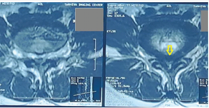
Figure 1: Axial T2-weighted magnetic resonance imaging showing a central disc bulge compressing the thecal sac.
T2-weighted axial images at the level of the L4-L5 disc showed a postero-central disc protrusion almost completely obliterating the canal with a canal diameter of 5 mm (Fig. 2).
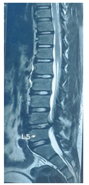
Figure 2: Sagittal T2-weighted magnetic resonance imaging showing a well-hydrated disc with a herniated nucleus pulposus compressing the thecal sac.
No apophyseal ring fracture was seen. The patient was counselled for emergency surgery and shifted to the operating room. In the prone position, the midline posterior approach was utilised, soft tissues were incised, and subperiosteal elevation of paraspinal muscles was performed. After confirming with fluoroscopy, central decompression with inferior laminectomy of L4 and complete laminectomy of L5 with preservation of bilateral pars interarticularis were performed. Ligamentum flavum was excised to expose the thecal sac. Intraoperatively, a bulged annulus at the level of L4-L5 was seen (Fig. 3).
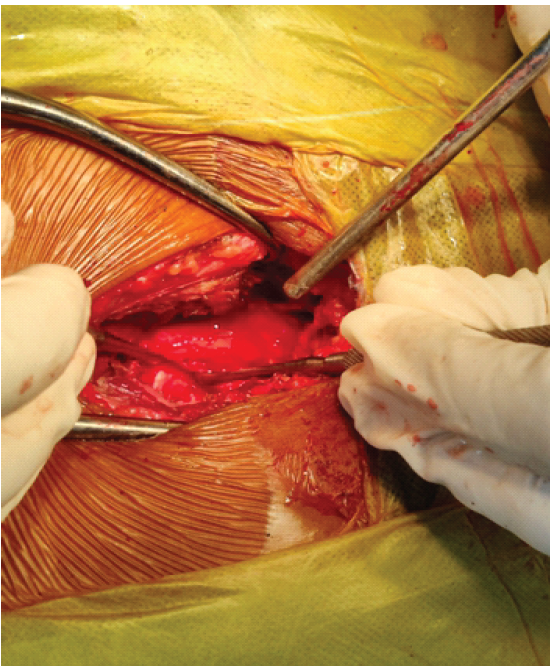
Figure 3: Intraoperative picture showing the post-laminectomy image with free central canal and L5 traversing root.
Disc space was explored by retracting the thecal sac and L5 nerve root for any fragments, but none were present. Annulotomy was done for any protruded disc particles, taking care not to damage the inner annulus and looking for any protruded disc fragments. Small disc fragments were removed piecemeal. Foraminotomy of the L5 nerve root was performed bilaterally, and the wound was closed in layers over a drain. The post-operative period was uneventful, and the suture was removed on post-operative day 12. The patient was started on active quadriceps and hamstring stretching, along with core-strengthening exercises. The patient showed neurological improvement from post-operative day 3, with recovery of bowel and bladder function and of both motor and sensory functions from post-operative day 5. The patient was followed up for 6 months and has a near-complete neurological recovery with full bowel and bladder recovery at present (Table 1). The boy was doing well on follow-up and had resumed his normal activities.
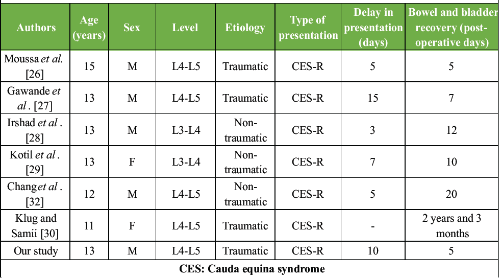
Table 1: Cases of pediatric or adolescent cauda equina syndrome to date
Lumbar disc herniation is a rare cause of back pain in children. It was first reported in the literature by Wahren in a 12-year-old child [4]. Subsequent studies by DeOrio and Bianco in 1982 showed an incidence of 0.5% in children <16 years [5]. A later study by Zitting et al. in 1998 showed no patients under 15 years and an incidence of 0.1–0.2% in 15–20-year-olds [6]. Several factors were identified as a potential cause of paediatric lumbar disc herniation, among which trauma is the most common and is seen in 30-60% of patients [3]. In recent studies, it has been established that pre-existing lesions, such as microdamage and degenerative changes, are present in these children, with trauma being an inciting event leading to exacerbation of symptoms [7,8,9]. Various genetic factors have also been studied as potential causes, with 13–57% of adolescents having a first-degree relative with the same disorder [10,11]. The presence of vertebral anomalies, such as scoliosis and transitional vertebrae, is also known to be associated with lumbar disc herniation in children [12]. Most of these children present with low back pain and restricted lumbar movements [13]. SLR is positive in 90% of children, as they have more nerve root tightness than adults [14]. The apophyseal ring remains attached to the outer annulus by means of Sharpey’s fibres and begins ossification by 6 years and fuses by 18 years [15]. The incidence of apophyseal ring fractures is higher in children and adolescent lumbar disc herniations (5–42%). Neurological deficits are rare in children, largely because a more mobile spine prevents nerve-root compression [16,17]. Paediatric lactate dehydrogenase does not respond well to conservative treatment, as the discs are well hydrated and do not resorb spontaneously. Traumatic events may cause significant annular damage; trauma can rupture the epiphyseal ring and lead to separation. Children are more mobile and do not observe strict bed rest [18,19,20,21]. Nevertheless, conservative treatment is the primary modality of treatment, and surgery is indicated in case of severe intractable pain not responding to 4–6 weeks of conservative treatment, CES, progressive neurological deficits, and correlating spinal deformities [22]. Surgery should not be delayed in these patients, as children have reported good long-term outcomes. Complete removal of the disc should be avoided, as it may result in stenotic changes at the level and degenerative changes at adjacent levels [2,23]. It is important to keep the inner annulus intact, as an intact annulus has been shown to facilitate the regeneration of the nucleus pulposus in the future [24]. The importance of early and prompt diagnosis of cauda equina is demonstrated by the fact that neurological recovery is good when decompression is performed within 48 h [25]. For this very reason, it has been subclassified into early, incomplete, and retention types, with the early and incomplete types having a better prognosis than the retention types. There are very few cases of CES due to lumbar disc herniation in adolescent children reported in the literature. We reviewed the available literature and identified 6 such cases, which we tabulated for comparison (Appendix, Table 1). All children were under 15 years of age. L4-L5, which was the most common level of disc herniation, was also the most common level leading to cauda equina, followed by L3-L4. All children were between the ages of 11 and 15 years, with a mean of 13 years. Four males and two females were reported in the literature to present with the above symptoms. Only 3 of the 6 cases were caused by traumatic events; therefore, factors other than trauma may play a significant role. All patients were operated on by open central decompression without instrumentation, and all regained full functional neurological recovery on follow-up [26,27,28,29,30,31]. Another interesting observation was that, unlike other cases, we did not find any extruded disc material. Our hypothesis in the above case is a subannular rupture and herniation of the nucleus pulposus, which may have caused the disc bulge leading to cauda equina [32]. An acute traumatic event in an overweight child might have led to a subannular rupture, which progressed to a cauda equina due to lack of proper rest and treatment, which might account for the delay in presentation of bowel and bladder symptoms in the child. The strengths of this report lie in its detailed documentation of an exceptionally rare case of paediatric CES, a condition found in fewer than 3% of children with slipped discs. Subannular rupture, where the soft inner core of the disc tears through the deep layers but remains trapped and pressurised under the outer ring and the surrounding ligament (contained herniation), might explain why serious symptoms, such as bladder and bowel problems, developed gradually as pressure built up behind that final layer, rather than happening instantly as they might with a full rupture. Mechanical failure may have been due to physical factors, such as heavy lifting and a BMI of 26 kg/m². This report provides a clear map of how the adolescent spine fails under stress and demonstrates that prompt surgery can lead to a full recovery. The primary limitation of this study is its single-case design, which limits the generalisability of the findings to the broader paediatric population. While we highlight a rare presentation of adolescent CES, the conclusions regarding management cannot be extrapolated to all cases of paediatric lumbar disc herniation. Furthermore, the follow-up period was limited to 6 months. While this was sufficient to document significant neurological and functional recovery, it is inadequate for assessing long-term complications, such as recurrent herniation, adjacent level degeneration, or potential spinal instability. In addition, our outcome assessment relied on clinical neurological examination rather than validated objective tools, such as the Oswestry disability index or specific paediatric functional scores. The radiological evaluation was also restricted to MRI; although this is the gold standard for soft tissue, the absence of a computed tomography scan (to avoid radiation exposure) might have limited our ability to detect subtle apophyseal ring injuries. Finally, while predisposing factors, such as a BMI of 26 kg/m² and genetic literature, were discussed, a systematic evaluation of these variables was not performed for our patient. Open central decompression remains a standard of care for acute CES. Due to the rarity of the condition in the paediatric or adolescent population, literature on MIS or endoscopy in CES for lumbar disc herniation was unavailable, and the urgency of the deficit dictated early open surgery after presentation for optimal decompression (allowing for maximum chance for neurological recovery). Thus, comparison with minimally invasive techniques or conservative protocols was not feasible for this specific patient. However, it might be explored in future cases as the technology advances.
A systematic search of the PubMed, Scopus and Cochrane Library databases was performed using suitable keywords, such as ‘cauda equina syndrome’, ‘lumbar disc herniation’, ‘pediatric’, ‘child’[ search strategy ((Cauda equina syndrome) AND (Lumbar disc herniation)) AND ((Pediatric*) OR (Child*))]. Reference lists of the articles were also screened. The last date on which the search was performed was 5th March, 2025. We did not contact the authors for any additional information.
The Preferred Reporting Items for Systematic reviews and Meta-Analyses flowchart is shown in Fig. 4. The preliminary search yielded 8 articles on PubMed and 16 on Scopus. No extra papers were found in the Cochrane Library (All Text). One additional paper was retrieved through a Google search. After screening duplicates and excluding unrelated articles based on the title, 8 papers were considered for assessment of full texts, and finally, 6 papers (case reports) were considered for qualitative summarisation (Fig. 4).
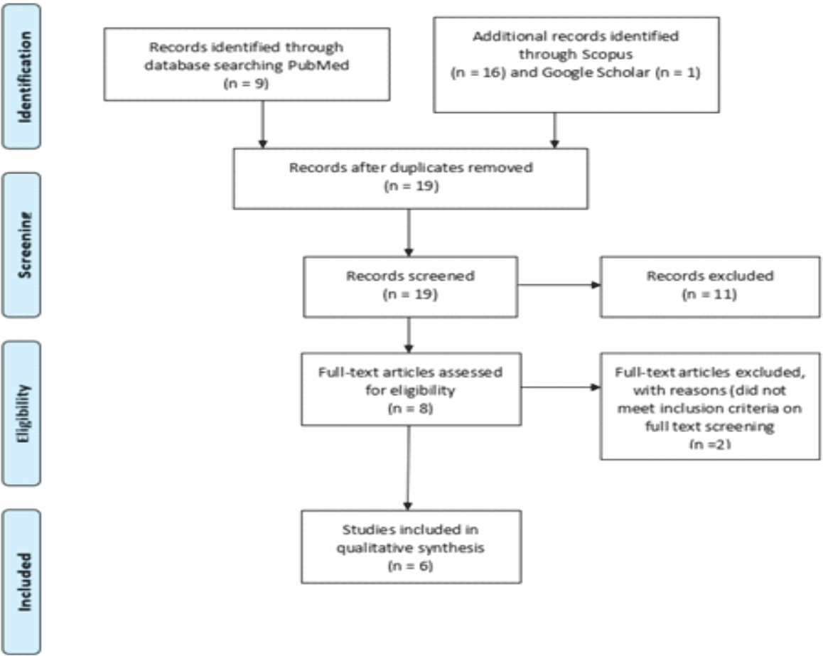
Figure 4: Preferred reporting items for systematic reviews and meta-analyses flow diagram.
We present a rare case of a 13-year-old boy with adolescent CES due to an acute lumbar disc herniation, which is under-reported in the literature. We have reviewed the available literature for similar cases. Cauda equina is a surgical emergency, and early and prompt diagnosis is imperative for good neurological recovery in these patients. Discectomy in children is a traumatic event which may accelerate the process of degeneration during growth. Careful handling and surgical expertise are required to prevent the activation of the degeneration cascade and to achieve excellent functional outcomes in these children.
Prompt diagnosis and early surgical intervention led to a good outcome in cauda equina syndrome. Long-term follow-up is required to see the disc degeneration cascade in adolescent patients.
References
- 1. Martínez-Lage JF, Fernández Cornejo V, López F, Poza M. Lumbar disc herniation in early childhood: Case report and literature review. Childs Nerv Syst 2003;19:258-60. [Google Scholar] [PubMed]
- 2. Luukkonen M, Partanen K, Vapalahti M. Lumbar disc herniations in children: A long-term clinical and magnetic resonance imaging follow-up study. Br J Neurosurg 1997;11:280-5. [Google Scholar] [PubMed]
- 3. Kurihara A, Kataoka O. Lumbar disc herniation in children and adolescents. A review of 70 operated cases and their minimum 5-year follow-up studies. Spine (Phila Pa 1976) 1980;5:443-51. [Google Scholar] [PubMed]
- 4. Wahren H. Herniated nucleus pulposus in a child of twelve years. Acta Orthop Scand 1945;16:40-2. [Google Scholar] [PubMed]
- 5. DeOrio JK, Bianco AJ Jr. Lumbar disc excision in children and adolescents. J Bone Joint Surg Am 1982;64:991-6. [Google Scholar] [PubMed]
- 6. Zitting P, Rantakallio P, Vanharanta H. Cumulative incidence of lumbar disc diseases leading to hospitalization up to the age of 28 years. Spine (Phila Pa 1976) 1998;23:2337-43. [Google Scholar] [PubMed]
- 7. Varlotta GP, Brown MD, Kelsey JL, Golden AL. Familial predisposition for herniation of a lumbar disc in patients who are less than twenty-one years old. J Bone Joint Surg Am 1991;73:124-8. [Google Scholar] [PubMed]
- 8. Silvers HR, Lewis PJ, Clabeaux DE, Asch HL. Lumbar disc excisions in patients under the age of 21 years. Spine (Phila Pa 1976) 1994;19:2387-91. [Google Scholar] [PubMed]
- 9. Russwurm H, Bjerkreim I, Ronglan E. Lumbar intervertebral disc herniation in the young. Acta Orthop Scand 1978;49:158-63. [Google Scholar] [PubMed]
- 10. Parisini P, Di Silvestre M, Greggi T, Miglietta A, Paderni S. Lumbar disc excision in children and adolescents. Spine (Phila Pa 1976) 2001;26:1997-2000. [Google Scholar] [PubMed]
- 11. Battié MC, Videman T. Lumbar disc degeneration: Epidemiology and genetics. J Bone Joint Surg Am 2006;88 Suppl 2:3-9. [Google Scholar] [PubMed]
- 12. Pinto FC, Poetscher AW, Quinhones FR, Pena M, Taricco MA. Lumbar disc herniation associated with scoliosis in a 15-year-old girl: Case report. Arq Neuropsiquiatr 2002;60:295-8. [Google Scholar] [PubMed]
- 13. Borgesen SE, Vang PS. Herniation of the lumbar intervertebral disk in children and adolescents. Acta Orthop Scand 1974;45:540-9. [Google Scholar] [PubMed]
- 14. Zhang Y, Sun Z, Liu J, Guo X. Advances in susceptibility genetics of intervertebral degenerative disc disease. Int J Biol Sci 2008;4:283-90. [Google Scholar] [PubMed]
- 15. Yen CH, Chan SK, Ho YF, Mak KH. Posterior lumbar apophyseal ring fractures in adolescents: A report of four cases. J Orthop Surg (Hong Kong) 2009;17:85-9. [Google Scholar] [PubMed]
- 16. Papagelopoulos PJ, Shaughnessy WJ, Ebersold MJ, Bianco AJ Jr., Quast LM. Long-term outcome of lumbar discectomy in children and adolescents sixteen years of age or younger. J Bone Joint Surg Am 1998;80:689-98. [Google Scholar] [PubMed]
- 17. Ozgen S, Konya D, Toktas OZ, Dagcinar A, Ozek MM. Lumbar disc herniation in adolescence. Pediatr Neurosurg 2007;43:77-81. [Google Scholar] [PubMed]
- 18. Villarejo-Ortega FJ, Torres Campa-Santamarina JM, Bencosme-Abinader JA, Alvarez- Sastre C, Pascual Martín-Gamero A, Pérez-Díaz C, et al. [Lumbar disc disease in adolescents]. Rev Neurol 2003;36:514-7. [Google Scholar] [PubMed]
- 19. Shillito J Jr. Pediatric lumbar disc surgery: 20 patients under 15 years of age. Surg Neurol 1996;46:14-8. [Google Scholar] [PubMed]
- 20. Baba H, Uchida K, Furusawa N, Maezawa Y, Azuchi M, Kamitani K, et al. Posterior limbus vertebral lesions causing lumbosacral radiculopathy and the cauda equina syndrome. Spinal Cord 1996;34:427-32. [Google Scholar] [PubMed]
- 21. Ebersold MJ, Quast LM, Bianco AJ Jr. Results of lumbar discectomy in the pediatric patient. J Neurosurg 1987;67:643-7. [Google Scholar] [PubMed]
- 22. Kumar R, Kumar V, Das NK, Behari S, Mahapatra AK. Adolescent lumbar disc disease: Findings and outcome. Childs Nerv Syst 2007;23:1295-9. [Google Scholar] [PubMed]
- 23. Bayliss MT, Johnstone B, O’Brien JP. 1988 Volvo award in basic science. Proteoglycan synthesis in the human intervertebral disc. Variation with age, region and pathology. Spine (Phila Pa 1976) 1988;13:972-81. [Google Scholar] [PubMed]
- 24. Ishihara H, Matsui H, Hirano N, Tsuji H. Lumbar intervertebral disc herniation in children less than 16 years of age. Long-term follow-up study of surgically managed cases. Spine (Phila Pa 1976) 1997;22:2044-9. [Google Scholar] [PubMed]
- 25. Ahn UM, Ahn NU, Buchowski JM, Garrett ES, Sieber AN, Kostuik JP. cauda equina syndrome secondary to lumbar disc herniation: A meta-analysis of surgical outcomes. Spine (Phila Pa 1976) 2000;25:1515-22. [Google Scholar] [PubMed]
- 26. Moussa MK, Alkefrawi P, Elkhalil JK. Large central disc herniation causing cauda equina syndrome in an adolescent. A case report. Int J Surg Case Rep 2021;79:119-22. [Google Scholar] [PubMed]
- 27. Gawande J, Verma PK, Mishra V, Chaurasia PK. Neglected cauda equina syndrome due to prolapsed lumbar intervertebral disc in an adolescent patient: A case report and review of literature. Orthop J MPC 2021;27:46-9. [Google Scholar] [PubMed]
- 28. Irshad M, Ahmad K, Malla HA. Lumbar disc herniation causing cauda equina syndrome in a paediatric patient. A case report. Ortop Traumatol Rehabil 2016;18:389-92. [Google Scholar] [PubMed]
- 29. Kotil K, Akçetin M, Bilge T. cauda equina compression syndrome in a child due to lumbar disc herniation. Childs Nerv Syst 2004;20:443-4. [Google Scholar] [PubMed]
- 30. Klug N, Samii M. Herniation of an intervertebral disc with cauda compression syndrome in childhood. J Neurol 1979;221:209-11. [Google Scholar] [PubMed]
- 31. Chang CY, Lai PH, Hsu SS. cauda equina syndrome in a pediatric patient due to lumbar disc herniation. J Med Sci 2011;31:275-8. [Google Scholar] [PubMed]
- 32. Kim HS, Syed I, Singh R, Jang IT. Subannular herniated nucleus pulposus with cauda equina syndrome treated by mobile outside-in, interannular transforaminal endoscopic lumbar discectomy (TELD)-a case report. Interdiscip Neurosurg 2021;24:101111. [Google Scholar] [PubMed]





Related Articles in Journal of Orthopaedic Case Reports
 October 1, 2026 Acute Cauda Equina Syndrome in Rapid Succession: A Case Series of Eight Consecutive Patients Undergoing Emergency Surgical Decompression Over One Week
 October 1, 2026 Efficacy of Transforaminal Nerve Root Block for Lumbar Disc Herniation
 September 1, 2026 Spinal Epidural Abscess Following Spinal Anesthesia in a Bacteremic Patient: A Case Report
 September 1, 2026 Direct Endoscopic Visualization of Gas Release in a Lumbar Disc Herniation: A Case Report