


Introduction
Massive rotator cuff tears (MRCTs), generally defined as tears exceeding 5 cm in diameter or involving two complete tendons, remain one of the most persistent challenges in orthopedic shoulder surgery [1,2]. These complex lesions typically feature significant tendon retraction, poor intrinsic tissue quality, altered glenohumeral kinematics, and muscle atrophy [3]. Successful surgical management requires optimal mechanical restoration and biological preservation of the healing environment.
The biomechanical superiority of double-row (DR) and transosseous-equivalent suture bridge repairs over single-row techniques is well documented in ex vivo models [4,5]. By maximizing the pressurized contact area between the tendon and the greater tuberosity footprint, DR constructs theoretically enhance tissue healing [6]. However, standard DR repairs frequently require four to six suture anchors [7].
The routine use of multi-anchor constructs in MRCTs introduces several critical clinical and economic concerns [8]. Using numerous anchors significantly increases procedural costs [9,10]. Furthermore, placing multiple large-diameter anchors into the osteoporotic tuberosity, common in older patients, can lead to “anchor crowding.” This phenomenon depletes trabecular bone stock, potentially creating stress risers and complicating future revision surgeries [11,12,13]. In addition, repairing degenerative tissue under high tension with a rigid multi-anchor construct may paradoxically induce localized ischemia at the tendon edge, impairing biological healing [14,15,16].
This study is based on the rationale that, following adequate soft tissue mobilization and capsular releases, the remaining tendon defect can be secured securely using a minimalist mechanical construct. We propose that a single-anchor DR (SADR) technique – utilizing exactly one medial anchor and one lateral anchor – provides sufficient stability while maximizing bone preservation and minimizing implant costs. This study aims to evaluate the 24-month patient-reported functional outcomes and clinical survivorship of the SADR technique applied to MRCTs, challenging the prevailing dogma that multi-anchor constructs are universally necessary [17].
Materials and Methods
Study design and patient selection
This retrospective, single-Center cohort study was conducted at the Department of Orthopaedics, Malabar Medical College Hospital and Research Centre. Institutional Ethics Committee approval was obtained on 13 November 2025 (Reference Number: MMCH&RC/IEC/2025/Nov/602) for the retrospective review of clinical records. The study evaluated patients treated between January 2021 and January 2023.
Inclusion criteria were
(1) Adults aged 45–80 years; (2) intraoperative arthroscopic confirmation of an MRCT (>5 cm or involving the complete supraspinatus and infraspinatus); (3) primary repair using the SADR technique; and (4) a minimum of 24 months of clinical follow-up.
Exclusion criteria included
(1) Prior ipsilateral shoulder surgeries; (2) concomitant labral pathology requiring independent repair; (3) advanced osteoarthritis (Kellgren-Lawrence grade III/IV); (4) partial footprint coverage; and (5) active systemic neurovascular or rheumatologic diseases. To evaluate purely functional clinical survivorship and avoid the confounding influence of asymptomatic imaging abnormalities, routine post-operative radiological imaging magnetic resonance imaging (MRI), was excluded from the outcome analysis.
Sample size calculation
An a priori power analysis was conducted based on the American Shoulder and Elbow Surgeons (ASES) score [18]. The established minimum clinically important difference (MCID) for the ASES score is 12.0–17.5 points [19]. Using a conservative MCID of 12.0 points, an estimated standard deviation (SD) of 15.0, an alpha level of 0.05, and a statistical power (1-β) of 0.80 for a paired t-test, the minimum required sample size was calculated at 26 patients. To account for potential data loss and support multivariate regression modeling, we included 37 patients (n = 37), yielding a statistical power exceeding 0.90 for the primary endpoint.
Surgical technique
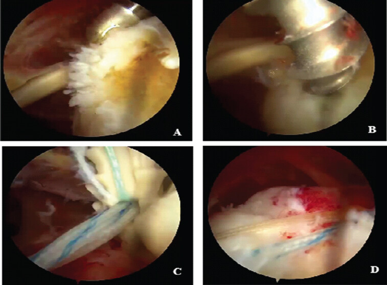
Surgeries were performed in the lateral decubitus position under general anaesthesia and an interscalene block. Following comprehensive diagnostic arthroscopy, soft tissue releases, and subacromial bursectomy, the tuberosity footprint was lightly decorticated [20]. Extensive capsular releases were performed to mobilize the retracted tendon. The SADR technique was then applied. One 5.5 mm double-loaded medial anchor was placed at the articular margin at the midpoint of the mobilized tendon. The suture limbs were passed through the tendon and tied. One limb from each knot was brought laterally, and a single lateral anchor was inserted into the lateral greater tuberosity, capturing and tensioning all bridging limbs to compress the tendon against the footprint (Fig. 1).
Post-operative rehabilitation
An early active rehabilitation protocol was utilized. Patients were provided an abduction sling for comfort but were encouraged to wean off its use as tolerated within the first 3–4 weeks. Immediate passive range of motion (ROM) and gentle active-assisted ROM exercises were initiated within the 1st week postoperatively. Active range of motion (AROM) was introduced early, starting at 3–4 weeks, progressing within pain-free limits. Light isotonic strengthening began at 8 weeks, with a return to heavy labor or overhead activities restricted until 6 months postoperatively.
Clinical evaluation and definition of failure
Patients were evaluated preoperatively and at 6, 12, and 24 months. The primary outcome was the ASES score (0–100). Secondary outcomes included the Constant-Murley score (0–100), Visual Analog Scale (VAS) for pain (0–10), and active forward flexion (FF) and external rotation (ER) in degrees.
Clinical failure was strictly defined as
(1) a post-operative drop in the ASES score of >20 points from the patient’s peak score, or (2) the necessity for revision shoulder arthroplasty due to intractable pain or pseudo-paralysis.
Statistical analysis
Continuous variables were reported as means ± SD. Paired t-tests were used to compare baseline and 24-month scores. Survivorship analysis utilized the Kaplan-Meier method. A multivariate Cox proportional hazards regression model was constructed to identify independent predictors of clinical failure, presenting hazard ratios (HR) and 95% confidence intervals [21]. A multiple linear regression model evaluated predictors of the final ASES score [22]. Significance was set at P < 0.05.
Results
Demographics and baseline characteristics
The final cohort consisted of 37 patients who completed the 24-month follow-up. The cohort included 21 males (56.8%) and 16 females (43.2%), with a mean age of 64.5 ± 6.5 years. The mean anteroposterior tear size was 5.3 ± 0.5 cm. Detailed patient demographics and pre-operative characteristics are summarized in Table 1.
Baseline demographics and clinical outcomes at 24 months (n=37)
| Variable | Baseline/pre-operative value | 24-month follow-up value | P-value (pre vs. 24 m) |
|---|---|---|---|
| Age (years) | 64.5±6.5 | – | – |
| Sex (male/female) | 21 (56.8%)/16 (43.2%) | – | – |
| BMI (kg/m2) | 28.2±4.0 | – | – |
| Dominant Arm involvement | 26 (70.3%) | – | – |
| Pre-operative tear size (AP, cm) | 5.3±0.5 | – | – |
| ASES score | 38.5±10.5 | 82.0±13.5 | <0.001 |
| Constant-Murley score | 35.0±10.0 | 78.0±12.5 | <0.001 |
| VAS pain (0–10) | 7.3±1.2 | 1.2±1.0 | <0.001 |
| Forward flexion (°) | 106±22 | 154±14 | <0.001 |
| External rotation (°) | 26±10 | 44±9 | <0.001 |
BMI: Body mass index, VAS: Visual Analog Scale, ASES: American Shoulder and Elbow Surgeons, AP: Anteroposterior
Longitudinal functional outcomes
Patients demonstrated significant functional improvements from baseline to the 24-month endpoint. The mean ASES score improved from a pre-operative 38.5 ± 10.5 to an excellent 82.0 ± 13.5 at 24 months (P < 0.001). The Constant-Murley score similarly increased from 35.0 ± 10.0 to 78.0 ± 12.5 (P < 0.001). VAS pain scores dropped from 7.3 ± 1.2 to 1.2 ± 1.0 (P < 0.001). AROM also recovered significantly. FF improved from 106° ± 22° to 154° ± 14° postoperatively (P < 0.001). ER increased from 26° ± 10° to 44° ± 9° (P < 0.001). These comprehensive clinical measures are documented in Table 1.
Clinical survivorship and predictors of failure
During the 24-month study period, 4 of the 37 patients (10.8%) met the strict criteria for clinical failure. Two patients required revision to a reverse total shoulder arthroplasty (RTSA), and two experienced a significant functional decline (ASES drop >20 points) without pursuing revision. Consequently, the overall clinical survivorship of the SADR technique at 2 years was 89.2%, as shown in Fig. 2.
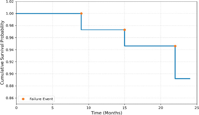
A multivariate Cox proportional hazards regression model identified that increasing patient age (HR = 1.07 per year, P = 0.038) and a higher pre-operative VAS pain score (HR = 1.22 per point, P = 0.025) were independent predictors of clinical failure (Table 2). Pre-operative tear size (HR = 1.10, P = 0.601) and body mass index (BMI) (HR = 1.04, P = 0.485) did not significantly predict clinical failure.
Multivariate and linear regression analyses for clinical failure and ASES scores
| Predictor variable | Hazard ratio (HR) for clinical failure (95% CI) | P-value (failure) | Linear beta (β) for final ASES score | P-value (ASES) |
|---|---|---|---|---|
| Age | 1.07 (1.01–1.14) | 0.038 | −0.14 | 0.510 |
| Sex (male vs. female) | – | – | 5.80 | 0.042 |
| Pre-operative VAS | 1.22 (1.02–1.45) | 0.025 | – | – |
| Initial tear size | 1.10 (0.75–1.50) | 0.601 | – | – |
| BMI | 1.04 (0.90–1.18) | 0.485 | −0.42 | 0.215 |
| Concomitant biceps tenodesis | – | – | 2.05 | 0.480 |
CI: Confidence interval, ASES: Active-assisted range of motion, BMI: Body mass index, VAS: Visual Analog Scale
Finally, a multiple linear regression analysis indicated that male sex was associated with a 5.8-point higher final ASES score (P = 0.042), whereas variables like age (P = 0.510), BMI (P = 0.215), and concomitant biceps tenodesis (P = 0.480) did not significantly predict the final score (Table 2).
Discussion
The primary finding of this retrospective analysis is that arthroscopic repair of MRCTs utilizing a minimalist SADR construct yields profound functional improvements and an 89.2% clinical survivorship rate at 2 years. This success highlights the viability of a bone-preserving, cost-effective alternative to multi-anchor repairs.
The debate over the optimal mechanical construct for MRCTs is ongoing [23]. While laboratory studies consistently favor rigid multi-anchor constructs for their high ultimate load to failure [4,6], translating this mechanical rigidity to clinical biology remains contentious. “Stress-shielding” suggests that overly rigid constructs might eliminate the mechanical micro-motion necessary to stimulate tenocyte proliferation [5,13,16]. Furthermore, inserting four to six anchors damages the tuberosity’s delicate micro-vascularity [11].
Our findings challenge purely mechanical dogmas, demonstrating that the biological environment can be optimized with minimal implants. Through extensive capsular releases and mobilization, our approach aims to eliminate the destructive tension associated with retracting massive tears. Once tension is managed, the SADR construct – one anchor approximating the tissue and one compressing it laterally – achieves “Goldilocks” fixation: Stable enough to prevent macroscopic failure but flexible enough to permit essential osteotendinous micro-motion and prevent anchor-induced ischemia [14].
Economically, the SADR technique holds substantial value [10]. Using strictly two anchors instead of four to six significantly reduces upfront procedural implant costs, an essential consideration in value-based healthcare [17].
Comparing our 89.2% survivorship to traditional multi-anchor cohorts [2] validates the SADR approach. The decision to exclude routine post-operative imaging aligns with historical observations that objective functional outcomes frequently do not correlate with structural MRI integrity. As noted by Denard et al., many patients with asymptomatic retears remain functionally satisfied [8]. Treating an asymptomatic MRI finding often leads to unnecessary revision surgery and patient anxiety [24]. By strictly defining failure through ASES score drops or the clinical need for RTSA, our data reflects genuine patient-centered value. However, we acknowledge our survivorship rate may appear high as benign, asymptomatic structural retears were excluded from the failure definition.
Our regression models identified advanced age and high pre-operative pain as significant predictors of failure, aligning with established literature linking age to diminished tissue regenerative capacity and high pain to chronicity and central sensitization [1,3]. Importantly, initial tear size did not predict failure, validating the mechanical efficacy of the minimalist construct in adequately mobilized tears.
This study has several limitations. It is a retrospective, single-arm study lacking a randomized, parallel control group. While the sample size (n = 37) was adequately powered for clinical outcomes, it remains relatively small for modeling rare complications. Most notably, the lack of post-operative radiologic imaging prevents assessment of exact structural healing or asymptomatic retear rates. Finally, a 24-month follow-up is sufficient to capture the initial functional plateau, but longer-term studies (5–10 years) are necessary to determine the long-term durability of this minimalist construct.
Conclusion
The use of a minimalist SADR mechanical construct is a highly effective evolutionary step in treating MRCTs. This approach preserves vulnerable tuberosity bone stock, substantially reduces implant costs, and prioritizes biological optimization through tension reduction over maximal biomechanical stiffness. The SADR technique yields excellent 2-year clinical survivorship and functional outcomes, offering a patient-Centered, cost-effective alternative that warrants further exploration in prospective, multi-Center trials.
Clinical Message
The management of massive rotator cuff tears can be highly successful without the traditional reliance on densely implanted surgical constructs. By prioritizing extensive soft-tissue mobilization and employing a minimalist, single-anchor DR technique, surgeons can establish a stable repair while actively preserving vulnerable tuberosity bone stock and mitigating implant-induced ischemia. This streamlined approach highlights that optimizing the biological healing environment through thoughtful implant conservation and tension reduction is just as critical as mechanical rigidity, offering a highly practical and cost-efficient strategy for everyday orthopedic practice.
Conflict of Interest:
Nil
Source of Support:
Nil
Consent:
The authors confirm that informed consent was obtained from the patient for publication of this article
References
- Guadagno K, Srikumaran U, Huish E. G Jr, Best M. J. (2023). Massive rotator cuff tears: algorithmic approach to surgical treatment. Annals of Joint, 8, 38-38. [Google Scholar] | [PubMed]
- Collin P, Betz M. A, Walch G, Mansat P, Favard L, Colmar M, François Kempf J, Thomazeau H, Gerber C. (2020). Clinical and structural outcome 20 years after repair of massive rotator cuff tears. Journal of Shoulder and Elbow Surgery, 29(3), 521-526. [Google Scholar] | [PubMed]
- Lo IK, Burkhart SS. Current concepts in arthroscopic rotator cuff repair. Am J Sports Med 2004;32:1084-9. [Google Scholar] | [PubMed]
- Meier SW, Meier JD. The effect of double-row fixation on initial repair strength in rotator cuff repair: A biomechanical study. Am J Sports Med 2006;34:1245-51. [Google Scholar] | [PubMed]
- Millett PJ, Warth RJ, Dornan GJ, Lee JT, Spiegl UJ. Clinical and structural outcomes after arthroscopic single-row versus double-row rotator cuff repair: A systematic review and meta-analysis of level I randomized clinical trials. J Shoulder Elbow Surg 2014;23:586-97. [Google Scholar] | [PubMed]
- Sugaya H, Maeda K, Matsuki K, Moriishi J. Repair integrity and functional outcome after arthroscopic double-row rotator cuff repair. A prospective outcome study. J Bone Joint Surg Am 2007;89:953-60. [Google Scholar] | [PubMed]
- Kim DH, Elattrache NS, Tibone JE, Jun BJ, DeLaMora SN, Kvitne RS. Biomechanical comparison of a single-row versus double-row suture anchor technique for rotator cuff repair. Am J Sports Med 2006;34:407-14. [Google Scholar] | [PubMed]
- Denard PJ, Jiwani AZ, Lädermann A, Burkhart SS. Long-term outcome of arthroscopic massive rotator cuff repair: The importance of double-row fixation. Arthroscopy 2012;28:909-15. [Google Scholar] | [PubMed]
- Marigi E. M, Kennon J. C, Dholakia R, Visscher S. L, Borah B. J, Sanchez-Sotelo J, Sperling J. W. (2021). Cost analysis and complication rate comparing open, mini-open, and all arthroscopic rotator cuff repair. JSES Reviews, Reports, and Techniques, 1, 84-89. [Google Scholar] | [PubMed]
- Mather RC, MacDermid JC, Papadonikolakis A, O'Toole RV. Societal and economic value of rotator cuff repair. J Bone Joint Surg Am 2013;95:1993-2000. [Google Scholar] | [PubMed]
- Erhart S, Schmoelz W, Blauth M. Biomechanical effect of bone defect size and anchor location on pullout strength in rotator cuff repair. Knee Surg Sports Traumatol Arthroscopy 2012;20:346-51. [Google Scholar] | [PubMed]
- Bedi A, Fox AJ, Kovacevic D, Deng XH, Warren RF, Rodeo SA. Doxycycline-mediated inhibition of matrix metalloproteinases improves healing after rotator cuff repair. Am J Sports Med 2010;38:308-17. [Google Scholar] | [PubMed]
- Cho NS, Lee BG, Rhee YG. Arthroscopic rotator cuff repair using a suture bridge technique: Is the repair integrity actually maintained? Am J Sports Med 2011;39:2108-16. [Google Scholar] | [PubMed]
- Boileau P, Brassart N, Watkinson DJ, Carles M, Hatzidakis AM, Krishnan SG. Arthroscopic repair of full-thickness tears of the supraspinatus: Does the tendon really heal? J Bone Joint Surg Am 2005;87:1229-40. [Google Scholar] | [PubMed]
- Park JY, Lhee SH, Choi JH, Park HK, Yu JW, Seo JB. Comparison of the clinical outcomes of single- and double-row repairs in rotator cuff tears. Am J Sports Med 2008;36:1310-6. [Google Scholar] | [PubMed]
- McElvany MD, McGoldrick E, Gee AO, Neradilek MB, Matsen FA. Rotator cuff repair: Published evidence on factors for success and structural integrity. J Bone Joint Surg Am 2015;97:163-72. [Google Scholar] | [PubMed]
- Wise K. L, Parikh H. R, Okelana B, Only A. J, Reams M, Harrison A, Braman J, Craig E, Cunningham B. P. (2022). Measurement of value in rotator cuff repair: patient-level value analysis for the 1-year episode of care. Journal of Shoulder and Elbow Surgery 2022;31:72-80. [Google Scholar] | [PubMed]
- Faul F, Erdfelder E, Lang AG, Buchner A. G*Power 3: A flexible statistical power analysis program for the social, behavioral, and biomedical sciences. Behav Res Methods 2007;39:175-91. [Google Scholar] | [PubMed]
- Tashjian RZ, DeLoach J, Porucznik CA, Powell AP. Minimal clinically important differences (MCID) and patient acceptable symptomatic state (PASS) for Visual Analog Scales (VAS) measuring pain in patients treated for rotator cuff disease. J Shoulder Elbow Surg 2009;18:927-32. [Google Scholar] | [PubMed]
- Galatz LM, Ball CM, Teefey SA, Middleton WD, Yamaguchi K. The outcome and repair integrity of completely arthroscopically repaired large and massive rotator cuff tears. J Bone Joint Surg Am 2004;86:219-24. [Google Scholar] | [PubMed]
- Harrell FE, Lee KL, Mark DB. Multivariable prognostic models: Issues in developing models, evaluating assumptions and adequacy, and measuring and reducing errors. Stat Med 1996;15:361-87. [Google Scholar] | [PubMed]
- Rabe-Hesketh S, Skrondal A. Multilevel and Longitudinal Modeling using Stata. 3rd ed. United States: Stata Press; 2012. [Google Scholar] | [PubMed]
- DeOrio JK, Cofield RH. Results of a second attempt at surgical repair of a failed initial rotator-cuff repair. J Bone Joint Surg Am 1984;66:563-7. [Google Scholar] | [PubMed]
- Chalmers PN, Ross H, Granger E, Presson AP, Zhang C, Tashjian RZ. Do arthroscopic clinical outcomes correlate with structural integrity? A systematic review and meta-analysis. J Shoulder Elbow Surg 2018;27:564-73. [Google Scholar] | [PubMed]
© 2026 Journal of Orthopaedic Case Reports - Published by Indian Orthopaedic Research Group
About the Authors
-

Dr. Amrith K Nath
-

Dr. Sandeep Vellarakkat
-

Dr. Jipin Gopi
-

Dr. Adarsh Rajan Sujatha
-

Dr. Abdulla Harafan
-

Dr. Shisham Hashim Roshan
Related Articles in Journal of Orthopaedic Case Reports
 August 1, 2026 Reverse Shoulder Arthroplasty with Virtual Implant Positioning System in the Management of Massive Rotator Cuff Tear with Rotator Cuff Arthropathy
 May 1, 2026 A Novel Failure Interface in Constrained Total Hip Arthroplasty: Dissociation at the Constrained Liner Insert-Bipolar Head Junction Following Periprosthetic Joint Infection
 May 1, 2025 Synovial Hemangioma of Knee in 4-Year-Old Girl Child – A Case Report
 November 1, 2024 Multifocal Epitheloid Hemangioma of the Bone – A Rare Entity