


Introduction
Clubfoot is one of the most common congenital foot deformities, affecting approximately 1.2/1,000 live births [1]. Since its introduction, the Ponseti method has revolutionised the management of idiopathic clubfoot, achieving reported success rates of 93–100% and markedly reducing the need for extensive surgical release [2,3,4]. This success has led to its increasing application in selected cases of non-idiopathic clubfoot, including those associated with arthrogryposis multiplex congenital (AMC), myelomeningocele (MMC), spina bifida, and syndromic disorders [5,6,7]. Historically, non-idiopathic clubfoot was managed primarily with extensive soft-tissue release, often resulting in residual deformity, stiffness, pain, functional limitation, and high recurrence rates [7,8]. Although modified Ponseti techniques have demonstrated encouraging outcomes in these complex deformities [6,7,8], variations in treatment protocols and the predominance of diagnosis-specific studies limit meaningful comparisons across heterogeneous non-idiopathic populations. These heterogeneous cohort subgroups (AMC, Spina bifida, and syndromic cohort) introduce clinical variability, but we had addressed this partially through the rigid versus flail sub-classification of non-idiopathic clubfoot based on soft-tissue phenotype in the literature.
Robust evidence evaluating the standard Ponseti protocol in non-idiopathic clubfoot remains limited. Therefore, this study aimed to compare the clinical outcomes of the standard Ponseti method in idiopathic and non-idiopathic clubfoot over a 4-year follow-up period, with minimally invasive adjunct procedures reserved exclusively for recurrent or residual cases.
Materials and Methods
This was a retrospective cohort study that used data from an institutional clubfoot registry that was kept up to date. Patients were enrolled consecutively and categorised according to etiology into idiopathic (Group I) and non-idiopathic (Group II) clubfoot. Randomization was not possible as the treatment allocation for both groups was determined by the underlying etiology and not by the investigator’s choice.[1.1][d1.2]
The results were compared between the two groups over a 4-year follow-up period.
The required sample size was calculated as 91 clubfeet based on a clubfoot incidence of 1/1,000 live births, with a 95% confidence interval (CI), and a 6.5% margin of error. Eighty patients were initially enrolled; five (6.25%) were excluded because of loss to follow-up, leaving a final cohort of 75 patients (105 clubfeet). Group I comprised 45 patients (63 clubfeet) with idiopathic clubfoot, whereas Group II comprised 30 patients (42 clubfeet) with non-idiopathic clubfoot. The total sample size (105 feet) was sufficient to meet the predetermined sample size (91 feet) with adequate post hoc power (>99% for the primary outcome). However, the individual non-idiopathic subgroups (21 Rigid vs. 21 Flail) remained relatively small and are acknowledged as a limitation for subgroup-level analyses only, separate from the adequately powered overall comparison. This study received Institutional ethics committee approval on January 28th, 2022.
Demographic variables including age at presentation, sex, and laterality were recorded. All children underwent evaluation by an orthopedic surgeon. Patients with postural clubfoot, metatarsus adductus, calcaneo-valgus foot, atypical clubfoot, or post-traumatic equinus deformity were excluded from the study. Children with suspected syndromic associations underwent multidisciplinary assessment, including pediatricians, neurologists, cardiologists, and genetic evaluation where indicated. Following clinical assessment, Pirani scoring was performed at presentation and every follow-up visit.
The Ponseti method was employed for manipulation and casting. During the first cast, the first metatarsal was elevated by supinating the forefoot to align it with the hindfoot, thereby correcting cavus deformity. In subsequent casts, pure abduction of the foot to 60–70° was achieved with counter-pressure applied over the head of the talus, as this manoeuvre alone results in complete correction of the subtalar joint and adequate stretching of the medial soft tissues. Each above-knee cast was changed on a weekly basis, and careful attention was paid to identify any cast slippage at subsequent visits. According to Ponseti’s recommendations, 60° of forefoot abduction and ≤15° of dorsiflexion were considered the threshold before performing tendo-Achilles tenotomy. This permits the calcaneus to swing completely beneath the talus, thereby correcting subtalar malalignment. Attempts to correct equinus before addressing heel varus and forefoot supination were avoided, as this may lead to rocker-bottom deformity. Particular care was taken to ensure appropriate alignment of the forefoot and midfoot before correcting hindfoot equinus.
Adequate coverage was confirmed by the absence of palpable talar head laterally after the cast, indicating adequate forefoot abduction. When the midfoot Pirani score dropped below 1 while the hindfoot score remained >1, tendo-Achilles tenotomy was indicated. Tenotomy was not performed when the hindfoot score was ≤1 either at presentation or following initial correction, provided dorsiflexion of up to 15–20° was achievable. All tenotomies were performed in the outpatient clinic under local anesthesia.
Following tenotomy, above-knee casts were applied in the maximum achievable corrected position and maintained for 3 weeks. After cast removal, Pirani scores were reassessed. Children with satisfactory correction received full-time bracing (23 h/day) for the initial 3 months, followed by night-time and nap-time bracing. Because bracing maintains rather than achieves correction, adequate correction was confirmed before brace initiation. During the first 2 weeks of FAO or AFO application, patients were monitored for midfoot instability, rocker-bottom deformity, heel slippage, loss of hindfoot correction, and skin-related complications such as blisters. AFO usage strictly related to the rigid variety was excessive when dorsiflexion was not needed; rather, co-contracture also required addressing.
Patients were reviewed monthly for 3 months, every 3 months during the 1st year, and every 6 months thereafter. At each follow-up visit, Pirani scores were assessed, and early heel rise during squatting, brace wear issues, and skin complications were meticulously evaluated. Recurrence was defined as a Pirani score >1 at any stage following final casting or during subsequent follow-up, attributable to excessive deformity components such as equinus, cavus, or heel varus, resulting in a midfoot or hindfoot score >1 and not amenable to casting. Recurrent equinus was treated with repeat casting and re-tenotomy, whereas recurrent heel varus or dynamic supination was managed by tibialis anterior transfer. No patient underwent postero-medial release, and no bony procedures.
All follow-up Pirani assessments were performed by the same orthopedic surgeon, reducing inter-observer variability, although inter-observer agreement was not formally evaluated using kappa statistics. Brace compliance was based on subjective parent-reported adherence recorded in the institutional clubfoot registry throughout the 4-year follow-up.
The primary outcome was successful correction, defined as a Pirani score ≤0.5 at completion of the casting phase (3 months). Secondary outcomes included number of casts, tenotomy rate, recurrence during 4 years, requirement for adjunct procedures, and subjective parent-reported brace compliance. The primary treatment objective was to achieve a stable, plantigrade, painless, and braceable foot after 4 years of follow-up.
Statistics
Data were entered into Microsoft Excel (Windows 10; Version 2021), and all statistical analyses were performed using R (version 4.4.0; R Foundation for Statistical Computing, Vienna, Austria) with the gtsummary , rstatix , and ggplot2 packages. Descriptive statistics, including median and interquartile range for continuous variables, and frequencies with percentages for categorical variables, were computed for each study group. The foot was used as the unit of analysis for casting-related outcomes (n = 105 feet: Group I = 63, Group II = 42), while the patient was the unit for brace compliance and surgical outcomes (n = 75 patients).
Between-group comparisons of continuous and ordinal variables (including Pirani score at each timepoint and number of casts) were performed using the Mann–Whitney U test (Wilcoxon rank-sum test), as these variables did not conform to a normal distribution. Associations between categorical variables were assessed using the Chi-square test. Spearman’s rank correlation (ρ) was used to assess the relationship between initial Pirani score and number of casts required, reported with 95% confidence intervals. A multivariable linear regression model was constructed with number of casts as the dependent variable; independent predictors included Pirani score at presentation, age at presentation, foot type (Idiopathic/Rigid/Flail), and virgin versus recurrent/residual status. Model fit was assessed using R2 and adjusted R2, with variance inflation factors examined to rule out multicollinearity. A post hoc power analysis was performed using observed effect sizes to confirm adequacy of the achieved sample following 17.6% attrition. The level of statistical significance was set at P < 0.05 (two-tailed) for all analyses.
Results
The median age at presentation in Group I (Idiopathic clubfoot) was 2.00 (0.8–4.0) months, and 5.5 (3.5–8.5) months in Group II (Non-idiopathic clubfoot). Two-thirds of patients in Group I (66.7%) presented before 3 months of age, whereas patients in Group II presented later, as illustrated in Fig. 1. Demographic characteristics, including age at presentation, sex, laterality, recurrence and subjective parent-reported brace compliance, are summarized in Table 1.
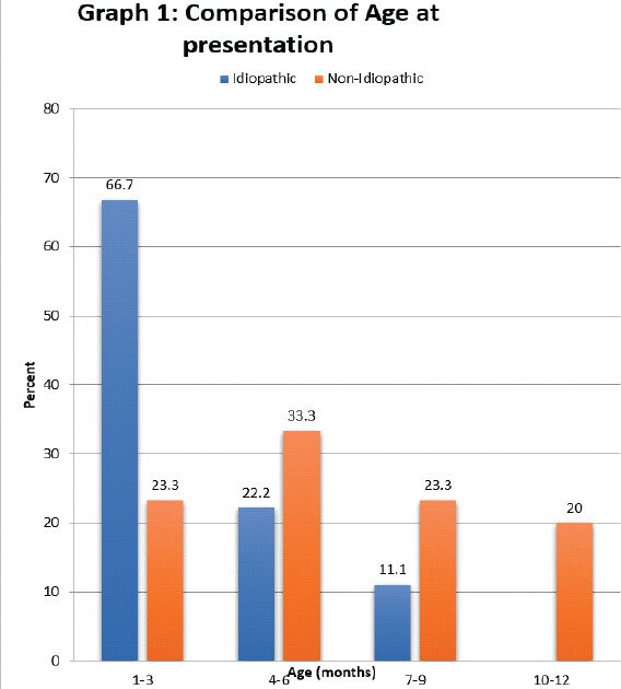
2/3rd of Idiopathic group presented within the first 3 month, whereas nearly half(43.3%) of the Non idiopathic ones presented later after 6 month.
Demographic details and comparison of various outcomes between Group(I) & Group(II), Total N= 75 Patients.
| Characteristic | Group I (Idiopathic) | Group II (Non-idiopathic) | Difference2 | Statistic2 | p-value2 |
|---|---|---|---|---|---|
| N = 451 | N = 301 | ||||
| AGE(MONTHS) | 2.00, 0.80 – 4.00 | 5.50, 3.50 – 8.50 | -3.000081 | 296 | <0.001 |
| SEX | 0 | >0.9 | |||
| F | 13 (29%) | 8 (27%) | |||
| M | 32 (71%) | 22 (73%) | |||
| UNI/ BIL | 0.682 | 0.7 | |||
| BIL | 18 (40%) | 12 (40%) | |||
| LT | 6 (13%) | 6 (20%) | |||
| RT | 21 (47%) | 12 (40%) | |||
| recurrence | 2.31 | 0.13 | |||
| No | 38 (84%) | 20 (67%) | |||
| Yes | 7 (16%) | 10 (33%) | |||
| BRACE COMPLIANCE | 5.92 | 0.015 | |||
| NO | 8 (18%) | 14 (47%) | |||
| YES | 37 (82%) | 16 (53%) |
1
Median, Q1 – Q3; n (%)
2
Wilcoxon rank sum test; Pearson’s Chi-squared test
Although the baseline Pirani score at presentation was comparable between groups (Table 2, P = 0.14), alleviating concern for gross baseline imbalance in deformity severity. The non-idiopathic group required a significantly greater number of casts (median 8 [6.0–12.0] per clubfeet of which median of 10 in the rigid type versus 6 in the flail type, as shown in Table 3). 40% (12/30) of patients in Group II required ≥10 casts, of whom approximately 83.3% (10/12) were non-compliant with bracing. Group I required a (median 5.0 [5.0–6.0] casts) per clubfoot; 30% of patients (15 patients) required more than the median number of casts. Among these 15 patients, 5 were non-compliant with bracing and subsequently required additional procedures due to recurrence.
Comparison of Various independent variables & Pirani score in both the groups (N=105 clubfeets)
| Characteristic | Overall | Idiopathic | Non-Idiopathic | Statistic2 | p-value2 |
|---|---|---|---|---|---|
| N = 1051 | N = 631 | N = 421 | |||
| PIRANI SCORE AT PRESENTATION | 5.00 (4.00 – 6.00) | 5.00 (4.00 – 6.00) | 5.00 (4.00 – 5.50) | 1,544 | 0.14 |
| 6WKS PIRANI SCORE | 0.50 (0.50 – 1.00) | 0.50 (0.50 – 1.00) | 1.25 (0.50 – 2.00) | 615 | <0.001 |
| 3MONTH | 0.50 (0.00 – 0.50) | 0.00 (0.00 – 0.50) | 0.50 (0.50 – 1.00) | 542 | <0.001 |
| 1YR | 0.50 (0.00 – 0.50) | 0.00 (0.00 – 0.50) | 0.50 (0.50 – 1.00) | 518 | <0.001 |
| 2-3YR | 0.00 (0.00 – 0.50) | 0.00 (0.00 – 0.00) | 0.50 (0.00 – 0.50) | 762 | <0.001 |
| 4YR | 0.00 (0.00 – 0.00) | 0.00 (0.00 – 0.00) | 0.00 (0.00 – 0.50) | 836 | <0.001 |
| NO OF CASTS | 6.00 (5.00 – 7.00) | 5.00 (5.00 – 6.00) | 8.00 (6.00 – 12.00) | 517 | <0.001 |
| TENOTOMY | 6.47 | 0.039 | |||
| 0 | 23 (22%) | 13 (21%) | 10 (24%) | ||
| 1 | 73 (70%) | 48 (76%) | 25 (60%) | ||
| 2 | 9 (8.6%) | 2 (3.2%) | 7 (17%) | ||
| extra surg | 8.20 | 0.042 | |||
| Both(Cav rel + tib ant) | 4 (3.8%) | 2 (3.2%) | 2 (4.8%) | ||
| Cavus release | 5 (4.8%) | 0 (0%) | 5 (12%) | ||
| No | 91 (87%) | 58 (92%) | 33 (79%) | ||
| Tibial Anterior | 5 (4.8%) | 3 (4.8%) | 2 (4.8%) |
1
Median (Q1 – Q3); n (%)
2
Wilcoxon rank sum test; Pearson’s Chi-squared test
Intra-subgroup comparison in Group II between Rigid type and Flail type clubfoot patients
| Characteristic | Overall | Rigid | Flail | Statistic2 | p-value2 |
|---|---|---|---|---|---|
| N = 421 | N = 211 | N = 211 | |||
| PIRANI SCORE AT PRESENTATION | 5.00 (4.00 – 5.50) | 5.00 (4.50 – 6.00) | 4.50 (3.50 – 5.00) | 322 | 0.010 |
| 6WKS PIRANI SCORE | 1.25 (0.50 – 2.00) | 1.50 (1.00 – 2.00) | 1.00 (0.50 – 1.50) | 290 | 0.080 |
| 3MONTH | 0.50 (0.50 – 1.00) | 1.00 (0.50 – 1.50) | 0.50 (0.50 – 0.50) | 323 | 0.008 |
| 1YR | 0.50 (0.50 – 1.00) | 0.50 (0.50 – 1.50) | 0.50 (0.50 – 0.50) | 260 | 0.3 |
| 2-3YR | 0.50 (0.00 – 0.50) | 0.50 (0.00 – 1.00) | 0.00 (0.00 – 0.50) | 279 | 0.12 |
| 4YR | 0.00 (0.00 – 0.50) | 0.50 (0.00 – 0.50) | 0.00 (0.00 – 0.50) | 309 | 0.013 |
| NO OF CASTS | 8.0 (6.0 – 12.0) | 10.0 (8.0 – 13.0) | 6.0 (5.0 – 7.0) | 378 | <0.001 |
| TENOTOMY | 5.25 | 0.073 | |||
| 0 | 10 (24%) | 2 (9.5%) | 8 (38%) | ||
| 1 | 25 (60%) | 14 (67%) | 11 (52%) | ||
| 2 | 7 (17%) | 5 (24%) | 2 (9.5%) | ||
| BRACE COMPLIANCE | 22 (52%) | 8 (38%) | 14 (67%) | 2.39 | 0.12 |
| extra surg | 9.76 | 0.021 | |||
| Both | 2 (4.8%) | 2 (9.5%) | 0 (0%) | ||
| Cavus release | 5 (12%) | 5 (24%) | 0 (0%) | ||
| No | 33 (79%) | 14 (67%) | 19 (90%) | ||
| Tibial Anterior | 2 (4.8%) | 0 (0%) | 2 (9.5%) |
1
Median (Q1 – Q3); n (%)
2
Wilcoxon rank sum test; Pearson’s Chi-squared test
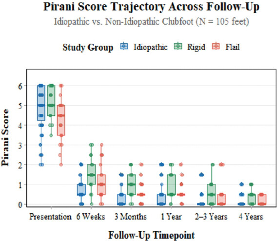
Table 2 demonstrates a statistically significant improvement in Pirani scores at 3 months, 1 year, and 4 years in both groups (P < 0.001*, 0.001*, and 0.001*, respectively). Fig. 2 illustrates a Box-whisker plot with dispersion of Pirani score correction in both groups, including various subgroups within Group II. Other demographic parameters such as sex and laterality did not differ significantly. The cumulative tenotomy rate was comparable (75.4% in Group I versus 73.3% in Group II). Notably, 8 patients (26.4% of Group II) in the Flail type sub-group did not require tenotomy, reflecting the greater flexibility observed in subgroups such as flail MMC, Down syndrome, Larsen syndrome and other syndromic feet.
Although there was no significant difference in brace compliance between groups, the recurrence was significantly lower in Group I than in Group II (5/63 clubfeet [7.9%] vs. 9/42 clubfeet [21.4%] P = 0.042* as shown in Table 2). Among the 14 recurrent clubfeet, 5 clubfeet in Group II required cavus release only. Tibialis anterior transfer was performed in 3 clubfeet in Groups I and 2 clubfeet in Group II, while the remaining 4 clubfeet required both procedures. Subgroup analysis in Table 3 showed that adjunct surgery was required more frequently in the rigid subgroup than in the Flail subgroup (7/21 [33.3%] vs. 2/21 [9.5%], P = 0.021*).
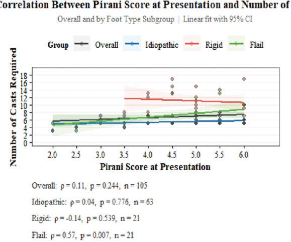
Spearman’s correlation demonstrated a significant association between the initial Pirani score and number of casts only in the flail subgroup (ρ = 0.57, P = 0.007), indicating that casting burden in rigid non-idiopathic feet is influenced by foot type rather than initial deformity severity, as shown in Fig. 3. At 4-year follow-up, final Pirani scores ≤0.5 were achieved in approximately 88.37% in recurred clubfeet Group I and 90.62% in Group II.
Multivariable linear regression (Table 4) identified four independent predictors of number of casts required (R2 = 0.707, F = 47.7, P < 0.001): Higher Pirani score at presentation (β = 0.87, 95% CI: 0.53–1.21, P < 0.001), older age at presentation (β = 0.43, 95% CI: 0.29–0.58, P < 0.001), rigid foot type compared to idiopathic (β = 3.93, 95% CI: 3.02–4.84, P < 0.001), and recurrent presentation (β = 1.41, 95% CI: 0.46–2.35, P = 0.004). In contrast, the Flail subtype did not differ significantly from idiopathic clubfeet in terms of casting burden (β = −0.45, P =0.392).
Multivariable linear regression analysis with the number of casts as dependent variable and other as independent variables.
| Predictor | β Coefficient | 95% CI | p-value |
|---|---|---|---|
| Pirani Score at Presentation | 0.401 | 0.092, 0.710 | 0.011 |
| Age at Presentation (months) | 0.291 | 0.163, 0.419 | <0.001 |
| Foot Type | |||
| Idiopathic | — | — | |
| Rigid | 3.80 | 3.05, 4.55 | <0.001 |
| Flail | 0.228 | -0.641, 1.10 | 0.604 |
| recurrence | |||
| No | — | — | |
| Yes | 1.13 | 0.354, 1.91 | 0.005 |
| TENOTOMY | 2.02 | 1.44, 2.60 | <0.001 |
Abbreviation: CI = Confidence Interval
R² = 0.803; Adjusted R² = 0.791; Residual SE = 1.35; F-statistic = 66.7; Model p-value = <0.001; N (feet) = 105
Post hoc power analysis confirmed adequate statistical power despite the 6.25% attrition. The primary outcome (number of casts; Cohen’s d = 1.34) achieved 99.98% power, while Pirani score at 4 years (Cohen’s d = 0.74) and brace compliance (Cohen’s h = 0.63) achieved powers of 87.40% and 76.69%, respectively.
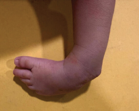
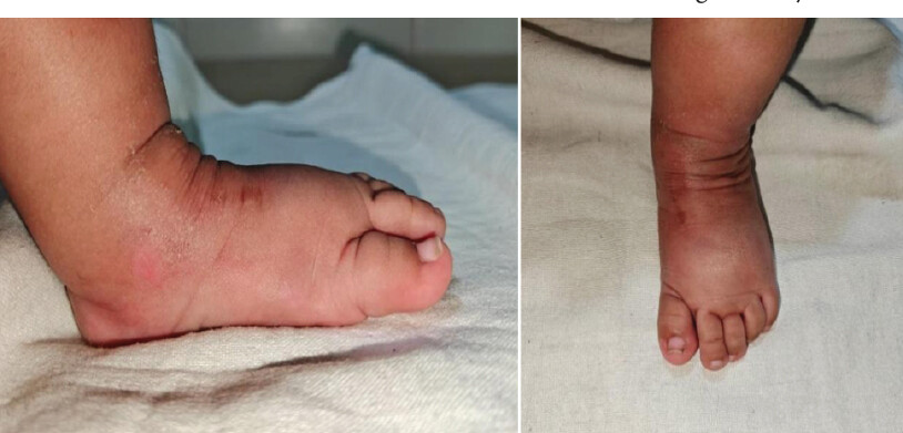
Fig. 4, 5, 6, 7, 8 demonstrate representative outcomes in a child with MMC and bilateral clubfoot, whereas Fig. 9 and 10 illustrate correction of idiopathic clubfoot from initiation till completion of Ponseti treatment.
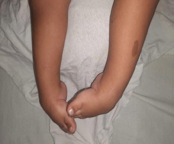
Left foot had Pirani score 4.5 and Right foot with score of 4 as depicted.
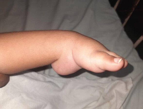
Child having Midfoot score of 2 which included medial crease score 1, curved lateral border 0.5, talar head coverage 0.5, and the Hindfoot score were 2.5 of which Equinus were 1, posterior crease 1 and empty heel 0.5.
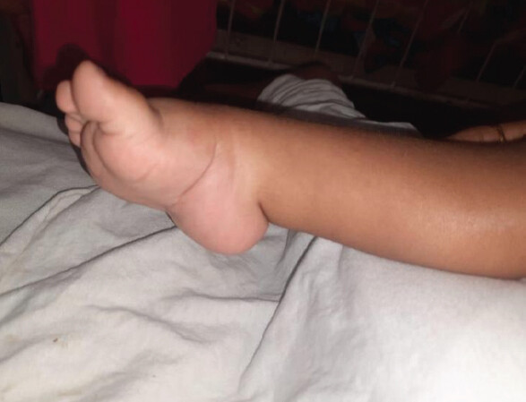
Child having a Midfoot score of 2 which included the Talar head coverage score 1, medial crease 0.5, curved lateral border 0.5 whereas the Hindfoot score were 2 in which equinus score was 1, posterior crease 0.5 and empty heel 0.5.
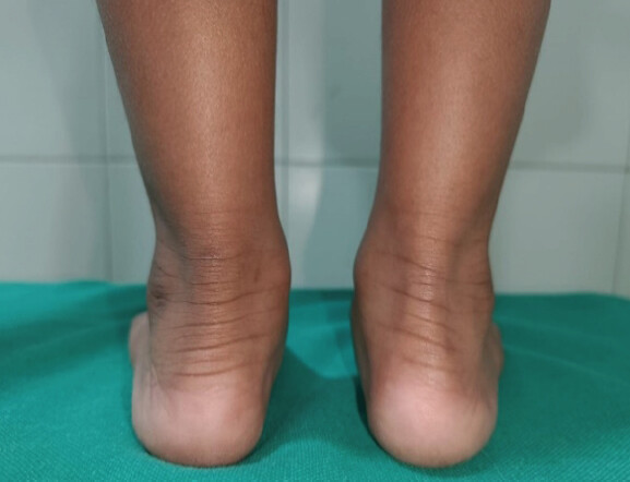
This patient did require an AFO (Ankle Foot Orthoses) for a longer period due to weakness of dorsiflexors in the foot.
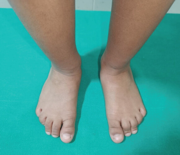
No Forefoot deformity and foot length compromise at the completion of treatment in the same MMC Child.
Discussion
The Ponseti method has consistently achieved excellent outcomes in idiopathic clubfoot, with reported success rates of 93–100% [2,3,4]. Historically, non-idiopathic clubfoot was managed primarily with extensive soft-tissue release, frequently resulting in residual deformity, stiffness, pain, functional limitation, and high recurrence rates [9,10,11,12,13,14]. Although favorable outcomes have recently been reported with the Ponseti method in non-idiopathic clubfoot, most studies have evaluated isolated etiologies or modified Ponseti protocols [5,6,7]. In contrast, the present study compared idiopathic and heterogeneous non-idiopathic clubfoot using the standard Ponseti protocol, reserving minimally invasive adjunct procedures exclusively for recurrent or relapsed cases.
The poor long-term outcomes associated with extensive soft-tissue release and bony procedures have led many authors to advocate Ponseti treatment even for complex clubfoot deformities [8,15,16,17,19,20,21,22]. Our demographic findings, including male predominance and the pattern of bilateral and right-sided involvement, are consistent with previous reports by Lochmiller et al. [23], Chung et al. [24], and others.
Ponseti [25,26] reported an average requirement of approximately 7.6 casts per foot. Consistent with previous literature, idiopathic clubfeet in our series required significantly fewer casts than non-idiopathic feet. Recurrence (11.1%) in Group I occurred only in children presenting after 6 months of age. Nearly one-quarter of idiopathic feet did not require tenotomy, and none of these patients’ developed recurrence, emphasising the importance of early presentation, meticulous casting technique, and strict brace compliance. Initial Pirani score correction at 3 months was achieved in 93.72% of idiopathic cases[27–29].
As expected, non-idiopathic clubfeet required significantly more casts, reflecting greater deformity rigidity. Despite this increased casting burden, tenotomy rates were comparable between groups, while recurrence remained within the range reported in previous studies [8,15,16,17,18,19,20,21,22]. Significant improvement in Pirani scores was observed throughout follow-up, with a final correction rate of 95.41% in non-idiopathic clubfoot, comparing favorably with published literature [15,30,31].
Janicki et al. [15] and Shah et al. [31] reported fewer casts but lower correction rates and higher recurrence than observed in our cohort. These findings suggest that a more persistent casting strategy, combined with strict brace adherence, may improve correction while reducing recurrence. Recurrent deformities in our series responded satisfactorily to repeat casting and minimally invasive adjunct procedures, avoiding extensive surgical release.
Final Pirani scores among recurrent feet were not uniformly zero, consistent with Khan et al. [32], who demonstrated that a clinically corrected clubfoot does not necessarily achieve a Pirani score of zero. This finding is particularly relevant because most recurrent feet in our study belonged to the heterogeneous non-idiopathic cohort.
Morcuende et al. [5] reported satisfactory correction of arthrogrypotic clubfeet using the Ponseti method, while Boehm et al. [6] demonstrated successful management of recurrence with repeat casting. Our findings support these observations. Based on our experience, rigid non-idiopathic clubfeet may require a realistic target of 30–40° forefoot abduction and 5–10° dorsiflexion before brace application. In patients with severe associated joint contractures, particularly those with AMC, ankle-foot orthoses may provide better maintenance than standard foot abduction orthoses.
Mehta and Gopinathan [33] proposed classification of non-idiopathic clubfoot into rigid and flail phenotypes, which was supported by our findings. Flexible deformities, including flail MMC, Down syndrome, Larsen syndrome, and other syndromic feet, required fewer casts, whereas rigid phenotypes required prolonged casting. The significant correlation between initial Pirani score and casting requirement observed only in the flail subgroup suggests that deformity severity predicts casting burden primarily in flexible feet, while rigidity itself determines treatment duration in rigid deformities. Nevertheless, variability in casting requirements remains inevitable, as highlighted by De Mulder et al. [22], and surgical intervention should be considered when further casting is unlikely to improve correction.
The heterogeneous non-idiopathic cohort (arthrogryposis, spina bifida, MMC, and syndromic clubfoot) introduces unavoidable clinical variability. We addressed this partially by analyzing the predefined rigid and flail subgroups based on the soft-tissue phenotype described by Mehta and Gopinathan [33]. Analysis at the level of the individual diagnostic subgroups (e.g. AMC alone, Spina bifida alone) were underpowered; therefore, diagnosis-specific conclusions cannot be drawn.
Brace compliance was assessed using serial Pirani scores during maintenance together with subjective parent-reported adherence. Overall compliance at 4 years was 73.3%, including 87.3% in Group I and 52.4% in Group II (P < 0.015), which compares favorably with the findings of Funk et al. [30]. However, these results should be interpreted cautiously because compliance was assessed subjectively, the sample size was modest, and the non-idiopathic cohort was heterogeneous. Objective monitoring using sensor-based brace compliance systems would provide more reliable assessment in future studies.
Limitation of study
This study has several limitations. Although data were derived from a prospectively maintained institutional clubfoot registry with predefined variables, its retrospective design cannot eliminate unmeasured confounding. As a single-center study, the findings may not be generalisable to institutions with different casting protocols, patient demographics, socioeconomic settings, brace-compliance practices, or surgical thresholds.
The heterogeneous non-idiopathic cohort and the relatively small size of individual diagnostic subgroups limited diagnosis-specific analyses. Consequently, the results should not be extrapolated to neglected or severely rigid clubfeet presenting late, particularly those with AMC or spina bifida, who may occasionally require extensive soft-tissue release or differential distraction techniques such as the JESS system. No such patients were encountered in the present series.
Brace compliance was assessed using subjective parent-reported adherence rather than objective monitoring, introducing the possibility of reporting bias. Furthermore, socioeconomic status, caregiver education, and access to healthcare or rehabilitation services, all recognised determinants of Ponseti success, were not systematically evaluated and should be incorporated into future prospective multicentre studies.
Conclusion
The Ponseti method is an effective and reliable treatment for both idiopathic and non-idiopathic clubfoot, providing excellent correction while substantially reducing the need for extensive soft-tissue release. Early initiation of treatment and meticulous adherence to the Ponseti protocol remain the key determinants of successful outcomes. In recurrent cases, timely minimally invasive procedures, including repeat tendo-Achilles tenotomy, cavus release, and tibialis anterior transfer, can effectively restore correction and minimise the need for major reconstructive surgery.
Long-term follow-up into adolescence is required to evaluate the durability of correction, functional outcomes, and late recurrence. Future studies should complement the Pirani score with validated functional outcome measures, such as the Ezra score and the Oxford foot and ankle questionnaire for children, to provide a more comprehensive assessment of gait, function, and quality of life. Although contemporary Ponseti guidelines emphasise clinical assessment over routine radiography, selective imaging remains valuable in resistant, residual, and syndromic clubfoot to supplement clinical evaluation.
Clinical Message
Meticulous Ponseti protocol fidelity and expeditious therapeutic initiation are the quintessential determinants of durable correction. Judiciously timed minimally invasive salvage interventions effectively mitigate recurrence, preserve musculoskeletal integrity, and circumvent extensive future reconstructive surgery.
Conflict of Interest:
Nil
Source of Support:
Nil
Consent:
The authors confirm that informed consent was obtained from the patient for publication of this article
References
- Smythe T, Kuper H, Macleod D, Foster A, Lavy C. Birth prevalence of congenital Talipes Equinovarus in low- and middle-income countries: A systematic review and meta-analysis. Trop Med Int Health 2017;22:269-85. [Google Scholar] | [PubMed]
- Ponseti IV. Congenital Clubfoot: Fundamentals of Treatment. Oxford: Oxford University Press; 1996. [Google Scholar] | [PubMed]
- Morcuende JA, Dolan LA, Dietz FR, Ponseti IV. Radical reduction in the rate of extensive corrective surgery for clubfoot using the Ponseti method. Pediatrics 2004;113:376-80. [Google Scholar] | [PubMed]
- Radler C. The Ponseti method for the treatment of congenital club foot: Review of the current literature and treatment recommendations. Int Orthop 2013;37:1747-53. [Google Scholar] | [PubMed]
- Morcuende JA, Dobbs MB, Frick SL. Results of the Ponseti method in patients with clubfoot associated with arthrogryposis. Iowa Orthop J 2008;28:22-6. [Google Scholar] | [PubMed]
- Boehm S, Limpaphayom N, Alaee F, Sinclair MF, Dobbs MB. Early results of the Ponseti method for the treatment of clubfoot in distal arthrogryposis. J Bone Joint Surg Am 2008;90:1501-7. [Google Scholar] | [PubMed]
- Van Bosse HJ, Marangoz S, Lehman WB, Sala DA. Correction of arthrogrypotic clubfoot with a modified Ponseti technique. Clin Orthop Relat Res 2009;467:1283-93. [Google Scholar] | [PubMed]
- Moroney PJ, Noël J, Fogarty EE, Kelly PM. A single-center prospective evaluation of the Ponseti method in nonidiopathic congenital talipes equinovarus. J Pediatr Orthop 2012;32:636-40. [Google Scholar] | [PubMed]
- Widmann RF, Do TT, Burke SW. Radical soft-tissue release of the arthrogrypotic clubfoot. J Pediatr Orthop B 2005;14:111-5. [Google Scholar] | [PubMed]
- Legaspi J, Li YH, Chow W, Leong JC. Talectomy in patients with recurrent deformity in club foot. A long-term follow-up study. J Bone Joint Surg Br 2001;83:384-7. [Google Scholar] | [PubMed]
- Van Bosse HJ. Syndromic feet: Arthrogryposis and myelomeningocele. Foot Ankle Clin 2015;20:619-44. [Google Scholar] | [PubMed]
- Flynn JM, Herrera-Soto JA, Ramirez NF, Fernandez-Feliberti R, Vilella F, Guzman J. Clubfoot release in myelodysplasia. J Pediatr Orthop B 2004;13:259-62. [Google Scholar] | [PubMed]
- De Carvalho Neto J, Dias LS, Gabrieli AP. Congenital Talipes Equinovarus in spina bifida: Treatment and results. J Pediatr Orthop 1996;16:782-5. [Google Scholar] | [PubMed]
- Hennigan SP, Kuo KN. Resistant Talipes Equinovarus associated with congenital constriction band syndrome. J Pediatr Orthop 2000;20:240-5. [Google Scholar] | [PubMed]
- Janicki JA, Narayanan UG, Harvey B, Roy A, Ramseier LE, Wright JG. Treatment of neuromuscular and syndrome-associated (nonidiopathic) clubfeet using the Ponseti method. J Pediatr Orthop 2009;29:393-7. [Google Scholar] | [PubMed]
- Dunkley M, Gelfer Y, Jackson D, Parnell E, Armstong J, Rafter C. Mid-term results of a physiotherapist-led Ponseti service for the management of non-idiopathic and idiopathic clubfoot. J Child Orthop 2015;9:183-9. [Google Scholar] | [PubMed]
- Gerlach DJ, Gurnett CA, Limpaphayom N, Alaee F, Zhang Z, Porter K. Early results of the Ponseti method for the treatment of clubfoot associated with myelomeningocele. J Bone Joint Surg Am 2009;91:1350-9. [Google Scholar] | [PubMed]
- Abo El-Fadl S, Sallam A, Abdelbadie A. Early management of neurologic clubfoot using Ponseti casting with minor posterior release in myelomeningocele: A preliminary report. J Pediatr Orthop B 2016;25:104-7. [Google Scholar] | [PubMed]
- Gelfer Y, Dunkley M, Jackson D, Armstrong J, Rafter C, Parnell E. Evertor muscle activity as a predictor of the mid-term outcome following treatment of the idiopathic and non-idiopathic clubfoot. Bone Joint J 2014;96-B:1264-8. [Google Scholar] | [PubMed]
- Matar HE, Beirne P, Garg N. The effectiveness of the Ponseti method for treating clubfoot associated with arthrogryposis: Up to 8 years follow-up. J Child Orthop 2016;10:15-8. [Google Scholar] | [PubMed]
- Jackson T, Jones A, Miller N, Georgopoulos G. Clubfoot and tethered cord syndrome: Results of treatment with the ponseti method. J Pediatr Orthop 2019;39:318-21. [Google Scholar] | [PubMed]
- De Mulder T, Prinsen S, Van Campenhout A. Treatment of non-idiopathic clubfeet with the Ponseti method: A systematic review. J Child Orthop 2018;12:575-81. [Google Scholar] | [PubMed]
- Lochmiller C, Johnston D, Scott A, Risman M, Hecht JT. Genetic epidemiology study of idiopathic Talipes Equinovarus. Am J Med Genet 1998;79:90-6. [Google Scholar] | [PubMed]
- Chung CS, Nemechek RW, Larsen IJ, Ching GH. Genetic and epidemiological studies of clubfoot in Hawaii. General and medical considerations. Hum Hered 1969;19:321-42. [Google Scholar] | [PubMed]
- Ponseti IV. Clubfoot management. J Pediatr Orthop 2000;20:699-700. [Google Scholar] | [PubMed]
- Ponseti IV. Relapsing clubfoot: Causes, prevention, and treatment. Iowa Orthop J 2002;22:55-6. [Google Scholar] | [PubMed]
- Agarwal RA, Suresh MS, Agarwal R. Treatment of congenital clubfoot with Ponseti method. Indian J Orthop 2005;39:244-7. [Google Scholar] | [PubMed]
- Gupta A, Singh S, Patel P, Patel J, Varshney MK. Evaluation of the utility of the Ponseti method of correction of clubfoot deformity in a developing nation. Int Orthop 2008;32:75-9. [Google Scholar] | [PubMed]
- Bhatiwal SK, Chopra BL, Khajotia BL, Chauhan S. Idiopathic clubfoot treated by Ponseti method: A series of 300 cases. Int J Res Orthop 2018;4:954-8. [Google Scholar] | [PubMed]
- Funk JF, Lebek S, Seidl T, Placzek R. Vergleich der behandlungsergebnisse bei kongenitalen idiopathischen und nichtidiopathischen Klumpfüßen: Prospektive evaluation der ponseti-therapie [Comparison of treatment results of idiopathic and non-idiopathic congenital clubfoot: Prospective evaluation of the Ponseti therapy. Orthopade 2012;41:977-83. [Google Scholar] | [PubMed]
- Shah A, Aroojis A, Mehta R. The Ponseti method of treatment for neuromuscular and syndromic (non-idiopathic) clubfeet: Evaluation of a programme-based approach at a mean follow-up of 5.8 years. Int Orthop 2021;45:155-63. [Google Scholar] | [PubMed]
- Khan MA, Chinoy MA, Moosa R, Ahmed SK. Significance of pirani score at bracing-implications for recognizing a corrected clubfoot. Iowa Orthop J 2017;37:151-6. [Google Scholar] | [PubMed]
- Mehta R, Gopinathan NR. Non-idiopathic clubfeet: A current concepts review. J Foot Ankle Surg (Asia Pacific) 2021;8:110-7. [Google Scholar] | [PubMed]
© 2026 Journal of Orthopaedic Case Reports - Published by Indian Orthopaedic Research Group
About the Authors
-

Dr. Sourav Lal Das
-

Dr. Binoti Sheth
-

Dr. Kiran Kumar R
-

Dr. Aditya Arjun Mugutrao
-

Dr. Pranav Balasaheb Jatkar
-

Dr. G Sai Abilash
Related Articles in Journal of Orthopaedic Case Reports
 April 20, 2015 Syringomyelia and Bone Malformations in the Setting of Hypertrichosis Cubiti (Hairy Elbow Syndrome)
 August 10, 2021 Management of Symptomatic Isolated Congenital Anterior Cruciate Ligament Deficiency with Gradual Correction of Biplanar Proximal Tibial Deformity: A Case Report
 March 10, 2024 Reviewers Acknowledgement & Photo Gallery March 2024
 February 10, 2021 Combined Pyogenic and Tuberculous Osteomyelitis of the Clavicle: A Case Report