


Introduction
Osteochondromas, also referred to as osteocartilaginous exostoses, represent the most frequently encountered benign bone tumors, accounting for approximately 35–40% of all benign bone neoplasms. They are characterized by a cartilage-capped bony projection arising from the surface of a bone, with cortical and medullary continuity between the lesion and the host bone being pathognomonic. While the majority originate in the metaphyseal regions of long bones most commonly the distal femur, proximal tibia, and proximal humerus, scapular involvement, though uncommon, is well described in the literature and carries distinct clinical implications owing to the unique anatomy of the scapulothoracic articulation [1].
Scapular osteochondromas may arise from either the dorsal or ventral surface of the bone. Ventral (subscapular) lesions are particularly troublesome, as they occupy the scapulothoracic space and can cause mechanical impingement against the posterior chest wall. This leads to the classic presentation of “snapping scapula” syndrome, pain, and, in advanced cases, scapular winging due to serratus anterior dysfunction [2]. Dorsal lesions, by contrast, may present as palpable posterior chest wall swellings that progressively enlarge and interfere with shoulder mechanics. This report presents two cases of symptomatic scapular osteochondroma, describes their clinical course, surgical management, and outcomes, and provides a focused discussion of the relevant literature.
Case Report
Case 1
A 27-year-old right-hand-dominant male, presented to the orthopaedic outpatient department with a 6-month history of swelling on right side of chest in the back with progressive right shoulder pain and mild restriction of movements of right shoulder. There was no history of trauma. Family history of multiple hereditary exostosis (MHE) was present. On physical examination, a massive, firm, non-tender bony swelling was palpable posterior to the right scapula as seen in Fig. 1. Shoulder range of motion had restriction of abduction and forward flexion beyond 90°. Neurological examination of the upper limb was normal. Scapular winging was absent.
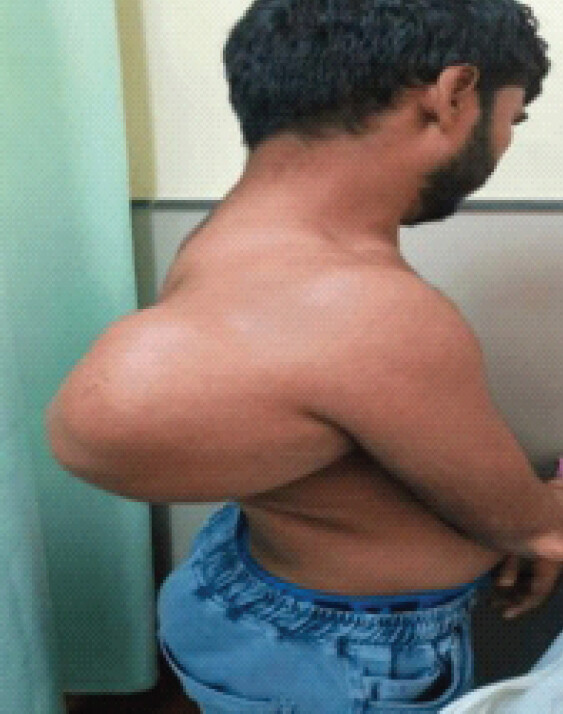
Plain radiographs of the right shoulder and chest demonstrated a bony excrescence on the dorsal surface of the scapula. magnetic resonance imaging (MRI) scan confirmed a sessile multiloculated lesion measuring 16 × 14.9 × 17.1 cm arising from the lateral border of the right scapula, with a chondroid matrix. A hyaline cartilage cap of approximately 4 mm thickness with abutment to dorsal scapular and suprascapular neurovascular bundle without any feature suggestive of malignant transformation. A diagnosis of dorsal scapular osteochondroma was established. Fig. 2 shows a 3D computed tomography (CT) Scan of the lesion to evaluate the extent of the tumor and helps in the preoperative planning.
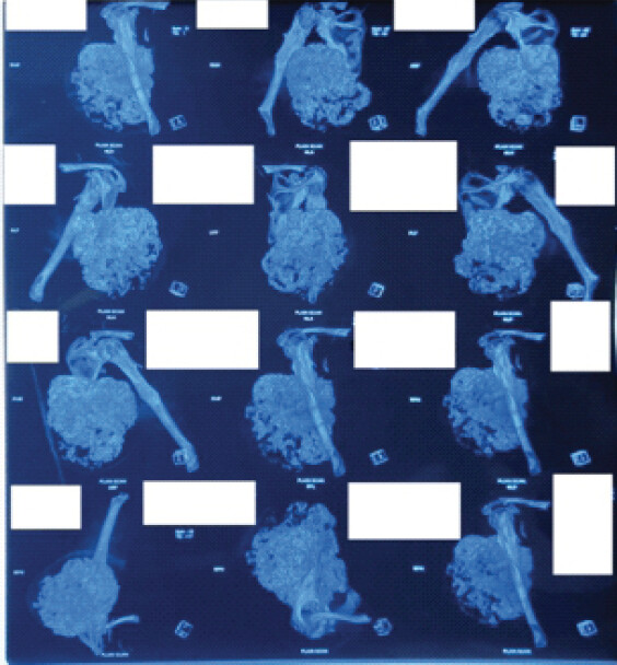
The patient was counselled regarding the diagnosis and surgical options. Under general anesthesia in the lateral decubitus position, a standard posterior approach was utilized. The trapezius and rhomboid muscles were reflected medially to expose the dorsal scapular surface. The serratus anterior was carefully detached from the medial border to access the subscapular space. The osteochondroma, along with its overlying reactive bursa, was identified and excised en-bloc at its base using an osteotome, ensuring no residual stalk tissue remained. The wound was closed in layers over a suction drain. The excised specimen measured 4.0 × 3.0 × 2.5 cm. Histopathological examination confirmed osteochondroma with a thin hyaline cartilage cap, orderly endochondral ossification, and no cytological atypia. Fig. 3 demonstrates the surgical excision of the tumor as detailed with the image. Fig. 3a shows the preoperative surgical marking. Fig. 3b and c show the exposure and Fig. 3d shows the excised tumor.
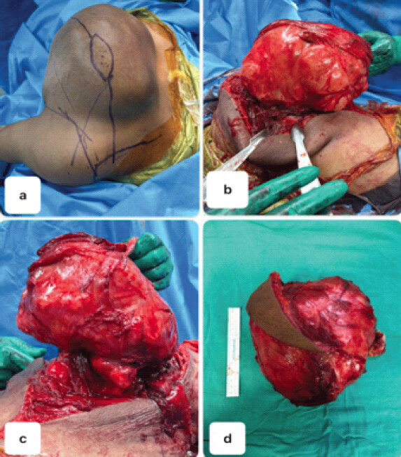
Postoperative recovery was uneventful. The drain was removed on day 2. Physiotherapy was commenced at 2 weeks, and the patient achieved full pain-free range of motion by 6 weeks. At 6-month follow-up, the patient was asymptomatic with no radiological evidence of recurrence. Shoulder function was excellent, and he had returned to his activities.
Case 2
A 25-year-old male student presented with an 8-month history of a progressively enlarging bony swelling over the posterior aspect of the left shoulder, associated with dull aching pain. There was no history of constitutional symptoms, trauma, or family history of similar lesions. On examination, a firm, immobile, non-tender bony prominence was palpable over the body of the left scapula. Shoulder abduction was restricted to 130° due to mechanical impingement. Distal neurovascular examination was normal. Fig. 4 shows the clinical photograph of the patient. Fig. (4a) from side, (4b) from front, (4c) from back.
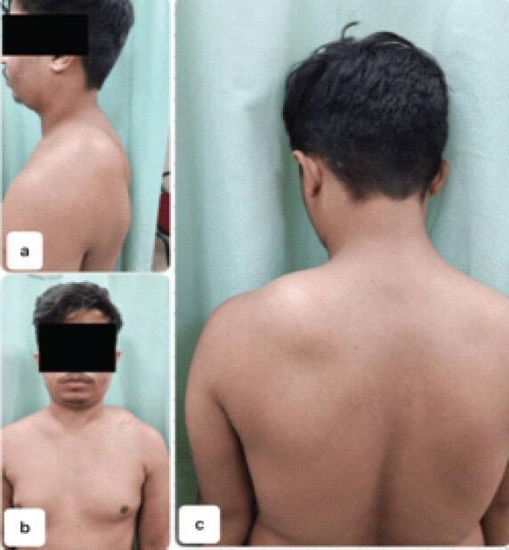
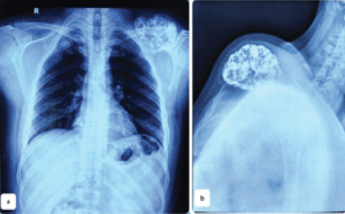
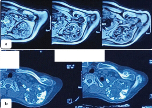
Plain radiographs revealed a sessile bony excrescence arising from the dorsal surface of the left scapular body. CT scan demonstrated a 6.7 × 6.5 × 5.5 cm lobulated osseous mass, chondroid matrix with cortical and medullary continuity with the scapula. MRI confirmed a cartilage cap of approximately 5 mm, abutting supraspinatus muscle with no signs of malignant transformation. The diagnosis of dorsal scapular osteochondroma was established. Fig. 5a shows the Antero-posterior view and Fig. 5b shows lateral view radiographs of the patient. Fig. 6a shows sections of MRI and Fig. 6b shows sections of CT scan of the site of swelling.
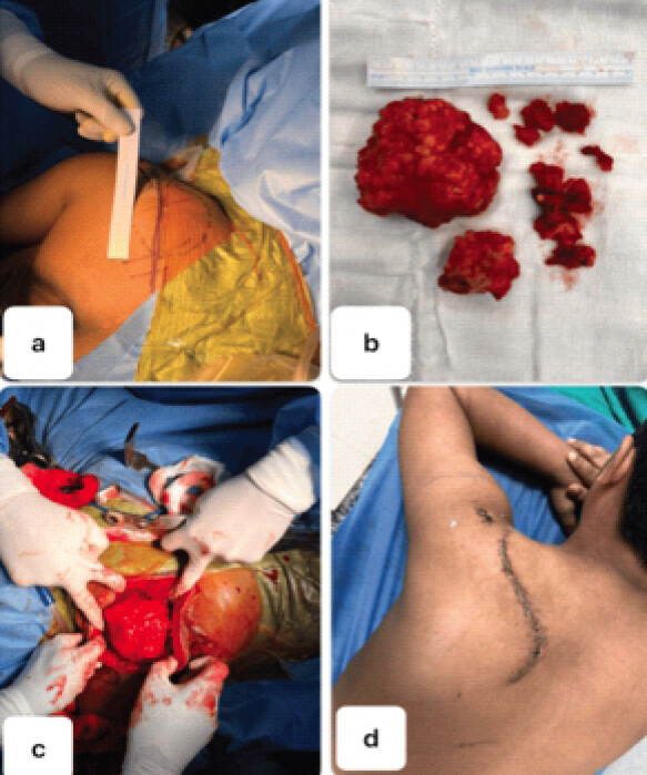
Surgical excision was performed under general anesthesia with the patient in the right lateral position. A curvilinear incision was made over the posterior aspect of the left scapula, centred over the mass. The deltoid was retracted superiorly and infraspinatus inferiorly with its fibers to expose the mass. The osteochondroma was dissected free from surrounding soft tissues, and the mass was removed from its base with an osteotome under direct visualization, ensuring complete excision. Histopathological examination confirmed the diagnosis of osteochondroma with no evidence of malignancy. Fig. 7a–c shows the preoperative surgical marking and surgical excision of the tumor. Fig. 7d shows the post-operative surgical site.
The postoperative course was unremarkable. Active-assisted shoulder physiotherapy was initiated at 2 weeks. By 8 weeks, the patient had regained full range of motion and was pain-free. At 6-month follow-up, clinical and radiological examination showed no recurrence, and the patient had returned to full activities of daily living without restriction.
Discussion
Scapular osteochondromas, account for only a small proportion of all osteochondromas. Both solitary and multiple (in the context of MHE) forms are reported.
The clinical presentations in our two cases are consistent with patterns described in the literature. In case 1, due to massive swelling, the symptoms were primarily due to mechanical impingement of mass on soft tissues. In Case 2, the dorsal lesion presented primarily as a visible and palpable posterior shoulder swelling with secondary mechanical restriction, a more typical presentation but one that can be easily misattributed to soft tissue tumors or lymphadenopathy if imaging is not obtained early. Both the tumors did not involve shoulder joint, but there was restriction of movements due to abutment of tumor to muscles responsible for shoulder movements.
MRI remains the gold standard for preoperative assessment of scapular osteochondromas, as it accurately delineates cartilage cap thickness, which is the most important imaging parameter for risk-stratifying malignant transformation. A cap thickness exceeding 1.5–2 cm in adults (up to 3 cm in skeletally immature patients) raises concern for secondary chondrosarcoma and mandates careful histopathological evaluation. In both our cases, cartilage cap thickness was well within benign limits [1, 3, 4].
Surgical excision is indicated for symptomatic lesions and is the definitive treatment. The key surgical principle is complete excision at the base of the stalk, as incomplete resection leaving residual periosteum or cartilage increases the risk of local recurrence. Dorsal lesions are more accessible via a posterior approach with modified Judet approach [1, 2, 5].
Both cases in the present report achieved excellent functional outcomes with no recurrence at 6 months. These findings reinforce the principle that timely and complete surgical excision is curative for scapular osteochondromas [2, 6, 7]. Delayed diagnosis, as documented in several series, may compound symptoms including progressive scapular winging, bursitis formation, and subscapular fibrosis, making dissection more complex and potentially jeopardizing functional recovery [5, 8, 9]. Orthopedic surgeons should maintain a high index of suspicion for scapular osteochondromas in younger patients presenting with mechanical shoulder symptoms, palpable posterior chest wall masses, or unexplained snapping scapula syndrome, and should not hesitate to obtain cross-sectional imaging when plain radiographs are inconclusive [9, 10].
Conclusions
Scapular osteochondroma, though uncommon, should be considered in young patients with posterior shoulder masses and mechanical symptoms. Although rare but it should be considered one of the differential diagnosis in cases of diagnostic dilemma. MRI assessment of cartilage cap thickness is central to malignancy risk stratification and thus detailed imaging is vital for reaching the diagnosis as well as decision making for further course of treatment. Complete excision at the base is curative for symptomatic lesions and yields excellent functional outcomes with low recurrence. Early recognition and treatment can avert secondary scapulothoracic pathology.
Clinical Message
Scapular osteochondroma should be suspected in patients presenting with persistent posterior shoulder swelling and mechanical shoulder symptoms. Advanced imaging is critical for diagnosis and assessment of malignant potential. Complete excision at the tumor base remains the definitive treatment and yields excellent outcomes.
Conflict of Interest:
Nil
Source of Support:
Nil
Consent:
The authors confirm that informed consent was obtained from the patient for publication of this article
References
- Vaishya R, Dhakal S, Vaish A. A solitary osteochondroma of the scapula. BMJ Case Rep 2014;21:2013202273. [Google Scholar] | [PubMed]
- Rustagi A, Agarwalla A, Agarwal S, Krishna L, Talwar J. Winging of scapula: An uncommon presentation of a common tumor. Cureus 2020;12:e12384. [Google Scholar] | [PubMed]
- Frost NL, Parada SA, Manoso MW, Arrington E, Benfanti P. Scapular osteochondromas treated with surgical excision. Orthopedics 2010;33:804. [Google Scholar] | [PubMed]
- Altwaijri NA, Fakeeha J, Alshugair I. Osteochondroma of the scapula: A case report and literature review. Cureus 2022;21:e30558. [Google Scholar] | [PubMed]
- Chillemi C, Franceschini V, Ippolito G, Pasquali R, Diotallevi R, Petrozza V. Osteochondroma as a cause of scapular winging in an adolescent: A case report and review of the literature. J Med Case Rep 2013;23:220. [Google Scholar] | [PubMed]
- Mohsen MS, Moosa NK, Kumar P. Osteochondroma of the scapula associated with winging and large bursa formation. Med Princ Pract 2006;15:387-90. [Google Scholar] | [PubMed]
- Kwon OS, Kelly JI. Delayed presentation of osteochondroma on the ventral surface of the scapula. Int J Shoulder Surg 2012;6:61-3. [Google Scholar] | [PubMed]
- Pérez D, Cano JR, Caballero J, López L. Minimally-invasive resection of a scapular osteochondroma. Interact Cardiovasc Thorac Surg 2011;5:468-70. [Google Scholar] | [PubMed]
- Gfrörer W, Seifert J, Matthes G, Ekkernkamp A, Hinz P. Das solitäre osteochondrom der skapula. Eine seltene differenzialdiagnose bei diffusen schulterbeschwerden [Solitary osteochondroma of the scapula. A rare differential diagnosis with unspecific shoulder pain]. Unfallchirurg 2005;108:684-6. [Google Scholar] | [PubMed]
- Tomo H, Ito Y, Aono M, Takaoka K. Chest wall deformity associated with osteochondroma of the scapula: A case report and review of the literature. J Shoulder Elbow Surg 2005;14:103-6. [Google Scholar] | [PubMed]
© 2026 Journal of Orthopaedic Case Reports - Published by Indian Orthopaedic Research Group
About the Authors
-

Prashant Sharma
-

Apurba Chowdhury
-

Ashish Rustagi
Related Articles in Journal of Orthopaedic Case Reports
 August 1, 2026 Vanishing Osteochondromas of the Distal Femur and Proximal Humerus: A Two-Case Report
 November 1, 2025 Distal Fibula Osteochondroma with Peroneal Tendon Subluxation – A Case Report
 October 1, 2026 Clinicopathological Spectrum and Management of Aneurysmal Bone Cysts in Atypical Anatomical Location: A Comprehensive Review of Demographics, Presentation, Diagnosis, and Treatment Outcomes
 October 1, 2026 Giant Cell Tumor of the Lateral Malleolus Treated by Tumor Excision and Reconstruction using A 180° Rotated Proximal Fibular Autograft – A Rare Case Report