

Solitary osteochondromas may regress spontaneously and should be monitored conservatively in asymptomatic patients.
Dr. Haya Alkuwari, Department of Orthopedic Surgery, Hamad General Hospital, Doha, Qatar. E-mail: halkuwari18@hamad.qa
Abstract
Introduction: Osteochondroma is the most common benign bone tumor, typically presenting during adolescence as a painless bony prominence. Although conservative management is commonly adopted for asymptomatic lesions, spontaneous regression of osteochondromas is considered rare and is sparsely reported in the literature.
Case Report: We report two cases of solitary osteochondromas demonstrating spontaneous regression without surgical intervention. The first case involved a 14-year-old female with a distal femoral osteochondroma, and the second involved a 16-year-old male with a proximal humeral osteochondroma. Both patients were managed conservatively with clinical and radiological follow-up, which showed progressive reduction and eventual regression of the lesions over time, with no development of alarming clinical or radiological features.
Conclusion: These cases highlight the potential for spontaneous regression of solitary osteochondromas in the pediatric and adolescent population. In the absence of concerning symptoms or radiological signs, conservative management with regular follow-up may be a safe and appropriate approach, helping to avoid unnecessary surgical intervention.
Keywords: Benign bone tumor, exostosis, osteochondroma, regression.

Osteochondroma is the most common benign bone tumor. Some scholars refer to it as a developmental anomaly rather than a true neoplasm. It accounts for 20–50% of benign bone tumors and 10–15% of all bone tumors [1]. These lesions grow on the bone surface, show cortical and medullary continuity with the overlying bone, and are covered by a hyaline cartilage cap [2]. Osteochondromas exhibit characteristic radiologic features that closely correlate with their histopathological appearance [1].
Osteochondromas may present as solitary lesions or as multiple growths, the latter typically associated with the autosomal dominant disorder known as hereditary multiple exostoses (HME) [1]. In children and adolescents, most lesions manifest as slow-growing, painless masses. Significant symptoms, however, may arise from complications such as fracture, bone deformity, mechanical joint issues, and vascular or neurologic damage, depending on where the osteochondroma is located [3].
Asymptomatic osteochondromas are managed conservatively through observation, while surgical excision is indicated for symptomatic lesions or those demonstrating suspicious radiological characteristics [4]. Spontaneous regression of osteochondroma is a rare clinical event [5]. We herein present two cases of spontaneous regression of solitary osteochondromas: one in a 14-year-old female involving the distal femur managed conservatively over 5.5 years, and the other in a 16-year-old male with a proximal humeral lesion that regressed following a traumatic fracture within 18 months.
Case 1
A 14-year-old previously healthy female presented with a painless bony swelling over the distal aspect of her right thigh. Notably, her mother had a similar lesion that was surgically excised during childhood, though no other swellings were detected in the patient. Clinical examination was unremarkable. Radiographs revealed a pedunculated bony projection arising from the lateral cortex of the distal metaphysis of the right femur, with corticomedullary continuity and orientation away from the adjacent joint – features consistent with osteochondroma (Fig. 1). Conservative management with routine clinical and radiologic follow-up was initiated.
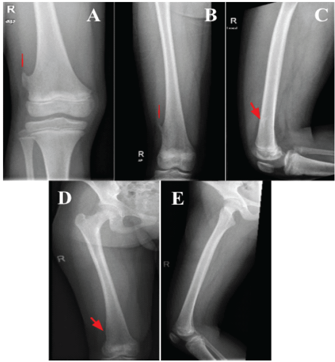
Figure 1: Serial anteroposterior and lateral radiographs of the right distal femur demonstrating a pedunculated osteochondroma arising from the lateral aspect, with corticomedullary continuity and projection directed away from the adjacent knee joint. (a) Initial presentation showing the lesion. (b and c) Follow-up radiographs at 3.5 years demonstrated interval reduction in size. (d and e) Follow-up radiographs at 5.5 years showed near-complete resolution of the lesion with only a small residual cortical projection at the same site.
The patient remained symptom-free for 3.5 years. After that, she presented with swelling and discomfort in the right thigh, particularly following physical activity. Examination showed mild local tenderness, and radiographs confirmed the persistence of the lesion with a slight reduction in size compared to previous imaging (Fig. 2). Skeletal survey ruled out HME. Given the absence of aggressive features, conservative management with serial follow-up was continued.
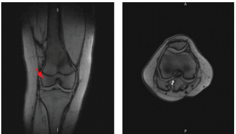
Figure 2: Magnetic resonance imaging of the right knee demonstrated interval disappearance of the previously noted distal femur osteochondroma, with only a small residual cortical projection and no evidence of a cartilaginous cap, soft tissue mass, or abnormal enhancement.
Serial imaging over the next 2 years demonstrated a gradual reduction in the size of the osteochondroma. Five and a half years after the first presentation, radiographs showed near-complete radiologic resolution of the lesion (Fig. 3).
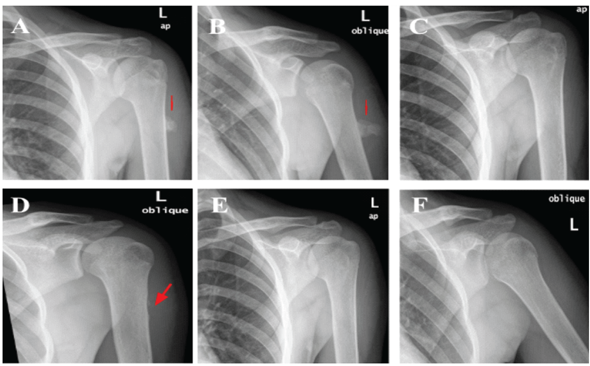
Figure 3: Serial anteroposterior and oblique radiographs of the left proximal humerus following trauma demonstrated progressive regression and complete resolution of a solitary pedunculated osteochondroma. (a and b) Radiographs at 2 weeks post-injury showed a pedunculated osteochondroma at the proximal third of the humerus with associated callus formation over the fracture stump. (c and d) Follow-up radiographs at 14 months demonstrated almost complete regression of the lesion with only a small bony remnant and scalloping of the posterolateral cortex at the site of the lesion. (e and f) Radiographs at 18 months showed complete resolution of the osteochondroma with full remodeling of the bone.
The patient continued to report mild, activity-related discomfort over the previously affected site. Further follow-up radiographs continued to show no evidence of recurrence. Magnetic resonance imaging (MRI) of the right knee was performed to assess soft tissue changes and confirmed the disappearance of the osteochondroma. The scan showed only a small residual cortical projection with no cartilaginous cap, soft tissue mass, or abnormal enhancement (Fig. 4). At the latest follow-up, the patient was clinically stable.
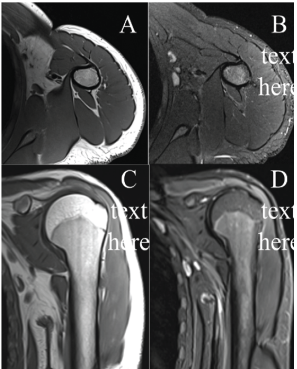
Figure 4: Magnetic resonance imaging of the left proximal humerus. (a and b) T1- and T2- weighted fat-sat axial cuts showing the almost complete resorption of the osteochondroma with only a bony remnant at the posterolateral aspect of the proximal third of the humerus. (c and d): T1 and T2 fat-sat coronal cuts showing almost complete resorption of the lesion.
Case 2
A 16-year-old boy, with no known co-existing medical conditions or family history of inherited diseases, presented to the emergency department complaining of localized pain and swelling in his left mid-arm for the past 2 weeks following a direct injury. Throughout this time, the arm was tender to the touch, and the swelling around the area progressively increased. There were no associated constitutional symptoms, and he had not experienced any issues in that arm prior to the injury.
A local assessment of the left arm revealed diffuse swelling in the proximal arm with soft compartments, while palpation indicated a tender, non-mobile, and firm mass at the lateral side of the left proximal humerus, situated just above the deltoid muscle insertion. The skin over the area appeared normal, and there were no indicators of any active infection. Nevertheless, the patient exhibited a full active and passive range of motion in the shoulder, with no evidence of shoulder instability and a complete intact distal neurovascular examination. No other areas showed similar masses or swellings.
Radiographic evaluation revealed a solitary pedunculated osteochondroma located at the proximal third of the humerus, along with signs of callus formation at the fracture site (Fig. 5). The patient was provided with an arm sling for comfort and was sent home with a follow-up appointment at the orthopedic oncology clinic. At the follow-up visit, the patient reported improved symptoms and had complete fracture healing within 4 months. It was decided to manage the tumor conservatively, with a plan to reassess the patient in 1 year through repeat radiographs to consider the possibility of surgical removal. Fourteen months after the injury, he showed no symptoms, and a new X-ray revealed nearly complete regression of the lesion, leaving only a small bony remnant and scalloping of the posterolateral cortex in the area of the lesion. A follow-up MRI of the humerus was performed to verify these findings. Finally, at 18 months post-fracture, another X-ray was obtained, which indicated total resorption of the lesion with no clinical symptoms.
Osteochondroma is the most prevalent benign bone tumor, typically presenting as a solitary, non-hereditary lesion in approximately 85% of cases [3]. The remaining 15% manifest as multiple lesions in HME [3,6]. Since many osteochondromas are asymptomatic and go undetected, the true incidence of these tumors remains unknown [4]. These lesions are most commonly identified within the first four decades of life, with approximately 75% occurring before the age of 20 [7,8] and exhibiting male predominance [9].
Nearly half of all osteochondromas develop in the lower limbs, with approximately 40% located around the knee. The humerus is affected in 10–20% of cases. Less common sites include the small bones of the hands and feet (10%), the scapula (4%), and the pelvis (5%) [1]. Solitary osteochondromas are rarely found in the spine. Clinically, osteochondromas are predominantly asymptomatic and are most often identified incidentally on radiographs performed for unrelated clinical indications [3]. When symptomatic, the most common presentation is a painless, palpable mass over the affected bone [10]. Symptoms may also develop due to mechanical compression of nearby structures, pathologic fracture, bursitis, or malignant transformation [11]. In certain cases, nerve impingement by an osteochondroma may manifest as limb numbness and tingling [12]. Furthermore, it may exert pressure on adjacent blood vessels, potentially leading to vascular complications such as aneurysm or pseudoaneurysm formation [13]. The most commonly affected structure is the popliteal artery [14]. Osteochondromas are benign tumors that typically have no impact on life expectancy. However, the risk of malignant transformation is estimated to be between 1% and 5% [6].
The pathogenesis of solitary osteochondromas was originally attributed to a developmental defect wherein a portion of the growth plate protruded through the periosteum and progressively enlarged to form a solitary lesion. This hypothesis is supported by documented cases of osteochondroma arising after trauma or radiation exposure [1]. However, recent studies have shown that heterozygous mutations in the exostosin (EXT)-1 gene are found equally in both solitary osteochondromas and HME, whereas EXT2 mutations are rarely identified in solitary lesions. These findings collectively suggest that solitary osteochondromas are true benign neoplasms [1,15]. Disruption of the Indian hedgehog pathway regulation has also been reported as a contributing factor in osteochondroma formation [2].
Conservative management primarily involves initial monitoring with plain radiographs, followed by routine clinical evaluations. Although no studies have confirmed the effectiveness of regular screening, annual or biennial follow-up may be considered, and MRI is recommended for further assessment if the tumor enlarges or pain arises after growth plate closure [6]. Surgical excision is generally indicated for lesions that are symptomatic, cosmetically concerning, or exhibit imaging features suggestive of malignancy – such as indistinct margins, areas of radiolucency, bone erosion or destruction, or when the diagnosis remains uncertain [2]. Incomplete excision of the perichondrium may lead to local recurrence, which has been documented in approximately 2–5% of cases [1].
Rarely, spontaneous resolution of osteochondroma may occur [16]. Most reported lesions in the literature were resolving before skeletal maturity and within 3 years of identification [17]. Tumors with sessile morphology were more prone to shrinkage compared to pedunculated morphology [18]. In addition, it has been shown that the thickness of the cartilage cap may be an important predictor of spontaneous regression in pediatric patients with osteochondroma, with a thinner cartilage cap seen on MRI being predictive of tumor shrinkage [19]. Valdivielso-Ortiz et al. [20] reported a case of a 9-year-old girl with a solitary sessile osteochondroma of the posterior distal femur that regressed almost completely within 4 years. Similarly, De Siqueira et al. [21] reported a case of spontaneous clinical and radiologic resolution of a sessile osteochondroma of the proximal humerus in a 3-year-old male, supporting the role of annual observation in asymptomatic pediatric patients.
Spontaneous regression of a solitary osteochondroma is an uncommon and poorly understood phenomenon. These cases highlight the efficacy of conservative management and regular follow-up in asymptomatic patients, especially when no concerning clinical or radiological signs are present. While most osteochondromas follow a benign course, clinicians should remain cautious for symptoms suggestive of neurovascular compromise, mechanical irritation, or malignant transformation. These cases reinforce the potential for a favorable natural history in selected patients and support individualized management strategies based on clinical presentation and imaging findings.
Solitary osteochondromas in children and adolescents may regress spontaneously; therefore, asymptomatic lesions without concerning clinical or radiological features can be safely managed with careful observation and regular follow-up, avoiding unnecessary surgical intervention.
References
- 1. Murphey MD, Choi JJ, Kransdorf MJ, Flemming DJ, Gannon FH. Imaging of osteochondroma: Variants and complications with radiologic-pathologic correlation. Radiographics 2000;20:1407-34. [Google Scholar] [PubMed]
- 2. Alabdullrahman LW, Mabrouk A, Byerly DW. Osteochondroma. In: StatPearls. Treasure Island, FL: StatPearls Publishing; 2025. Available from: https://www.ncbi.nlm.nih.gov/books/nbk544296/?utm_source=chatgpt.com [Google Scholar] [PubMed]
- 3. Kitsoulis P, Galani V, Stefanaki K, Paraskevas G, Karatzias G, Agnantis NJ, et al. Osteochondromas: Review of the clinical, radiological and pathological features. In Vivo 2008;22:633-46. [Google Scholar] [PubMed]
- 4. Tepelenis K, Papathanakos G, Kitsouli A, Troupis T, Barbouti A, Vlachos K, et al. Osteochondromas: An updated review of epidemiology, pathogenesis, clinical presentation, radiological features and treatment options. In Vivo 2021;35:681-91. [Google Scholar] [PubMed]
- 5. Heyworth PB, Rashid M. Regression of a solitary osteochondroma of the distal humerus in a toddler following trauma. Radiol Case Rep 2018;14:187-9. [Google Scholar] [PubMed]
- 6. Bovée JV. Multiple osteochondromas. Orphanet J Rare Dis 2008;3:3. [Google Scholar] [PubMed]
- 7. Subbarao K. Benign tumors of bone. Nepal J Radiol 2012;2:1-12. [Google Scholar] [PubMed]
- 8. Hakim DN, Pelly T, Kulendran M, Caris JA. Benign tumours of the bone: A review. J Bone Oncol 2015;4:37-41. [Google Scholar] [PubMed]
- 9. Wootton-Gorges SL. MR imaging of primary bone tumors and tumor-like conditions in children. Magn Reson Imaging Clin N Am 2009;17:469-87, vi. [Google Scholar] [PubMed]
- 10. Motamedi K, Seeger LL. Benign bone tumors. Radiol Clin North Am 2011;49:1115-34, v. [Google Scholar] [PubMed]
- 11. Gavanier M, Blum A. Imaging of benign complications of exostoses of the shoulder, pelvic girdles and appendicular skeleton. Diagn Interv Imaging 2017;98:21-8. [Google Scholar] [PubMed]
- 12. Mnif H, Koubaa M, Zrig M, Zammel N, Abid A. Peroneal nerve palsy resulting from fibular head osteochondroma. Orthopedics 2009;32:528. [Google Scholar] [PubMed]
- 13. Perez-Burkhardt JL, Gómez Castilla JC. Postraumatic popliteal pseudoaneurysm from femoral osteochondroma: Case report and review of the literature. J Vasc Surg 2003;37:669-71. [Google Scholar] [PubMed]
- 14. Vasseur MA, Fabre O. Vascular complications of osteochondromas. J Vasc Surg 2000;31:532-8. [Google Scholar] [PubMed]
- 15. Pacifici M. Hereditary multiple exostoses: New insights into pathogenesis, clinical complications, and potential treatments. Curr Osteoporos Rep 2017;15:142-52. [Google Scholar] [PubMed]
- 16. Hoshi M, Takami M, Hashimoto R, Okamoto T, Yanagida I, Matsumura A, et al. Spontaneous regression of osteochondromas. Skeletal Radiol 2007;36:531-4. [Google Scholar] [PubMed]
- 17. Passanise AM, Mehlman CT, Wall EJ, Dieterle JP. Radiographic evidence of regression of a solitary osteochondroma: A report of 4 cases and a literature review. J Pediatr Orthop 2011;31:312-6. [Google Scholar] [PubMed]
- 18. Aiba H, Yamada S, Yamamoto N, Hayashi K, Miwa S, Tsuchiya H, et al. Spontaneous shrinkage of solitary osteochondromas. Skeletal Radiol 2018;47:61-8. [Google Scholar] [PubMed]
- 19. Adachi R, Nakamura T, Asanuma K, Hagi T, Uchiyama T, Sudo A. Thin cartilage cap may be related to the spontaneous regression in pediatric patients with osteochondroma. Curr Oncol 2022;29:9884-90. [Google Scholar] [PubMed]
- 20. Valdivielso-Ortiz A, Barber I, Soldado F, Aguirre-Canyadell M, Enriquez G. Solitary osteochondroma: Spontaneous regression. Pediatr Radiol 2010;40:1699-701. [Google Scholar] [PubMed]
- 21. De Siqueira AL, Abd-Elmaged HM, Salih D, Mustafa ME. Spontaneous resolution of proximal humerus sessile osteochondroma: A report of case and literature review. Clin Case Rep 2023;12:e8372. [Google Scholar] [PubMed]





Related Articles in Journal of Orthopaedic Case Reports
 November 1, 2025 Distal Fibula Osteochondroma with Peroneal Tendon Subluxation – A Case Report
 September 1, 2026 Management of Scapular Osteochondroma: An Uncommon Report of Two Cases
 July 1, 2026 Posterior Scapular Osteochondroma in a Pediatric Patient: An Uncommon Presentation of a Common Benign Tumor
 June 1, 2026 Sequential Development and Staged Management of Distal Femoral Osteochondroma with Progressive Bilateral Genu Valgum: A Case Report