


Introduction
Meniscal injuries are among the most common intra-articular knee pathologies encountered in orthopedic practice, frequently presenting with pain, mechanical symptoms, and functional limitation [1]. While bucket-handle tears are more commonly described in the medial meniscus, they also occur in the lateral meniscus, often in association with acute traumatic events and anterior cruciate ligament (ACL) injuries. Acute lateral meniscus tear patterns are generally longitudinal vertical tears (including bucket-handle), radial tears, and flap tears which are common after trauma [2]. Lateral meniscus bucket-handle tears are clinically significant due to their impact on knee stability, load transmission, and long-term joint health.
Bucket-handle tears represent a subtype of longitudinal vertical meniscal tears characterized by displacement of the inner fragment, which may migrate into the intercondylar notch or remain within the compartment [3]. Compared to medial meniscal tears, lateral meniscus bucket-handle tears may demonstrate greater mobility due to the relatively increased laxity and anatomical characteristics of the lateral compartment, potentially resulting in variable imaging appearances [4].
Magnetic resonance imaging (MRI) remains the gold standard modality for evaluating meniscal pathology, with high sensitivity and specificity. Classical MRI signs described for bucket-handle tears include the double posterior cruciate ligament (PCL) sign, the absent bow tie sign, and the flipped meniscus sign and head in the sand sign. However, these signs are not always present, particularly in cases where the displaced fragment does not occupy the intercondylar notch or when displacement is partial.
The diagnosis of lateral meniscus bucket-handle tears can be particularly challenging due to their variable displacement patterns and less predictable imaging features compared to medial meniscal injuries. Subtle or atypical presentations may lead to underdiagnosis or delayed treatment, which can adversely affect clinical outcomes [5].
In this context, identification of additional MRI features is essential to improve diagnostic accuracy. We describe a novel radiological sign – the Meniscal Duplication Sign – characterized by the presence of two low-signal meniscus-like structures within the same sagittal plane, corresponding to the native meniscus and a displaced fragment. This configuration produces a distinctive visual analogy resembling a “cement mixing trowel,” which may facilitate recognition, particularly in cases of lateral meniscus bucket-handle tears where classical signs are inconspicuous.
Case Report
An 18-year-old male presented with complaints of pain in the right knee for 1 month, following a twisting injury sustained while playing football. The patient reported episodes of intermittent locking, instability, clicking, and difficulty in achieving full extension of the knee. There was no history of prior knee surgery or chronic instability.
Clinical examination
On physical examination, there was mild joint effusion with localized tenderness along the lateral joint line. Range of motion was restricted, particularly terminal extension, with a mechanical block noted.
Special tests revealed:
-
McMurray test: Positive for lateral meniscus (painful click on external rotation and valgus stress)
-
Thessaly test: Positive at 20° of flexion
-
Apley grinding test: Positive.
Ligamentous examination:
-
Lachman test: Positive
-
Anterior drawer test: Positive
-
Pivot shift test: Couldn’t be evaluated because of the possibility of tear progression
-
No significant varus or valgus instability.
These findings were suggestive of a lateral meniscal injury, likely a bucket-handle tear.
MRI findings
MRI of the right knee was performed using standard proton density and T2-weighted sequences.
Sagittal images demonstrated:
-
Two distinct low-signal intensity meniscus-like structures within the lateral compartment on the same slice (Fig. 1)
-
One structure corresponded to the residual native meniscus, while the second represented a displaced meniscal fragment
-
The fragment was located adjacent to the native meniscus rather than within the intercondylar notch.
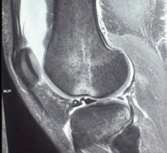
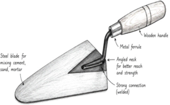
This produced a characteristic configuration termed the Meniscal Duplication Sign, with a morphology resembling a “Trowel” (Fig. 2), where:
-
The displaced fragment formed the “blade”
-
The residual meniscus formed the “handle”
Additional findings:
-
Subtle truncation/irregularity of the normal meniscal body
-
Possible early loss of normal bow-tie appearance (inconsistent across slices)
-
No obvious double PCL sign visualized.
Arthroscopic findings and management
Diagnostic arthroscopy of the knee was performed under spinal/combined anesthesia.
Intraoperative findings revealed:
-
A longitudinal vertical tear of the lateral meniscus extending from the posterior horn to the body
-
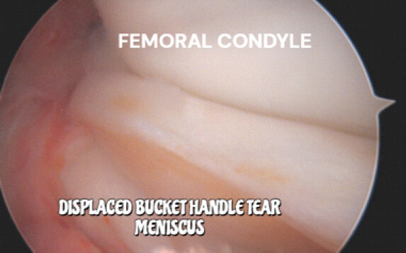
A displaced inner fragment (bucket-handle fragment) lying within the lateral compartment, confirming MRI findings (Fig. 3)
-
The fragment was mobile but remained partially attached at the anterior and posterior horns.
The displaced fragment corresponded to the structure identified on MRI as the second meniscus-like band, confirming the Meniscal Duplication Sign.
Articular cartilage surfaces were inspected and found to be normal.
The ACL was torn.
Plan of repair at our center
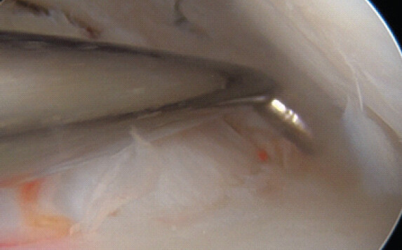
A structured repair strategy was followed based on tear location and morphology after temporary reduction of the tear (Fig. 4):
-
Mid-third tear: Repaired using inside-out (Sironix- Healthium, Peenya, Bengaluru) or outside-in sutures, ensuring robust fixation of the central segment.
-
Posterior root/Posterior horn region: Secured using all-inside vertical sutures, providing stable fixation in the posterior compartment with minimal risk to neurovascular structures (Sironix/smith and Nephew).
-
Meniscal body: Reinforced using continuous running sutures, restoring hoop stress continuity along the length of the body (Conmed-sequent).
-
Anterior root component: A complete anterior root tear was repaired using an outside-in technique or an all-inside device based on accessibility and construct stability.
At our center, meniscal tear fixation is performed using a structured and reliable technique. In cases of bucket-handle tears, an initial inside-out suture is placed to provisionally hold the displaced meniscal fragments in a reduced position. Subsequently, all-inside sutures are passed in a horizontal mattress configuration to achieve stable fixation along the tear. Finally, the reduction sutures placed using the inside-out technique are securely tied over the capsule to maintain anatomical alignment and ensure optimal healing.
Following completion of the repair, the microfracture procedure was performed to promote healing, and then, the meniscus was probed and taken through a full range of motion to confirm stability, anatomical contour restoration, and absence of displacement. The articular cartilage and both cruciate ligaments were visualized and found to be intact [6].
Postoperative rehabilitation
Postoperative rehabilitation was initiated immediately following surgery. The patient was placed in a hinged knee brace locked at 60° of flexion, which was maintained for the first 6 weeks to protect the repair and minimize shear forces across the meniscus. Early passive range-of-motion exercises within the safe arc were permitted under supervision, avoiding deep flexion [7].
Weight-bearing was restricted to toe-touch/partial weight-bearing during the first 6 weeks, followed by progressive weight-bearing between 6 and 12 weeks, guided by pain, quadriceps control, and clinical assessment. Strengthening of the quadriceps and hip stabilizers was gradually introduced, along with closed-chain kinetic exercises, after adequate healing was established.
By the end of 6 weeks, the patient regained a full range of motion, and functional rehabilitation continued with a focus on proprioception, neuromuscular control, and return-to-sport conditioning.
Discussion
Bucket-handle tears of the meniscus represent a clinically important subset of meniscal injuries that often necessitate prompt surgical intervention, particularly in young and active individuals. Accurate preoperative diagnosis is essential for appropriate surgical planning, especially with regard to meniscal preservation and repair strategies. MRI plays a central role in this diagnostic pathway, with several well-established primary and secondary signs aiding in the identification of displaced meniscal fragments [8].
The Meniscal Duplication Sign described in this report represents a novel addition to the spectrum of MRI findings associated with bucket-handle tears. Unlike the classical double PCL sign, which depends on displacement of the meniscal fragment into the intercondylar notch, this sign is based on the simultaneous visualization of both the native meniscus and the displaced fragment within the same sagittal imaging plane. This results in the appearance of two parallel low-signal structures resembling duplicated meniscal tissue [9].
The pathoanatomical basis of this sign lies in the longitudinal vertical tear pattern of bucket-handle injuries. Following disruption, the inner fragment may displace but remain within the same compartment rather than migrating into the notch. When imaged in the sagittal plane, this leads to a “double contour” or duplication appearance. This is particularly relevant in early displacement, incomplete tears, or cases where the fragment is constrained by surrounding structures such as intact meniscocapsular attachments [10].
An important advantage of the Meniscal Duplication Sign is its potential utility in scenarios where classical signs are absent or equivocal. For instance, the absent bow tie sign requires evaluation across multiple sequential slices and may be influenced by slice thickness and positioning. Similarly, the double PCL sign is only present when the fragment is displaced centrally into the notch. In contrast, the duplication sign may be appreciated on a single sagittal image, potentially facilitating quicker recognition during routine MRI interpretation.
The associated “Trowel appearance” provides a valuable visual analogy, with the displaced fragment forming the “blade” and the residual meniscus representing the “handle.” While such analogies are not typically used as formal nomenclature in radiological literature, they can significantly enhance pattern recognition and teaching, particularly for trainees and early learners.
From a clinical perspective, recognition of this sign may improve diagnostic confidence and reduce the likelihood of missed bucket-handle tears. Early diagnosis is critical, as delayed treatment can lead to further meniscal damage, chondral injury, and progression to osteoarthritis. Moreover, timely identification allows for consideration of meniscal repair, which is increasingly favored over meniscectomy in suitable cases for lateral meniscus tears for good healing and biomechanical reasons.
However, this report has certain limitations. As a single case observation, the sensitivity, specificity, and reproducibility of the Meniscal Duplication Sign cannot be established. Larger studies and retrospective imaging reviews are necessary to validate this finding and determine its diagnostic accuracy in comparison with established MRI signs. Interobserver reliability and applicability across different MRI protocols also warrant further evaluation.
Despite these limitations, the Meniscal Duplication Sign represents a simple, intuitive, and potentially valuable addition to the radiological assessment of meniscal injuries. Its recognition may complement existing diagnostic criteria and contribute to improved detection of bucket-handle tears in routine clinical practice.
Limitations
Being a single case report, the findings cannot be generalized, and further studies with larger sample sizes are required to establish the diagnostic utility of this MRI sign.
Conclusion
The Meniscal Duplication Sign is a novel and useful MRI feature for diagnosing bucket-handle tears of the lateral meniscus. Recognition of this sign may improve diagnostic confidence, particularly in atypical presentations.
Clinical Message
Visualization of two meniscus-like structures in the same sagittal plane should raise suspicion for a displaced bucket-handle tear, even when classical signs are absent.
Conflict of Interest:
Nil
Source of Support:
Nil
Consent:
The authors confirm that informed consent was obtained from the patient for publication of this article
References
- Pereira RR, Cabral J, Janeiro J, Padín J, Brito JS, Goes RA. Buffalo Horn Sign - A New Finding on MRI for Meniscal Bucket-Handle Tears. Rev Bras Ortop (Sao Paulo). 2025;60 2. s00451809514 doi:10.1055/s-0045-1809514. [Google Scholar] | [PubMed] | [CrossRef]
- Chen KH, Lee WC, Huang YC. Deep learning for detection of bucket-handle meniscal tears on MRI: A diagnostic accuracy study. Eur Radiol 2025;35:1123-32. [Google Scholar] | [PubMed]
- Simonetta R, Palco M, Giuca G, Leonetti D, Russo R, Familiari F. Anatomical repair of bucket-handle medial meniscal tears combined with a ramp lesion: a case report and surgical technique. AME Surg J. 2025;5:51 doi:10.21037/asj-25-20. [Google Scholar] | [PubMed] | [CrossRef]
- Sajeev J, Rajendrababu AR, Jeevo J, Babu VM, Thomas R, Amaravati RS. Bucket handle tear of the medial meniscus with a new magnetic resonance imaging sign: The “Head in the sand sign” - a case report. J Orthop Case Rep 2026;16:9-13. [Google Scholar] | [PubMed]
- Gaillard F, Jones J, Weerakkody Y. Bucket-Handle Meniscal Tear. Radiopaedia; 2026 Available from: https://radiopaedia.org/articles/bucket-handle-meniscal-tear. [cited 2026 Aug 20]. [Google Scholar] | [PubMed]
- Toye L. Bucket-handle Tear of the Meniscus. Radsource MRI Web Clinic. 2021 Jan Available from: https://radsource.us/bucket-handle-tear-of-the-meniscus/ [cited 2026 Aug 20]. [Google Scholar] | [PubMed]
- Pujol N, Beaufils P. The natural history of meniscal lesions. Orthop Traumatol Surg Res 2021;107:S237-44. [Google Scholar] | [PubMed]
- Abram SG, Beard DJ, Price AJ, Hopewell S, Monk AP, Bayliss LE. Arthroscopic meniscal surgery: A systematic review and meta-analysis of outcomes. Br J Sports Med 2020;54:652-63. [Google Scholar] | [PubMed]
- Krych AJ, Reardon PJ, Johnson, Mohan R, Peter L, Levy BA. Nonoperative management of meniscal tears: Predictors of failure and clinical outcomes. Am J Sports Med 2020;48:1737-44. [Google Scholar] | [PubMed]
- De Smet AA, Tuite MJ. Use of the “two-slice-touch” rule for the MRI diagnosis of meniscal tears. AJR Am J Roentgenol 2020;215:605-10. [Google Scholar] | [PubMed]
© 2026 Journal of Orthopaedic Case Reports - Published by Indian Orthopaedic Research Group
About the Authors
-

Joseph Sajeev
-

Anand Raj Rajendrababu
-

Raphael Thomas
-

Ronnie George
-

Manikya Ramesh
-

Rajkumar S Amaravati
Related Articles in Journal of Orthopaedic Case Reports
 April 1, 2026 Bucket Handle Tear of the Medial Meniscus with a New Magnetic Resonance Imaging Sign: The “Head in the Sand Sign” – A Case Report
 April 1, 2026 Arthroscopic Management of a Bucket-Handle Acetabular Labral Tear in a Pediatric Patient: A Rare Case Report with Associated Fleck Sign
 October 1, 2025 Lax Medial Patellofemoral Ligament Syndrome as a Cause of Recurrent Patellar Instability: A Radiologic Case Series
 September 1, 2025 An Anomalous Attachment of the Medial Meniscus Posterior and Anterior Roots Mimicking a Bucket Handle Meniscus Tear: A Case Report and Review of the Literature