


Introduction
First metatarsophalangeal (MTP) arthrodesis is a standard procedure for end-stage hallux rigidus and a reconstructive option in severe deformity, inflammatory arthritis, neuromuscular disease, and failed forefoot surgery [1].
Although union rates are high, non-union can cause pain, swelling, transfer metatarsalgia, implant fatigue, and loss of correction [2,3]. Revision is challenging because bone loss, osteolysis, sclerosis, shortening, malalignment, scarring and failed implants may coexist [3,4].
This case illustrates a multifactorial failed MTP-I fusion treated by biological, mechanical and whole-forefoot reconstruction, followed by a focused literature review and revision framework.
Case Report
Patient information and initial presentation
A 62-year-old woman presented with progressive right MTP-I pain, stiffness and walking limitation after failed conservative treatment.
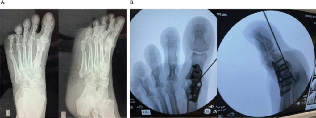
Examination showed advanced hallux rigidus with hallux varus and second-toe varus/claw deformity. Weight-bearing radiographs confirmed end-stage MTP-I arthritis (Fig. 1).
She had a 45 pack-year smoking history. Distal pulses were palpable and arterial duplex ultrasonography was normal. MTP-I arthrodesis was recommended.
Index arthrodesis and early post-operative course
The index procedure used cup-and-cone preparation and low-profile dorsal plate fixation. Intraoperative alignment was considered satisfactory, with dorsiflexion relative to the floor and neutral transverse alignment relative to the second ray.
Postoperatively, she remained non-weight-bearing for 4 weeks before progressive loading. At 3 months, alignment and fixation were maintained, but osseous consolidation was incomplete; retrospectively, dorsiflexion appeared slightly greater than ideal.
Diagnosis of failure
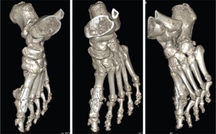
At 4 months, weight-bearing pain and swelling increased. Radiographs were initially inconclusive, but at 6 months showed dorsal plate fracture and persistent fusion line. Computed tomography (CT) confirmed pseudarthrosis, absent bridging trabecular bone, and proximal phalangeal osteolysis (Fig 2). There were no clinical signs of infection; white blood cell, erythrocyte sedimentation rate, and C-reactive protein were normal. Symptomatic aseptic non-union with implant fatigue failure was diagnosed.
Failure analysis
Failure was considered multifactorial. Persistent smoking was the main modifiable biological risk. CT showed local biological failure with pseudarthrosis, sclerosis, and osteolysis. Plate fracture was interpreted as the endpoint of micromotion and cyclic loading, not the primary cause. Subtle sagittal malposition and untreated second-ray deformity may have increased forefoot loading.
Revision surgical technique
Revision was performed through the previous dorsal approach. Failed hardware was removed, fibrous pseudarthrosis and sclerotic margins were excised, intraoperative cultures were obtained, and all non-viable bone was debrided to bleeding cancellous surfaces. Cultures were negative.
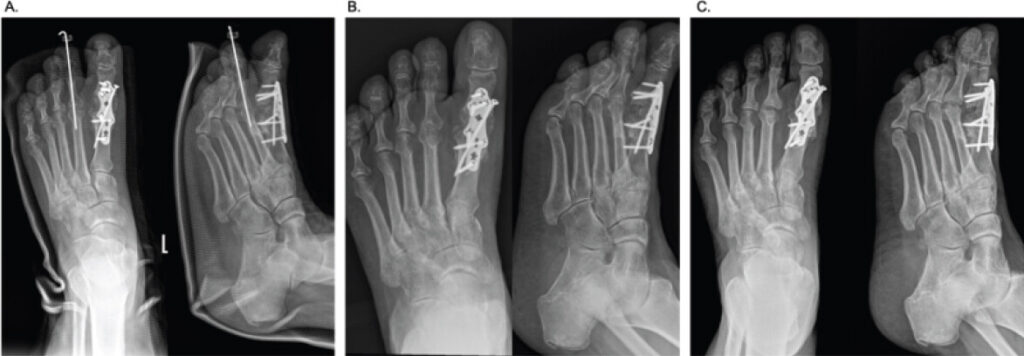
Because compression alone would have shortened the first ray, a tricortical iliac crest graft of approximately 1.8 × 1.2 cm was used to restore length and bone stock. Fixation used an interfragmentary compression screw and a stronger dorsal revision plate.
The second-ray deformity was corrected with subcapital second metatarsal osteotomy and proximal interphalangeal arthrodesis stabilized by K-wire to improve forefoot balance.
A conservative post-operative protocol was used: Eight weeks non-weight-bearing in a boot followed by gradual loading. Smoking cessation was achieved and maintained during healing.
Outcome and follow-up
Recovery was uncomplicated. At 3 months, she walked fully weight-bearing in normal footwear without pain or swelling, and radiographs showed maintained alignment with progressive graft incorporation (Fig. 3).
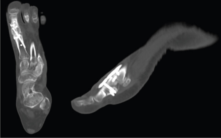
At 6 months, CT showed bridging trabecular bone, graft incorporation, closure of the fusion gap, and stable fixation without recurrent osteolysis (Fig. 4). At 1 year, she remained asymptomatic in regular footwear. The American Orthopaedic Foot and Ankle Society hallux metatarsophalangeal-interphalangeal score improved from 29/100 to 85/100 (Table 1).
Clinical timeline
| Time point | Clinical event | Interpretation |
|---|---|---|
| Pre-operative | Advanced hallux rigidus with hallux varus and second-/third-toe deformity; 45 pack-year smoker; normal arterial duplex. | Complex forefoot deformity with major modifiable biological risk factor. |
| Index surgery | First MTP arthrodesis using cup-and-cone preparation and low-profile dorsal plate fixation. | Primary fusion with standard preparation and plate fixation. |
| 4 weeks | Non-weight-bearing followed by progressive weight-bearing. | Routine primary rehabilitation protocol. |
| 3 months | Alignment maintained; fixation intact; complete osseous consolidation not evident. | Delayed or incomplete early fusion. |
| 4 months | Increasing pain and swelling during weight-bearing. | Symptomatic mechanical activity at the fusion site suspected. |
| 6 months | Radiographs demonstrated dorsal plate breakage; CT confirmed pseudarthrosis, absent bridging bone and proximal phalangeal osteolysis. WBC, ESR and CRP were normal before revision. | Aseptic symptomatic non-union with implant fatigue failure suspected. |
| Revision | Hardware removal, pseudarthrosis debridement, tricortical iliac crest graft, compression screw, stronger dorsal revision plate, second-ray correction and intraoperative cultures. | Biology, length, compression, fixation stability and forefoot loading addressed; intraoperative cultures were negative. |
| Post-revision | Eight weeks non-weight-bearing; smoking cessation maintained. | Risk-adjusted protection and host optimization. |
| 3 months after revision | Full weight-bearing in regular footwear without pain or swelling. | Clinical recovery. |
| 6 months after revision | CT-confirmed graft incorporation, bridging trabecular bone and stable fixation; pain-free walking in regular footwear; AOFAS-HMI improved from 29/100 to 85/100. | Radiographic union with major clinical and functional improvement. |
CT: Computed tomography, WBC: White blood cell, ESR: Erythrocyte sedimentation rate, CRP: C-reactive protein, MTP: Metatarsophalangeal, AOFAS-HMI: American Orthopaedic Foot and Ankle Society hallux metatarsophalangeal-interphalangeal score
Patient perspective and functional assessment
The patient reported resolution of weight-bearing pain and swelling, return to regular footwear, and improved walking tolerance.
Discussion
Why this case is more than an isolated hardware failure
Plate breakage should not be treated as a diagnosis. It is usually the visible endpoint of an unhealed fusion exposed to repetitive loading. Implant exchange alone may miss the underlying causes: Impaired biology, insufficient compression, bone loss, malalignment, premature loading, or adjacent-ray pathology.
Here, incomplete consolidation preceded symptoms, and symptoms preceded radiographic plate failure. CT then confirmed pseudarthrosis and osteolysis, supporting biological failure and micromotion before fatigue fracture.
Biological risk: Smoking and host factors
Smoking is a major modifiable risk factor for impaired osseous healing through vasoconstriction, reduced tissue oxygenation, impaired osteoblast activity, diminished collagen synthesis, and reduced angiogenesis [5,6]. Even when MTP-I-specific studies vary, broader foot-and-ankle fusion data support cessation before elective arthrodesis.
Normal pulses and duplex findings reduced concern for major arterial disease but did not exclude smoking-related microvascular or cellular impairment.
Local biology and joint preparation
Fusion requires cartilage and sclerotic bone removal, viable cancellous surfaces, and stable contact. Cup-and-cone preparation preserves bone stock and assists alignment, whereas planar cuts may improve cancellous apposition and compression [7,8,9,10]. In revision, the specific preparation method is less important than complete debridement to bleeding bone. In this case, excision of fibrous tissue and sclerosis converted an inactive pseudarthrosis into a viable graft bed.
Fixation and compression in revision arthrodesis
Stable fixation and compression are central to MTP-I fusion. Evidence generally supports constructs combining compression with dorsal plating, particularly in revisions or biologically compromised patients [11,12]. The revised construct used an interfragmentary compression screw and stronger dorsal plate to compress the graft interfaces and resist bending during rollover.
Structural grafting: Restoring biology, bone stock and length
Revision often involves bone loss, osteolysis, shortening and sclerosis. When compression would shorten the first ray, structural grafting is required. Tricortical iliac crest autograft provides support and osteogenic, osteoconductive and osteoinductive properties [13]. CT-confirmed incorporation and bridging trabeculation showed that the graft served both structural and biological roles.
Forefoot biomechanics and second-ray deformity
MTP-I arthrodesis should be assessed within the whole forefoot. Adjacent lesser-ray deformity may alter rollover mechanics and plantar pressure, although direct causality cannot be proven from a single case. Correcting the second-ray deformity was intended to improve balance and reduce abnormal loading across the revised first ray.
Post-operative protection
Although early protected weight-bearing may be safe after selected primary fusions [14,15], this evidence should not be applied uncritically to high-risk revisions. Smoking history, pseudarthrosis, bone loss, and structural grafting justified 8 weeks of non-weight-bearing before gradual loading.
Proposed revision framework
On the basis of the present case and the available literature, failed MTP-I arthrodesis should be assessed using a structured framework. The surgeon should determine whether failure is symptomatic, confirm non-union with CT when radiographs are equivocal, exclude infection, evaluate host biology, assess bone loss and first-ray length, analyze fixation and alignment, identify adjacent-ray deformity, and individualize the post-operative protocol. Revision should be designed to restore both biology and mechanics (Table 2).
Failure analysis and revision response
| Problem identified | Evidence in the present case | Revision response |
|---|---|---|
| Host biological risk | 45 pack-year smoking history and continued smoking after primary surgery. | Smoking cessation after revision and prolonged protection. |
| Local biological failure | CT-confirmed pseudarthrosis, sclerosis and osteolysis. | Excision of fibrous tissue and sclerotic bone to bleeding cancellous surfaces. |
| Structural bone loss | Osteolysis involving the base of the proximal phalanx. | Tricortical iliac crest graft to restore bone stock and length. |
| Mechanical instability | Persistent symptoms followed by dorsal plate breakage. | Interfragmentary compression screw and stronger dorsal revision plate. |
| Possible sagittal malposition | Retrospective concern for excessive dorsiflexion. | Realignment during graft insertion and revision fixation. |
| Adjacent forefoot deformity | Second-ray varus/claw-toe deformity. | Second metatarsal osteotomy and PIP arthrodesis. |
| Rehabilitation risk | Primary protocol used 4 weeks non-weight-bearing; delayed consolidation became evident. | Eight weeks non-weight-bearing after revision before gradual loading. |
| Need for objective union assessment | Radiographs were initially inconclusive. | CT used to confirm both non-union and later union. |
| Infection as differential diagnosis | No clinical infection; pre-revision WBC, ESR, and CRP were normal; no intraoperative purulence; intraoperative cultures negative. | Single-stage aseptic revision rather than staged septic reconstruction. |
| Functional impairment | Pre-revision AOFAS-HMI 29/100 (pain 0, function 29, alignment 0). | Post-revision improvement to 85/100 (pain 30, function 40, alignment 15). |
CT: Computed tomography, WBC: White blood cell, ESR: Erythrocyte sedimentation rate, CRP: C-reactive protein, MTP: Metatarsophalangeal, AOFAS-HMI: American Orthopaedic Foot and Ankle Society hallux metatarsophalangeal-interphalangeal score, PIP: Proximal interphalangeal
Comparison with published revision literature
Literature on failed MTP-I arthrodesis is limited. Takács and Swierstra used inlay grafting for pseudoarthrosis, supporting biological augmentation and stable graft-host contact [16]. Larger revision cohorts by Waizy et al. and Gaudin et al. show that revision is clinically important and carries higher complication risk than primary fusion [4,17].
Saccomanno and Bitterman treated infected non-union with staged revision, antibiotic spacer placement, and tricortical iliac crest autograft [18]. Their report supports structural grafting but differs because infection required staging. In the present aseptic case, normal inflammatory markers, absence of purulence, and negative cultures supported single-stage revision.
Malunion is another failure mode. Hirao et al. used CT-based planning and a custom guide for malunited pronation deformity after MTP-I arthrodesis, emphasizing three-dimensional alignment assessment [19]. In this case, suspected excessive dorsiflexion prompted careful sagittal restoration during grafting and fixation.
For recalcitrant non-union with hostile biology, Kaiser and Levin reported medial femoral condyle free-flap reconstruction [20]. By contrast, this case united with non-vascularized tricortical iliac crest autograft because infection was excluded, macrovascular supply was preserved, bone loss was reconstructable and stable compression fixation was achievable.
Salvage arthrodesis after failed MTP-I arthroplasty also informs bone-loss reconstruction. Mao et al., Yurteri et al., Johnson et al. and Adamson et al. highlight strategies including structural grafting, custom implants and length maintenance when bone stock is compromised [21,22,23,24] (Table 3).
Selected literature relevant to failed or revised first MTP arthrodesis and salvage reconstruction
| Author/year | Type of problem | Revision or salvage strategy | Relevance to present case |
|---|---|---|---|
| Prat et al., 2023 [10] | Revision-first MTP arthrodesis cohort. | Revision arthrodesis with variable techniques. | Demonstrates that revision first MTP fusion has higher complication risk than primary fusion. |
| Takács and Swierstra, 2011 [16] | Pseudoarthrosis after failed MTP-1 arthrodesis. | Inlay bone grafting technique for repair. | Directly supports graft-based revision for failed first MTP fusion. |
| Gaudin et al., 2018 [17] | Multicenter series of 158 revision procedures after first MTP arthrodesis. | Revision for hardware problems, non-union, malunion, and related complications. | Shows that revision after first MTP fusion is not negligible and may address more than one failure mechanism. |
| Saccomanno and Bitterman, 2023 [18] | Infected non-union after first MTP arthrodesis. | Staged revision with antibiotic spacer and tricortical iliac crest autograft. | Closest comparator for structural iliac crest autograft in failed fusion, although infected and staged. |
| Hirao et al., 2014 [19] | Malunited pronation deformity after first MTP arthrodesis. | 3D-CT planning, custom guide, deformity correction and re-fusion. | Highlights the importance of three-dimensional alignment analysis in revision. |
| Kaiser and Levin, 2023 [20] | Persistent/recalcitrant osseous non-union of the first MTP joint. | Medial femoral condyle free flap. | Represents advanced vascularized biological salvage when local biology is severely compromised. |
| Mao et al., 2020 [21] | Failed first MTP arthroplasty. | Systematic review of salvage arthrodesis with or without structural graft. | Supports the role of salvage arthrodesis in bone-loss revision settings. |
| Yurteri et al., 2024 [22] | Failed first MTP total arthroplasty. | Arthrodesis using iliac crest autograft. | Contemporary series supporting iliac crest grafting in revision first MTP reconstruction. |
| Johnson et al., 2023 [23] | Osseous defects in revision first MTP fusion. | Custom three-dimensional printed implant. | Modern comparator for reconstructing bone loss and first-ray length when standard grafting is insufficient. |
| Adamson et al., 2021 [24] | Failed first MTP arthroplasty with bone loss preventing fixation. | Arthrodesis using allograft struts without hardware. | Demonstrates the centrality of length and position restoration when fixation options are limited by bone loss. |
This case was distinct because aseptic failed primary arthrodesis occurred in a heavy smoker with CT-confirmed pseudarthrosis, osteolysis, and plate fatigue and second-ray deformity. Union was achieved without staged infection treatment, vascularized bone transfer, or arthroplasty conversion by combining debridement to viable bone, structural autograft, compression, enhanced fixation, adjacent deformity correction, smoking cessation, and prolonged protection.
Revision series support a whole-forefoot, stepwise escalation approach (Table 4). Gaudin et al. identified revision indications beyond hardware problems, including non-union, malunion, metatarsalgia, claw-toe deformity, and interphalangeal disorders [17]. Takács, Swierstra, and Malhotra et al. support graft-based repair and length-preserving bone-block arthrodesis [16,25], whereas Kaiser and Levin represent escalation to vascularized bone for rare hostile non-unions [20] (Table 4).
Revision series and case-series evidence supporting the reconstruction strategy
| Study | Design/population | Main contribution | Relevance to present case |
|---|---|---|---|
| Takacs and Swierstra, 2011 [16] | Technical note/case series of 26 patients with pseudoarthrosis after failed MTP-1 arthrodesis. | Described a turnaround inlay graft bridging the pseudoarthrosis, with fixation and protected heel weight-bearing. | Directly supports graft-based biological reconstruction for established first MTP pseudarthrosis. |
| Gaudin et al., 2018 [17] | Multicenter study of 158 revision procedures after first MTP arthrodesis. | Showed that revision indications include hardware discomfort, non-union, malunion, metatarsalgia/claw-toe deformity, and IP-joint disorders. | Supports analyzing failed fusion as a spectrum of mechanical, alignment, and adjacent-ray problems rather than only broken hardware. |
| Kaiser and Levin, 2023 [20] | Level IV preliminary case series for persistent osseous non-union of the first MTP joint. | Reported medial femoral condyle free-flap reconstruction as a vascularized biological salvage option. | Defines the upper end of biological escalation for recalcitrant non-union; the present case achieved union without vascularized transfer. |
| Malhotra et al., 2015 [25] | Retrospective case series: 24 patients/25 feet undergoing interposition bone-block arthrodesis. | Used non-vascularized tricortical autologous iliac crest graft to maintain first-ray length in failed hallux MTP surgery. | Strongly supports the use of structural iliac crest autograft when bone loss or shortening would make simple compression inadequate. |
MTP-1: First metatarsophalangeal
Conclusion
Non-union after MTP-I arthrodesis is usually multifactorial. Revision should restore viable bone surfaces, bone stock, first-ray length, alignment, compression, and fixation stability while correcting modifiable host and biomechanical risk factors. In the present case, successful union was achieved after pseudarthrosis excision, structural tricortical iliac crest autografting, compression screw fixation, revision dorsal plating, smoking cessation, correction of associated second-ray deformity and prolonged post-operative protection.
Clinical Message
Revision should not be limited to hardware exchange. Surgeons should systematically assess infection, biological risk factors, bone loss, fixation stability, alignment, post-operative loading and adjacent forefoot deformity. In high-risk cases, successful revision may require structural autografting, stable compression fixation, deformity correction, and smoking cessation and prolonged protected weight-bearing.
Conflict of Interest:
Source of Support:
Nil
Consent:
The authors confirm that informed consent was obtained from the patient for publication of this article
References
- Coughlin MJ, Shurnas PS.Hallux rigidus. Grading and long-term results of operative treatment. J Bone Joint Surg Am 2003;85:2072-88. [Google Scholar] | [PubMed]
- Roukis TS.Nonunion after arthrodesis of the first metatarsal-phalangeal joint: A systematic review. J Foot Ankle Surg 2011;50:710-3. [Google Scholar] | [PubMed]
- Myerson MS, Cerrato R.Current management of complications after first metatarsophalangeal joint arthrodesis. Foot Ankle Clin 2011;16:105-20. [Google Scholar] | [PubMed]
- Prat D, Haghverdian BA, Pridgen EM, Lee W, Wapner KL, Chao W, Farber DC.High complication rates following revision first metatarsophalangeal joint arthrodesis: a retrospective analysis of 79 cases. Arch Orthop Trauma Surg 2023;143:1799-1807. [Google Scholar] | [PubMed]
- Patel RA, Wilson RF, Patel PA, Palmer RM.The effect of smoking on bone healing: A systematic review. Bone Joint Res 2013;2:102-11. [Google Scholar] | [PubMed]
- Haverstock BD, Mandracchia VJ.Cigarette smoking and bone healing: implications in foot and ankle surgery. J Foot Ankle Surg 1998;37:69-74. [Google Scholar] | [PubMed]
- Füssenich W, Seeber GH, Van Raaij TM, Van Lingen CP, Zuurmond RG, Stevens M.Factors associated with nonunion in arthrodesis of the first metatarsophalangeal joint: A multicenter retrospective cohort study. Foot Ankle Int 2023;44:508-15. [Google Scholar] | [PubMed]
- Füssenich W, Seeber GH, Zwoferink JR, Somford MP, Stevens M.Non-union incidence of different joint preparation types, joint fixation techniques, and postoperative weightbearing protocols for arthrodesis of the first metatarsophalangeal joint in moderate-to-severe hallux valgus: A systematic review. EFORT Open Rev 2023;8:101-9. [Google Scholar] | [PubMed]
- Korim MT, Mahadevan D, Ghosh A, Mangwani J.Effect of joint pathology, surface preparation and fixation methods on union frequency after first metatarsophalangeal joint arthrodesis: A systematic review of the English literature. Foot Ankle Surg 2017;23:189-94. [Google Scholar] | [PubMed]
- Prat D, Sourugeon Y, Haghverdian BA, Pridgen EM, Lee W, Wapner KL.“In situ” joint preparation technique for first metatarsophalangeal arthrodesis: A retrospective comparative review of 388 cases. J Foot Ankle Surg 2023;62:855-61. [Google Scholar] | [PubMed]
- Kang YS, Bridgen A.First metatarsophalangeal joint arthrodesis/fusion: A systematic review of modern fixation techniques. J Foot Ankle Res 2022;15:30. [Google Scholar] | [PubMed]
- Balu AR, Baumann AN, Tsang T, Talaski GM, Anastasio AT, Walley KC.Evaluating the biomechanical integrity of various constructs utilized for first metatarsophalangeal joint arthrodesis: A systematic review. Materials (Basel) 2023;16:6562. [Google Scholar] | [PubMed]
- Brewster M.Revision first metatarsophalangeal arthrodesis with structural bone grafting. Foot Ankle Clin 2018;23:95-108. [Google Scholar] | [PubMed]
- Crowell A, Van JC, Meyr AJ.Early weight-bearing after arthrodesis of the first metatarsal-phalangeal joint: A systematic review of the incidence of non-union. J Foot Ankle Surg 2018;57:1200-3. [Google Scholar] | [PubMed]
- Schwab A, Rose G, Meyr A, Ramdass R.Nonunion incidence following differing weightbearing protocols after first metatarsal phalangeal joint arthrodesis: A systematic review. J Foot Ankle Surg 2026;65:69.e1-6. [Google Scholar] | [PubMed]
- Takács IM, Swierstra BA.Pseudoarthrosis repair after failed metatarsophalangeal 1 arthrodesis. Acta Orthop 2011;82:114-5. [Google Scholar] | [PubMed]
- Gaudin G, Coillard JY, Augoyard M, Tourné Y, Meusnier T, Beaudet P.Incidence and outcomes of revision surgery after first metatarsophalangeal joint arthrodesis: Multicenter study of 158 cases. Orthop Traumatol Surg Res 2018;104:1221-6. [Google Scholar] | [PubMed]
- Saccomanno R, Bitterman AD.Staged revision arthrodesis with interposition of tricortical iliac crest autograft to address an infected nonunion of the first metatarsophalangeal joint: A case report. J Am Podiatr Med Assoc 2023;113:21-149. [Google Scholar] | [PubMed]
- Hirao M, Ikemoto S, Tsuboi H, Akita S, Ohshima S, Saeki Y.Computer assisted planning and custom-made surgical guide for malunited pronation deformity after first metatarsophalangeal joint arthrodesis in rheumatoid arthritis: A case report. Comput Aided Surg 2014;19:13-9. [Google Scholar] | [PubMed]
- Kaiser D, Levin LS.Medial femoral condyle free flap for persistent osseous nonunion of the first metatarsophalangeal joint: A preliminary report of a new surgical indication for the medial femoral condyle free flap. Foot Ankle Orthop 2023;8:24730114231191135. [Google Scholar] | [PubMed]
- Mao DW, Zheng C, Amatullah NN, Kam C, Kon Kam King C.Salvage arthrodesis for failed first metatarsophalangeal joint arthroplasty: A network meta-analysis. Foot Ankle Surg 2020;26:614-23. [Google Scholar] | [PubMed]
- Yurteri A, Mercan N, Kekeç AF, Yıldırım A.Arthrodesis using Iliac Crest autograft in revision surgery of the first metatarsophalangeal joint total arthroplasty for the treatment of Hallux Rigidus: A retrospective study. J Orthop Surg Res 2024;19:508. [Google Scholar] | [PubMed]
- Johnson LJ, Leatham PR, Clougherty CO, Highlander PD.Salvage first metatarsophalangeal joint fusion with a three-dimensional-printed implant for osseous defects: A case series. J Am Podiatr Med Assoc 2023;113:21-148. [Google Scholar] | [PubMed]
- Adamson P, Janney C, Chen J, Panchbhavi V.First metatarsal phalangeal joint arthrodesis without the use of hardware after failed arthroplasty: A case report. J Orthop Case Rep 2021;11:63-6. [Google Scholar] | [PubMed]
- Malhotra K, Nunn T, Qamar F, Rao V, Shanker J.Interposition bone block arthrodesis for revision hallux metatarsophalangeal joint surgery: A case series. Foot Ankle Int 2015;36:556-64. [Google Scholar] | [PubMed]
© 2026 Journal of Orthopaedic Case Reports - Published by Indian Orthopaedic Research Group
About the Authors
-

Dr. Georgios Renieris
-

Dr. Natalia Renieri
-

Dr. Athanasios Georgokostas
Related Articles in Journal of Orthopaedic Case Reports
 March 1, 2026 Clavicle Fracture Non-Union in a 3-Year-Old Child: A Case Report and Literature Review
 October 1, 2026 Real-Time Coagulation Monitoring via Thromboelastography and Platelet Mapping in Total Hip Arthroplasty for a Patient with Factor XI Deficiency: A Case Report
 October 1, 2026 Posterior Fibular Entrapment in a Bosworth Fracture-Dislocation: A Case Report and Review of Diagnostic and Operative Considerations
 October 1, 2026 Humeral Fractures in Metabolically Compromised Bone: Outcomes of Structural Strut Onlay Allograft with Open Reduction and Internal Fixation – A Case Series of Three Patients