


Introduction
Human hydatid disease is a zoonotic infection by Echinococcus granulosus, acquired through ingestion of egg-contaminated food or water. The liver and lungs are the predominant sites of cyst formation, whereas musculoskeletal involvement is rare. Osseous hydatidosis represents only 0.2–3% of all reported cases [1]. In bone hydatidosis, pelvic sites are less common than vertebral or long-bone involvement. Primary osseous disease arises from hematogenous spread of scolices bypassing hepatic and pulmonary filters, explaining its rarity. Unlike soft-tissue cysts, bone lesions lack pericyst formation, infiltrate trabeculae, show irregular margins without sclerosis or periosteal reaction, and rarely calcify [2, 3]. Osseous cystic echinococcosis presents considerable therapeutic difficulties owing to its high recurrence rate, particularly in sites such as the ilium and hip, where complete surgical excision is technically challenging.
Case Report
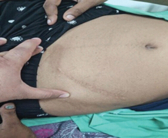
A 25-year-old female from a rural background presented to our outpatient department with complaints of recurrent swelling in the left lower abdominal quadrant for the past 4 months, which had been gradually increasing in size. There was no history of associated fever, urinary or bowel disturbances, significant weight loss, discharge, or preceding trauma. She reported a similar episode 3 years earlier, for which she underwent evaluation and surgical excision of a cystic mass at another center; however, detailed medical records were unavailable. A healed surgical scar mark of 8 × 1 cm was present over the left iliac region extending along the iliac crest (Fig. 1), and 1 scar mark of 2 × 2 cm was present over the left gluteal region. The patient remained asymptomatic for approximately 3 years following the initial procedure.
Clinical examination
Abdominal examination revealed a soft, non-tender, palpable mass measuring approximately 7 × 7 cm in the left iliac fossa. The mass was firm in consistency, non-mobile, and appeared fixed to the underlying structures.
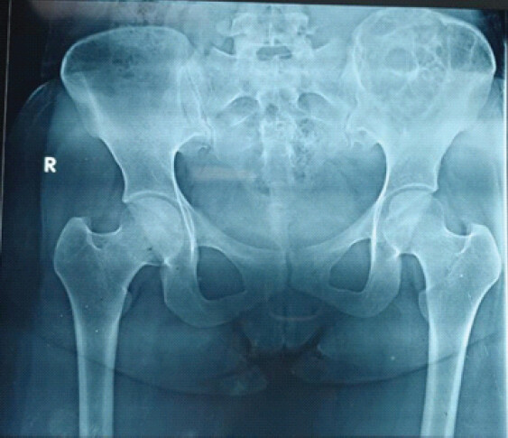
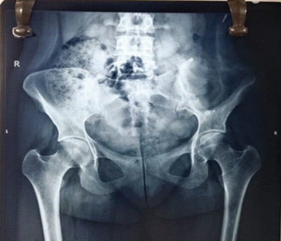
Plain radiography demonstrated osteolytic lesions involving the left iliac wing with extension into a portion of the sacrum (Fig. 2).
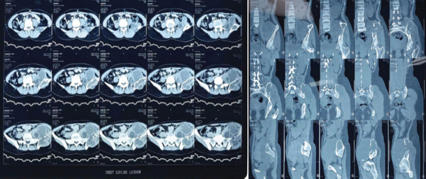
Contrast-enhanced computed tomography revealed an expansile osteolytic lesion in the left iliac bone, accompanied by a surrounding multiloculated lesion involving the iliopsoas and gluteus medius muscles, with multiple thin-walled cystic components (Fig. 3), whereas the magnetic resonance imaging (MRI) revealed a well-defined multiloculated cystic lesion with lobulated margins in the left iliac bone, breaching the cortex and extending into the iliopsoas and gluteus medius, with additional smaller lesions in adjacent muscles. Follow-up MRI at 3 months showed progression, with a multiloculated cystic mass involving the iliac bone, cortical disruption, anterior extension to the abdominal wall, and inferior spread to the level of the hip joint. Pre-operative planning was done involving the team of a gastrosurgeon, orthopedic surgeon, and a plastic surgeon. Thorough surgical debridement was planned, along with careful exploration of the neurovascular bundle.
Positioning
The patient was taken inside the operating theater and was positioned in the right lateral decubitus position.
Surgical steps
Endopelvic exploration was performed via an anterior approach, with an incision from the pubic symphysis to the anterior superior iliac spine, extended along the iliac crest. The femoral neurovascular bundle was preserved, and the anterior abdominal wall muscles were reflected from the pubic ramus and inguinal ligament. Multiple cysts were observed protruding from the left iliac blade into the extraperitoneal space. Hip exposure was achieved through a Tshaped incision, beginning at the posterior inferior iliac spine, extending along the iliac crest to join the anterior incision, and continuing distally lateral to the femoral vessels before curving around the thigh. Dissection proceeds more deeper meticulously and under loupe magnification to preserve the neurovascular bundles of the Gluteus maximus muscle and the fibers of the gluteus maximus and gluteus medius were separated, multiple cysts were encountered within the gluteus medius and minimus muscle. After meticulous dissection and confirmation of the cyst boundaries, careful excision of the cyst was performed along with a Type I left hemipelvectomy using a bone saw Fig 4.
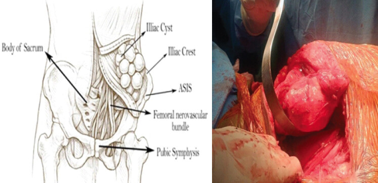
During the procedure, the cyst was removed in toto, no anaphylactic reaction was observed intraoperatively. Thorough irrigation was carried out using 3% hypertonic saline followed by 10% povidone-iodine solution for 20–30 minutes. The cyst was also found to involve the body of the illium and was removed using a curette, followed by additional irrigation with 10% povidone-iodine solution. All involved muscles were excised, including a major portion of the gluteus medius, gluteus minimus, and quadratus lumborum, while the gluteus maximus was preserved.
Wound closure was performed in layers with two drains left in situ. Endoprosthetic reconstruction was avoided to reduce infection risk, and postoperative albendazole was maintained for six months.
Post-operative period
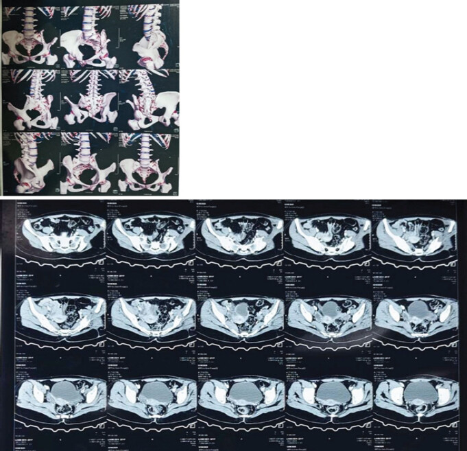
The post-operative period was uneventful; however, the patient had developed numbness over the anterolateral aspect of the left thigh extending up to the left ankle, which was attributed to neurapraxia. Drains and sutures were removed on post-operative day 14, with the surgical wound noted to be healthy and dry; also, numbness had reduced, and was localized over the anterolateral aspect of left thigh. A post-operative radiograph and CT scan were done, which showed no evidence of any remnant lesion (Fig. 5 and 6).
Complications
The complications we encountered in the post-operative period were numbness over the anterolateral aspect of the left thigh and abductor muscle weakness, which manifested as an abductor lurch gait.
Rehabilitation
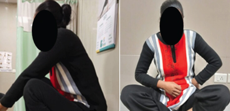
Gentle hip abduction and adduction exercises were initiated 2 weeks after surgery. At 4 weeks postoperatively, the patient was able to ambulate with support, exhibiting an abductor lurch attributable to post-operative abductor muscle weakness. She was started on hip abduction exercises. At 6 weeks postoperatively, the numbness was limited to the anterolateral aspect of the proximal third of the left thigh, and range of movement had improved; she was able to do ground-level activities. Comprehensive gait training and rehabilitation were initiated, leading to a satisfactory functional recovery. At 3 months following surgery, the patient was able to sit cross-legged and squat comfortably (Fig. 7), and could ambulate independently without external support, though a mild residual lurch persisted.
Discussion
Osseous hydatid disease is a rare parasitic infection caused by the larval stage of Echinococcus tapeworm, with skeletal involvement reported in only 0.2–3% of hydatid cyst cases [1]. Osseous hydatidosis predominantly affects the vertebrae, with pelvic sites less frequent than vertebral and long-bone involvement. E. granulosus completes its cycle in dogs and other carnivores (definitive hosts), with sheep and herbivores as intermediates; humans are accidental hosts. Transmission occurs via the fecal–oral route, with larvae entering the portal circulation and primarily involving the liver. Hematogenous spread accounts for dissemination: ~90% of cysts occur in the liver and lungs, 2–3% in kidneys, spleen, and muscles, and <1% in heart, brain, or bone. Among skeletal cases, vertebrae are most common (~50%), followed by pelvis (16–28%) [3].
The disease often remains asymptomatic until advanced, when bone pain or deformity may develop. Physical findings are typically minimal. Given its non-specific presentation, a high index of suspicion is warranted in at-risk populations such as sheepherders and butchers.
Differential diagnoses include infectious conditions, particularly tuberculosis, and neoplasms, with additional considerations depending on lesion location [4].
Hydatid cysts may cause symptoms due to mass effect, obstruction, or allergic reactions. Management typically combines surgical excision with anthelminthic therapy to reduce recurrence [2]. Musculoskeletal hydatidosis can mimic infections or tumoral lesions, making diagnosis challenging. Although MRI and CT are the most accurate imaging modalities, ultrasound and plain radiography remain widely used in endemic, resource-limited settings [3, 5]. Plain radiographs in skeletal hydatidosis often show non-specific expansile radiolucent lesions with cortical thinning, mimicking spinal tuberculosis or primary bone tumors. Pelvic involvement, particularly of the iliac bone, may display a characteristic “waffle-like” osteolytic pattern. CT typically reveals well-defined ovoid cystic lesions, sometimes with “double-layered arcuate” calcifications, and is useful for evaluating cortical erosion and pathological fractures [1, 6, 7]. Surgery remains the primary treatment, supplemented by albendazole (± praziquantel), though radical bone resection is technically challenging and prosthetic implantation carries high infectious risk [8].
Verghese et al. described a scapular case of hydatid bone disease, emphasizing that primary osseous involvement is rare and frequently misdiagnosed on initial clinical and radiological evaluation due to non-specific features. Recognition of this entity is essential in the differential diagnosis of bony lesions [9]. Osseous hydatid cysts are rare but potentially serious lesions that exhibit behavior similar to locally aggressive malignancies. While immunofluorescent assays can aid in diagnosis, definitive confirmation relies on histopathological examination. Surgical excision remains the cornerstone of treatment, although recurrence is frequent, particularly in anatomically challenging locations [10].
Reported complications include pathological fractures, secondary infection, fistula formation, and frequent recurrence [11]. Hydatid bone disease mimics tumors; the parasite spreads along paths of least resistance, producing progressive erosion with fracture risk, and recurs in up to 48% due to incomplete eradication, as shown in Table 1 [12].
| Author year | Lesions | Initial diagnosis | Management | Outcome |
|---|---|---|---|---|
| Belzunegui et at. (1997) [13] | Hemipelvis and proximal femur | – | Girdlestone arthroplasty followed by 10-year chemotherapy | Recurrence, managed by chemotherapy |
| Siwach et at. (2009) [14] | Pelvis and femur (pathological fracture) | – | Hindquarter amputation followed by chemotherapy | Death due to sepsis |
| Nath et at. (2009) [15] | Illiac bone, acetabulum, ischial tuberosity and pubic rami | ABC, recurrence was also labeled as ABC | Wide excision | – |
| Neelapal et at. (2010) [16] | Hip joint and ilium | Propionibacterial infection | 2 years of chemotherapy followed by cemented THR | Recurrence, managed by customized hemipelvic replacement |
| Bhatnagar et at. (2017) [3] | Acetabulum, pubic rami and femoral head | – | Surgical excision followed by chemotherapy | No recurrence and good functional outcome |
ABC: Aneurysmal bone cyst, THR: Total hip replacement
Conclusion
Osseous hydatid disease is a rare manifestation of echinococcal infection that presents major diagnostic and therapeutic challenges. Surgical excision remains the cornerstone of management, yet complete eradication is often hindered by proximity to vital neurovascular structures and the need to preserve function. Consequently, residual disease and high recurrence rates are common. Radical resection may also result in post-operative muscle weakness requiring intensive rehabilitation. Intraoperative cyst rupture and local dissemination further compound recurrence risk despite scolicidal measures and adjuvant anti-helminthic therapy. These factors underscore the necessity of thorough debridement, extended medical therapy, and vigilant long-term follow-up.
Clinical Message
Optimal functional outcomes in musculoskeletal hydatid cyst disease depend on meticulous surgical planning and a multidisciplinary approach; while radical resection remains key to preventing recurrence, it is also the most common complication.
Conflict of Interest:
Source of Support:
Nil
Consent:
The authors confirm that informed consent was obtained from the patient for publication of this article
References
- Papanikolaou A.Osseous hydatid disease. Trans R Soc Trop Med Hyg 2008;102:233-8. [Google Scholar] | [PubMed]
- Rammeh S, Romdhane E.Pathological Diagnosis of Appendicular Musculoskeletal Infections Peh WC, Ladeb MF..Imaging of Appendicular Musculoskeletal Infections Medical Radiology. Cham, Switzerland: Springer Nature; 202533-74. Available from: https://link.springer.com/10.1007/978-3-031-93638-8_3. [Last accessed on 2026 Jan 24] [Google Scholar] | [PubMed] | [CrossRef]
- Bhatnagar N, Kishan H, Sura S, Lingaiah P, Jaikumar K.Pelvic hydatid disease: A case report and review of literature. J Orthop Case Rep 2017;7:25-8. [Google Scholar] | [PubMed]
- Inayat F, Azam S, Baig AS, Nawaz G, Ullah W.Presentation patterns, diagnostic modalities, management strategies, and clinical outcomes in patients with hydatid disease of the pelvic bone: A comparative review of 31 cases. Cureus 2019;11:e4178. [Google Scholar] | [PubMed]
- Ammar A, Riahi H, Chaabouni M, Venturelli N, Renault V, Dray B.The multifaceted musculoskeletal hydatid disease. Skeletal Radiol 2024;53:2181-94. [Google Scholar] | [PubMed]
- Govindasamy A, Bhattarai PR, John J.Lytic parasitic: A case of bone destructing echinococcosis. Ther Adv Infect Dis 2021;8:20499361211047664. [Google Scholar] | [PubMed]
- Boussaid S, Daldoul C, Hassayoun M, Rekik S, Jammali S, Sahli H.Primitive pelvic bone hydatidosis: What an amazing extension. Clin Case Rep 2021;9:e05054. [Google Scholar] | [PubMed]
- Laasri K, Naggar A, Bahlouli N, Chait F, Ahallat I, Boufettal M.Osseous hydatid disease: A mimic of other skeletal pathologies. Radiol Case Rep 2023;18:3145-51. [Google Scholar] | [PubMed]
- Verghese SC, George ST, Duraisamy P, Karthikeyan TM, Kumar SA, Arasu S.A rare case report of primary hydatid disease of the scapula - an unforeseen diagnosis!. J Orthop Case Rep 2022;12:98-101. [Google Scholar] | [PubMed]
- Loudiye H, Aktaou S, Hassikou H, El-Bardouni A, Manouar ME, Fizazi M.Hydatid disease of bone. Review of 11 cases. Joint Bone Spine 2003;70:352-5. [Google Scholar] | [PubMed]
- Kalinova K, Proichev V, Stefanova P, Tokmakova K, Poriazova E.Hydatid bone disease: A case report and review of the literature. J Orthop Surg (Hong Kong) 2005;13:323-5. [Google Scholar] | [PubMed]
- Atalay İB, Ulucaköy C, Seyhoğlu MC, Yükrük FA, Mısırlıoğlu M, Güngör BŞ.A rare lesion that mimics a bone tumor: Osseous hydatidosis. Jt Dis Relat Surg 2022;33:435-9. [Google Scholar] | [PubMed]
- Belzunegui J, Maiz O, Lopez L, Plazaola I, Gonzalez C, Figueroa M.Hydatid disease of bone with adjacent joint involvement. A radiological follow-up of 12 years. Rheumatology 1997;36:133-5. [Google Scholar] | [PubMed]
- Siwach R, Singh R, Kadian VK, Singh Z, Jain M, Madan H.Extensive hydatidosis of the femur and pelvis with pathological fracture: A case report. Int J Infect Dis 2009;13:e480-2. [Google Scholar] | [PubMed]
- Nath P, Bhattacharya S, Dutta V, Joshi GR, Patel M.Primary iliac bone hydatid disease: An unusual presentation. Med J Armed Forces India 2009;65:180-1. [Google Scholar] | [PubMed]
- Neelapala VS, Chandrasekar CR, Grimer RJ.Revision hip replacement for recurrent Hydatid disease of the pelvis: A case report and review of the literature. J Orthop Surg 2010;5:17. [Google Scholar] | [PubMed]
© 2026 Journal of Orthopaedic Case Reports - Published by Indian Orthopaedic Research Group
About the Authors
-

Dr. Supriya Thakur
-

Dr. Anurag Baghel
-

Dr. Ashish Singh
-

Dr. Rajiv Kumar Bharti
-

Mr. Hamza Ali
Related Articles in Journal of Orthopaedic Case Reports
 October 1, 2026 Recurrent Ventral Scapular Osteochondroma Presenting with Pseudo-winging in an Adolescent: A Case Report and Follow-up of a Previously Published Case
 September 1, 2025 A Case of Atlanto-Axial Rotatory Fixation Requiring Internal Fixation
 August 1, 2025 Intra-Articular Steroid Hip Injections Association with Fracture: A Case Series
 August 10, 2022 A Rare Case Report of Primary Hydatid Disease of the Scapula – an Unforeseen Diagnosis!