


Introduction
Osteoarthritis of the knee is a common disease, with approximately 25 million people estimated to have it in Japan based on domestic epidemiological studies [1]. Osteophyte is osteogenic changes that appear and enlarge as osteoarthritis progresses. Osteophytes may irritate surrounding tissues, causing various symptoms. Typically, they compress the medial collateral ligament [2] and the posterior joint capsule [3,4], often leading to pain and restricted range of motion. Anterior osteophytes have been reported to cause knee pain by impingement on the quadriceps tendon [5], but this report selected conservative treatment. Regarding surgical treatment for symptomatic osteophytes, while there is a report of favorable outcomes with surgery for popliteus tendon impingement [6], there are no reports on surgical treatment for quadriceps tendon impingement. The author experienced a case who had left knee pain with snapping due to interference of an osteophyte with the quadriceps tendon, and the symptoms resolved following arthroscopic osteophyte resection. This is the first report of a surgical procedure of osteophyte impingement to quadriceps tendon.
Case Report
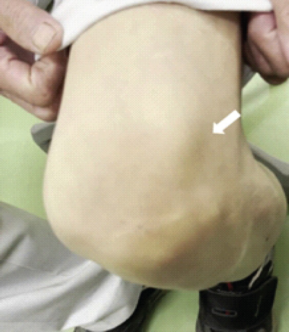
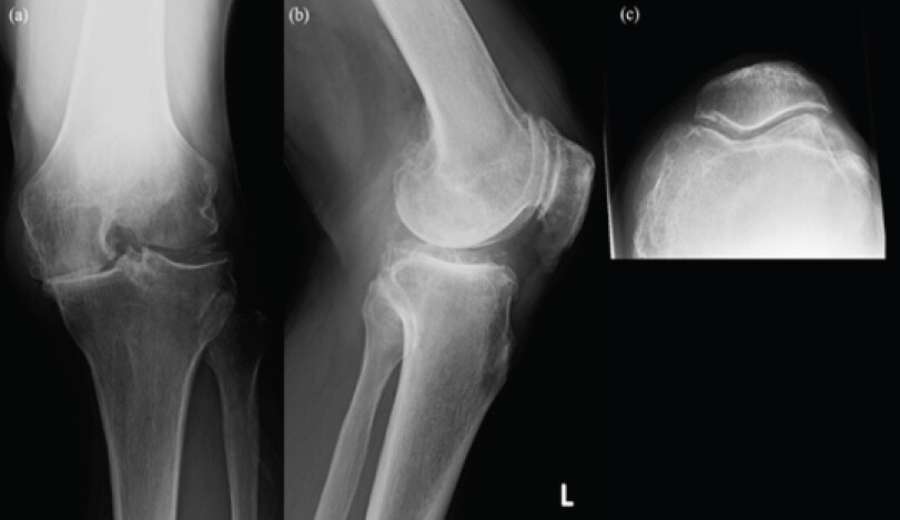
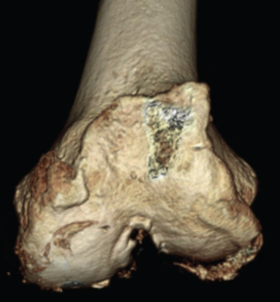
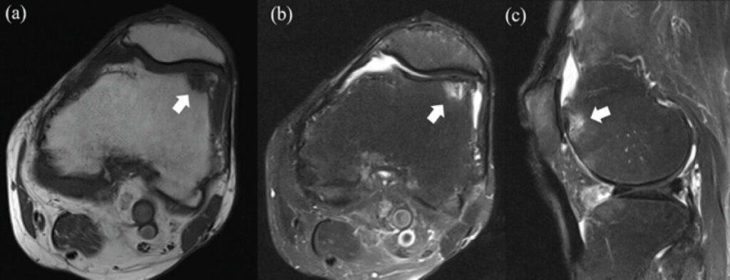
An 82-year-old Asian man had become aware of left knee pain with snapping a few years before. The knee pain hadn’t arisen during gait. He came to our hospital because the symptom continued. There was previous medial collateral injury which was repaired with surgery and sudden deafness as a history. The range of motion of the left knee was almost full, but the pain and snapping happened at a range of 90–120°. The tenderness located in superolateral from patella, and the tactile sensation was bone by palpation (Fig. 1). The joint stability was normal in either the anterior, posterior, varus, or valgus. The joint space was narrow at the medial femorotibial joint and patellofemoral joint; osteophytes were formed medial, lateral, and patellofemoral joint in a plain radiography (Fig. 2). The knee was diagnosed with knee osteoarthritis of Kellgren-Lawrence classification grade 4. In a computed tomography (CT), the osteophyte at the lateral femoral trochlea was confirmed at sites corresponding to the areas of tenderness (Fig. 3). Magnetic resonance imaging (MRI) showed a change of intensity, low signal in T1-weighted image and a high signal in fat-suppressed T2-weighted image, occurred at the osteophyte (Fig. 4).
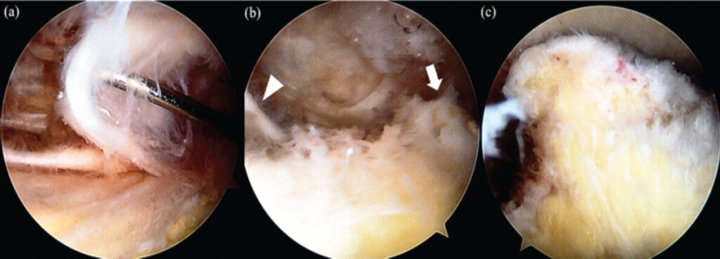
The primary cause of pain was diagnosed as impingement of the quadriceps tendon by osteophytes, not osteoarthritis. After several months of conservative treatment, including quadriceps exercises and pain reliever, no improvement was achieved, arthroscopic osteophyte resection was planned. Observation from the superomedial portal revealed a cord-like structure continuous with the quadriceps tendon on the lateral part of the superior pouch, which was in contact with the planned osteophyte resection during flexion (Fig. 5). Both of these were excised until no contact confirmed.
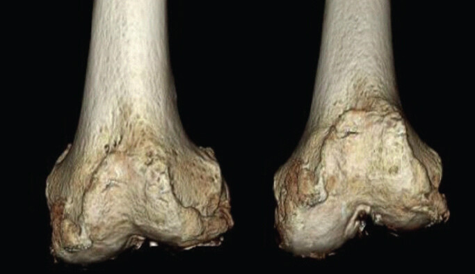
After surgery, snapping and pain resolved. CT also showed that the anterolateral osteophytes were adequately resected (Fig. 6). At the 6-month post-operative follow-up, the patient remains pain-free and is doing well.
Discussion
Osteophytes proliferate as osteoarthritis progresses, interfering with surrounding soft tissues and potentially contributing to pain and functional impairment such as joint contracture. Osteophytes are thought to be an adaptation process arising in response to joint deformity, but their function and mechanism of formation remain largely unclear[7,8].
Dughhbaj had reported a case similar to the present one in which symptomatic osteophytes causing quadriceps impingement. In the report, the diagnosis was based on physical findings and ultrasonography. Treatment consisted of rest and physical therapy, which had been shown to improve symptoms [5]. However, in our case, conservative treatment, such as quadriceps exercises, failed to produce improvement over several months needed to surgery.
A report on popliteal tendon impingement noted that in 2 out of 3 cases, patients complained of symptoms indicative of mechanical irritation, such as a click or locking in the posterolateral region during flexion [6]. Such mechanical symptoms are useful for diagnosis, and in fact, we were able to diagnose this disease based on the snapping sensation complained by the patient. In this case, MRI revealed signal intensity changes at the site of the osteophyte, which supported the diagnosis. However, one report found no association between signal intensity changes within osteophytes and knee pain; therefore, physical findings should be given greater weight than imaging findings in diagnosis. In addition, osteophyte impingement of the popliteus tendon may occur even after total knee arthroplasty; therefore, residual osteophytes should be considered as a possible cause if pain persists after surgery [9].
Quadriceps fat pad impingement syndrome is one of the differential diagnoses for this condition. This condition is characterized by the presence of edematous changes in the suprapatellar fat pad as seen on MRI. However, while the reports on this condition use the term “impingement,” they did not refer exactly where the impingement occurs [10,11].
Gaine reported three cases in which symptoms improved following surgery for osteophytes causing popliteal tendon impingement [6]. According to their report, osteophyte resection was performed in two cases, and popliteus tendon resection in one case. In. our case, pre-operative imaging suggested that the impingement could be resolved by osteophyte resection alone. However, arthroscopic examination revealed a cord-like structure extending from the quadriceps tendon at the contact site. The snapping was presumed to result from interference between this structure and the osteophyte; therefore, the soft-tissue cord was also resected. Although the symptoms may have improved solely due to osteophyte resection, careful assessment of the sites where the osteophytes come into contact is necessary during surgery for this condition to determine whether additional treatment is required.
In this case, surgical treatment resulted in immediate improvement in symptoms, and the patient has remained symptom-free to date.
Conclusion
We experienced a case that an osteophyte formed on the lateral femoral trochlea caused impingement to the quadriceps tendon, resulting in left knee pain. Because the conservative treatment was ineffective, the patient underwent arthroscopic surgery and achieved a favorable result.
Clinical Message
When examining patients presenting with superior knee pain, it is necessary to suspect osteophyte impingement to the quadriceps tendon. Physical examination and medical history are most important for diagnosis, and surgical treatment is considered in cases resistant to conservative therapy.
Conflict of Interest:
Source of Support:
Nil
Consent:
The authors confirm that informed consent was obtained from the patient for publication of this article
References
- Yoshimura N, Akune T, Fujiwara S, Shimizu Y, Yoshida H, Omori G.Prevalence of knee pain, lumbar pain and its coexistence in Japanese men and women: The longitudinal cohorts of motor system organ (LOCOMO) study. J Bone Miner Metab 2014;32:524-32. [Google Scholar] | [PubMed]
- Asavanapakas P, Pariyakhup W, Boonriong T, Boonsri P, Laohawiriyakamol T, Chuaychoosakoon C.Osteophyte-related medial collateral ligament injury in asymptomatic osteoarthritis following minor trauma: A case report. Ann Med Surg (Lond) 2023;85:2014-6. [Google Scholar] | [PubMed]
- Leie MA, Klasan A, Oshima T, Putnis SE, Yeo WW, Luk L.Large osteophyte removal from the posterior femoral condyle significantly improves extension at the time of surgery in a total knee arthroplasty. J Orthop 2019;19:76-83. [Google Scholar] | [PubMed]
- Yau WP, Chiu KY, Tang WM, Ng TP.Residual posterior femoral condyle osteophyte affects the flexion range after total knee replacement. Int Orthop 2005;29:375-9. [Google Scholar] | [PubMed]
- Dughhbaj M, Shehab D, Ozcakar L.Dynamic ultrasound imaging for quadriceps tendon impingement in a patient with anterior knee pain. Pain Med 2017;18:2047-8. [Google Scholar] | [PubMed]
- Gaine WJ, Mohammed A.Osteophyte impingement of the popliteus tendon as a cause of lateral knee joint pain. Knee 2002;9:249-52. [Google Scholar] | [PubMed]
- Kraan PM, Berg WB.Osteophytes: Relevance and biology. Osteoarthritis Cartilage 2007;15:237-44. [Google Scholar] | [PubMed]
- Sengupta M, Zhang YQ, Niu JB, Guermazi A, Grigorian M, Gale D.High signal in knee osteophytes is not associated with knee pain. Osteoarthritis Cartilage 2006;14:413-7. [Google Scholar] | [PubMed]
- Geannette C, Sahr M, Mayman D, Miller TT.Ultrasound diagnosis of osteophytic impingement of the popliteus tendon after total knee replacement. J Ultrasound Med 2018;37:2279-83. [Google Scholar] | [PubMed]
- Bas A, Tutar O, Yanki I, Samanci C.Quadriceps fat-pad impingement syndrome: MRI findings. BMJ Case Rep 2012;2012:bcr2012007643. [Google Scholar] | [PubMed]
- Yaşar E, Adıgüzel E, Kesikburun S, Demir Y, Ilica T.Quadriceps fat pad impingement and synovial hypertrophy with mild to moderate lipoma arborescens. Am J Phys Med Rehabil 2015;94:101-1. [Google Scholar] | [PubMed]
© 2026 Journal of Orthopaedic Case Reports - Published by Indian Orthopaedic Research Group
About the Authors
-

Dr. Hiroya Akase
-

Dr. Masashi Hirakawa
-

Dr. Nobuhiro Kaku
Related Articles in Journal of Orthopaedic Case Reports
 September 1, 2026 Comparative Study of Post-operative Pain and Functional Outcomes in Robotic-assisted Total Knee Replacement versus Cruciate-retaining Manual Total Knee Replacement
 September 1, 2026 A Prospective Study of Functional Outcome of Total Knee Arthroplasty in Kellgren–Lawrence Grade IV Osteoarthritis
 July 1, 2026 Extradigital Glomus Tumor of the Knee Mimicking Osteoarthritis: A Case Report
 March 1, 2026 Comparative Analysis of Suprapatellar and Infrapatellar Nailing in Tibial Fracture: A Randomized Clinical Study