


Introduction
Spinal epidural abscess (SEA) is an uncommon but potentially life-threatening infection characterized by the accumulation of purulent material within the spinal epidural space. Despite advances in diagnostic imaging and antimicrobial therapy, SEA remains a neurosurgical emergency because delayed diagnosis can result in irreversible neurological deficits, paralysis, septic shock, and death. The reported incidence ranges from approximately 0.2 to 2 cases/10,000 hospital admissions, although recent studies suggest the incidence is increasing due to an ageing population, greater use of spinal instrumentation and neuraxial procedures, improved magnetic resonance imaging (MRI) availability, intravenous drug use, diabetes mellitus, chronic kidney disease, malignancy, and immunosuppressive conditions [1, 2, 3, 4].
Staphylococcus aureus is the predominant pathogen, accounting for approximately 60–70% of all SEA cases, with methicillin-sensitive Staphylococcus aureus (MSSA) remaining the most frequently isolated organism worldwide [2,5]. Infection usually occurs through hematogenous spread from a distant focus, although direct inoculation following spinal surgery, epidural catheterization, lumbar puncture, or spinal anesthesia has also been reported. Patients with concurrent bacteremia are considered to be at increased risk of developing spinal infections because circulating organisms may seed the epidural space, particularly following disruption of normal tissue barriers during neuraxial procedures [2, 6].
Spinal anesthesia is widely regarded as a safe and effective technique with an extremely low incidence of infectious complications. Published literature suggests that SEA following single-shot spinal anesthesia is exceptionally rare, with only isolated case reports and small case series available. Most reported cases have occurred in patients with significant predisposing factors such as diabetes mellitus, prolonged epidural catheterization, immunosuppression, or active systemic infection [6, 7, 8]. Consequently, the decision to perform neuraxial anesthesia in bacteremic patients remains controversial, as current evidence is limited and largely based on observational studies and expert opinion rather than randomized trials.
The diagnosis of SEA is frequently delayed because the classical triad of back pain, fever, and neurological deficit is present in only a minority of patients at initial presentation. MRI with gadolinium contrast remains the gold standard investigation because of its excellent sensitivity for detecting epidural collections, vertebral osteomyelitis, and associated paraspinal abscesses. Early recognition, prompt surgical decompression when indicated and targeted antimicrobial therapy are the cornerstones of management and are associated with significantly improved neurological recovery [1, 2, 5].
To the best of our knowledge, reports describing extensive lumbar SEA developing after spinal anesthesia in a patient with active MSSA bacteremia secondary to necrotizing fasciitis are exceedingly uncommon, and no similar case has been reported from Fiji. We present this case to highlight the potential risk of neuraxial anesthesia in the presence of bacteremia, the importance of maintaining a high index of suspicion for SEA in patients who develop persistent back pain and neurological symptoms after spinal anesthesia, and the favorable outcome that can be achieved with timely MRI diagnosis, urgent surgical decompression, and culture-directed antibiotic therapy.
Case Report
A 31-year-old female was admitted to Labasa Hospital, Fiji, with necrotizing fasciitis requiring multiple surgical debridements. Blood cultures obtained during admission demonstrated MSSA bacteremia with Gram-positive cocci in clusters identified on microscopy. She underwent operative management under spinal anesthesia.
After discharge, the patient developed progressively worsening lower back pain associated with lower limb weakness. She presented to Lautoka Hospital this time. The pain became severe and persistent, raising concern for spinal pathology.
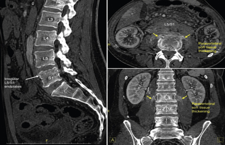
Initial contrast-enhanced computed tomography (CT) of the spine demonstrated irregularity of the L5/S1 endplates with adjacent paravertebral soft-tissue thickening suggestive of spondylodiscitis; however, no definite drainable abscess was identified. Due to persistent neurological symptoms and ongoing severe back pain, MRI of the lumbar spine was subsequently performed (Fig. 1).
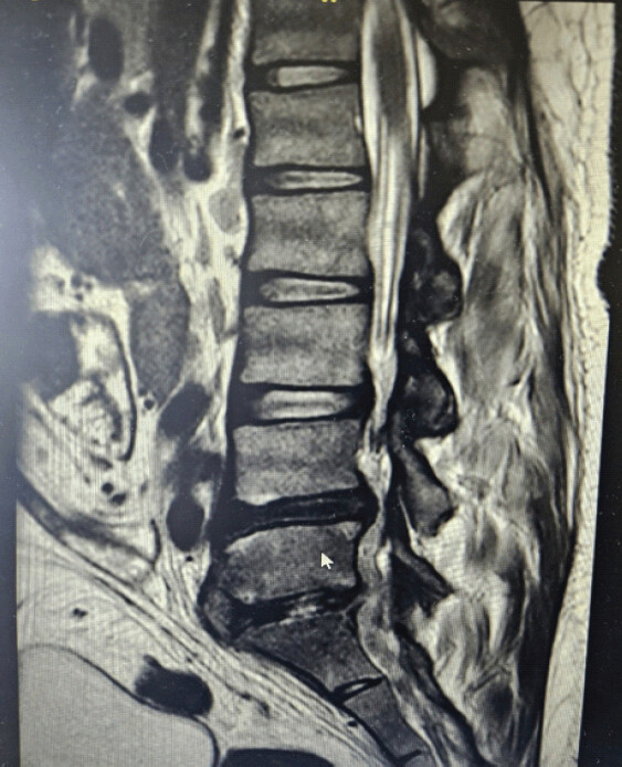
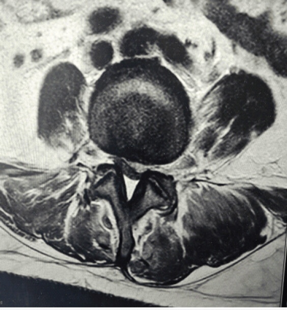
MRI demonstrated an elongated epidural collection extending from L4 to S1 with post-contrast rim enhancement causing significant thecal sac compression. Associated L5/S1 spondylodiscitis, marrow signal abnormalities, prevertebral and paravertebral soft-tissue enhancement, and left paravertebral as well as erector spinae abscesses were identified (Fig. 2 and 3).
Inflammatory markers were elevated with a C-reactive protein level of 123 mg/L. Microbiological investigations confirmed MSSA sensitive to cloxacillin, erythromycin, and gentamicin.
Given the radiological evidence of epidural abscess with neurological deterioration, urgent surgical decompression was undertaken. The patient underwent lumbar laminectomy and spinal canal decompression under general anesthesia.
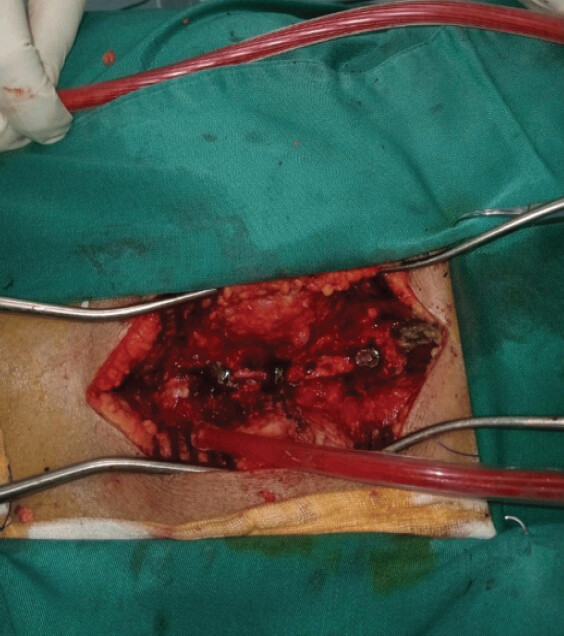
Intraoperatively, frank purulent material was identified within the epidural space at S1 with extensive inflammatory pannus and infected tissue extending from L2 to L5. Subperiosteal dissection was performed, followed by laminectomy using Kerrison rongeurs. The ligamentum flavum and inflammatory tissue occupying the epidural space were excised. Extensive irrigation and debridement were performed, and tissue specimens were sent for microbiological analysis (Fig. 4).
Postoperatively, the patient was managed with targeted intravenous antibiotic therapy based on culture sensitivities. Neurological function and pain improved significantly following decompression. The patient regained ambulation and continued recovery with multidisciplinary management and follow-up.
Written informed consent for publication and use of radiological and intraoperative images was obtained from the patient.
Discussion
SEA is rare globally but carries high morbidity because diagnosis is often delayed. Reviews describe back pain as the most common presenting symptom, while the classical triad of back pain, fever, and neurological deficit is present in only a minority of patients, making early recognition difficult [6, 8]. S. aureus remains the leading causative organism, and recognized risk factors include diabetes mellitus, bacteremia, intravenous drug use, spinal procedures, and immunosuppression.
Worldwide, SEA has been reported after neuraxial procedures, although this remains uncommon [2, 8]. A published case report described meningitis and epidural abscess after spinal anesthesia in a bacteremic diabetic patient undergoing amputation for infected gangrene, which is clinically similar to this case because both involved active infection, bacteremia, diabetes, neuraxial anesthesia, and subsequent epidural infection. Reviews on regional anesthesia in infected patients note that available data are limited, but spinal or epidural anesthesia during bacteremia may be a risk factor for central neuraxial infection [2].
SEA is a neurosurgical and orthopedic emergency requiring high clinical suspicion and prompt intervention. The classical triad of fever, back pain, and neurological deficit is inconsistently present, often contributing to delayed diagnosis [6, 8].
Several risk factors for SEA have been described, including diabetes mellitus, immunosuppression, intravenous drug use, spinal instrumentation, and bacteremia [6, 8]. In this case, active MSSA bacteremia secondary to necrotizing fasciitis likely predisposed the patient to epidural infection following spinal anesthesia.
The role of neuraxial anesthesia in bacteremic patients remains controversial. While transient bacteremia may not absolutely contraindicate spinal anesthesia, active systemic infection increases the risk of hematogenous spread and inoculation into the epidural space [2]. This case highlights the importance of careful risk assessment before performing neuraxial procedures in septic patients.
MRI remains the gold standard imaging modality for diagnosis of SEA due to its superior sensitivity in detecting epidural collections, spinal cord compression, and associated osteodiscitis [6, 8]. In our patient, CT imaging demonstrated only subtle suspicious findings, whereas MRI clearly identified extensive epidural abscess formation and neural compression.
Management of SEA typically involves a combination of surgical decompression and prolonged intravenous antibiotic therapy, particularly in patients presenting with neurological deficits [4]. Early surgical intervention is associated with improved neurological recovery and reduced morbidity [3, 4].
This case reinforces several important clinical lessons. Persistent or worsening back pain following neuraxial anesthesia in patients with active systemic infection should not be considered an expected post-operative symptom. The development of neurological deficits warrants urgent MRI because CT may fail to detect early epidural abscess formation. In patients with MSSA bacteremia, clinicians should maintain a high index of suspicion for SEA, as timely surgical decompression combined with culture-directed antimicrobial therapy can result in excellent neurological recovery (Table 1).
Conclusion
SEA following spinal anesthesia is rare but potentially catastrophic, particularly in bacteremic patients. Persistent or progressive back pain with neurological deficits following neuraxial procedures should prompt urgent MRI evaluation. Early recognition, surgical decompression, and targeted antimicrobial therapy are essential to optimize neurological recovery and reduce morbidity.
Severe or worsening back pain following spinal anesthesia in patients with systemic infection should raise suspicion for SEA. Prompt MRI evaluation and urgent decompression are critical to prevent irreversible neurological injury.
Clinical Message
Spinal epidural abscess should be suspected in bacteremic patients who develop persistent or worsening back pain following spinal anesthesia. Early MRI, prompt surgical decompression, and culture-directed antimicrobial therapy are essential to prevent permanent neurological deficits and improve clinical outcomes.
Conflict of Interest:
Source of Support:
Nil
Consent:
The authors confirm that informed consent was obtained from the patient for publication of this article
References
- Reihsaus E, Waldbaur H, Seeling W.Spinal epidural abscess: A meta-analysis of 915 patients. Neurosurg Rev 2000;23:175-204 discussion 205. [Google Scholar] | [PubMed]
- Horlocker TT, Wedel DJ.Neuraxial block and infection. Reg Anesth Pain Med 2006;31:311-23. [Google Scholar] | [PubMed]
- Curry WT Jr , Hoh BL, Amin-Hanjani S, Eskandar EN.Spinal epidural abscess: Clinical presentation, management, and outcome. Surg Neurol 2005;63:364-71. discussion 371. [Google Scholar] | [PubMed]
- Arko L 4th, Quach E, Nguyen V, Chang D, Sukul V, Kim BS.Medical and surgical management of spinal epidural abscess: A systematic review. Neurosurg Focus 2014;37:E4. [Google Scholar] | [PubMed]
- Vakili M, Crum-Cianflone NF.Spinal epidural abscess: A series of 101 cases. Am J Med 2017;130:1458-63. [Google Scholar] | [PubMed]
- Darouiche RO.Spinal epidural abscess. N Engl J Med 2006;355:2012-20. [Google Scholar] | [PubMed]
- Sendi P, Bregenzer T, Zimmerli W.Spinal epidural abscess in clinical practice. QJM 2008;101:1-12. [Google Scholar] | [PubMed]
- Davis DP, Wold RM, Patel RJ, Tran AJ, Tokhi RN, Chan TC.The clinical presentation and impact of diagnostic delays on emergency department patients with spinal epidural abscess. J Emerg Med 2004;26:285-91. [Google Scholar] | [PubMed]
- Chae HJ, Kim J, Kim C.Clinical characteristics of spinal epidural abscess accompanied by bacteremia. J Korean Neurosurg Soc 2021;64:88-99. [Google Scholar] | [PubMed]
- Babic M, Simpfendorfer CS, Berbari EF.Update on spinal epidural abscess. Curr Opin Infect Dis 2019;32:265-71. [Google Scholar] | [PubMed]
© 2026 Journal of Orthopaedic Case Reports - Published by Indian Orthopaedic Research Group
About the Authors
-

Dr. Mohammed Zoheb Iqbal
-

Dr. Mark Rokubuli
-

Dr. Reshwin Rajan
-

Dr. Manoa Valeniyasana
Related Articles in Journal of Orthopaedic Case Reports
 August 1, 2026 Adolescent Cauda Equina Secondary to Traumatic Lumbar Disc Herniation: A Rare Case Report with Review of Literature
 August 1, 2026 Back to Basics at the Craniovertebral Junction: Occipito-Cervical Fusion for Basilar Invagination with Atlantoaxial Instability
 July 1, 2026 Role of Surgical Stabilization in the Management of Post-discectomy Septic Spondylodiscitis: Two Successful Case Reports
 June 1, 2026 Recurrent Vasovagal Syncope Triggered by Procedural Pain and Post-operative Analgesia during Spinal Anesthesia for High Tibial Osteotomy: A Case Report