


Introduction
A variety of intricate injuries are represented by carpus fracture-dislocations. There are not many documented instances of a displaced lunate and a scaphoid fragment being moved close to the radiocarpal joint [1,2,3,4]. The viability of the proximal pole of the scaphoid and lunate is at high risk due to the rupture of vascular systems and the extension of soft-tissue lesions in these types of injuries [5]. If the blood supply is not restored, scaphoid non-union, lunate avascular necrosis, scaphoid proximal pole avascular necrosis, and future carpal instability are unavoidable [6]. We describe a young patient who experienced severe proximal volar dislocation of a complete lunate complex and hemi-scaphoid, around 5 cm from its anatomical placement.
Case Report
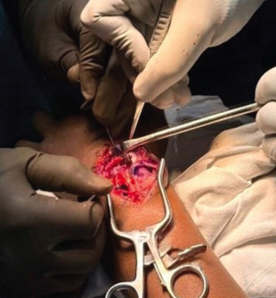
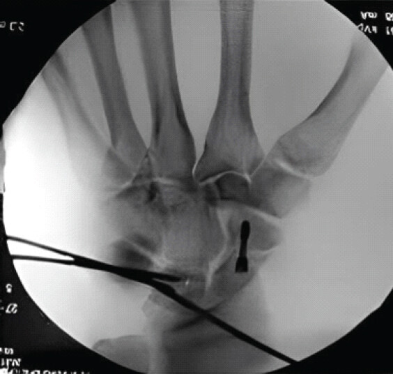
A 16-year-old right-handed male experienced an isolated closed injury to his left wrist following a fall from the rooftop of a one-story building. On examination, the left wrist exhibited swelling and tenderness on palpation. Neurovascular assessment of the hand revealed no abnormalities, and there were no sensory deficits in the distribution of the median nerve. Range of motion in the wrist was limited, and a palpable mass measuring 3 × 3 × 2 cm was noted in the mid-portion of the volar forearm. Imaging studies, including X-ray and computed tomography (CT), indicated a volar trans-scaphoid perilunate fracture-dislocation that had migrated into the forearm (Figs. 1 and 2). The patient underwent surgical fixation 2 weeks after injury, once the soft-tissue envelope was assessed as suitable for open reduction. Under general anesthesia, a volar approach was utilized to release the lunate and the proximal pole of the scaphoid, which were connected by the scapho-lunate ligament. Intraoperatively, this scapholunate ligament was found intact and was, therefore, preserved rather than repaired; the torn volar (palmar) radiocarpal capsule and palmar radiocarpal ligaments were identified, tagged, and protected for later repair, and the median nerve and flexor tendons were retracted and decompressed during this exposure. Following this release, reduction to their anatomical positions was achieved through a dorsal 3–4 carpal approach (Fig. 3). Reduction was performed sequentially: the lunate was first reduced into the lunate fossa under fluoroscopic guidance to restore the radiolunate relationship, after which the hemi-scaphoid fragment, still tethered to the lunate by the intact scapholunate ligament, was reduced and provisionally pinned to re-establish the scapholunate and midcarpal relationships. The torn dorsal radiocarpal, capitolunate, and triquetrolunate ligaments identified through the dorsal approach were repaired primarily using non-absorbable sutures passed through drill holes in the lunate and triquetrum, and the previously tagged volar capsule and palmar radiocarpal ligaments were then repaired through the volar window to reconstitute the palmar carpal stabilizers. The scaphoid fracture was stabilized using a 2.4 mm Herbert screw, while the lunate was secured with three 1.0 mm Kirschner wires (Fig. 4). Three Kirschner wires, rather than a single wire, were chosen for the lunate because its small size and largely cartilage-covered surface precluded screw fixation; a triangulated, multiplanar wire configuration engaging the lunate, capitate, and triquetrum was used to neutralize rotational and shear forces on the reduced lunate and protect the ligament repairs during healing. The procedure lasted approximately 1 h 50 min, with negligible (nil) blood loss under pneumatic tourniquet control maintained for almost 2 h. A dorsal forearm splint was applied, allowing for free movement of the elbow and fingers. The Kirschner wires were removed 8 weeks post-surgery, and the splint was subsequently replaced with an orthosis for wrist immobilization. A structured hand-therapy program, focusing on edema control and active range-of-motion exercises for the fingers and elbow, was initiated at 3 weeks post-surgery while the wrist remained protected in the splint; protected active and active-assisted wrist range-of-motion exercises were then introduced following Kirschner-wire removal and transition to an orthosis at 8 weeks, with progressive strengthening exercises added once radiographic union and adequate soft-tissue healing were confirmed.
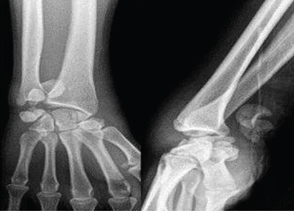
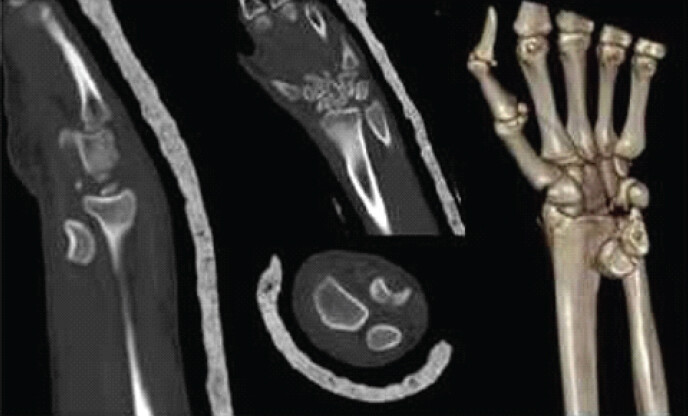
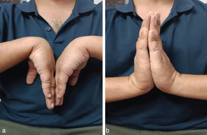
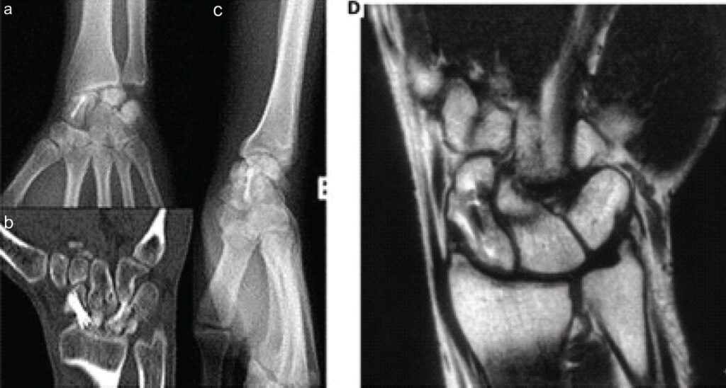
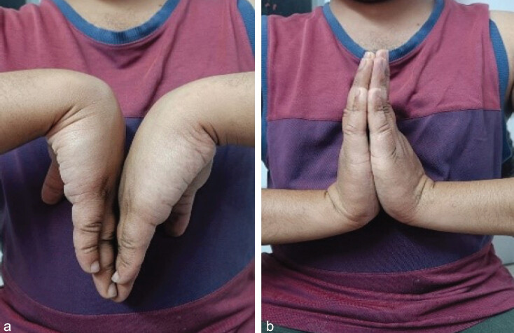
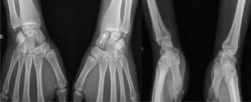
At the 12-month follow-up, the patient reported experiencing pain in the left wrist, quantified as 5 on the visual analog scale (VAS). The range of motion in the wrist was restricted, with flexion at 60°, extension at 80° (Fig. 5), ulnar deviation at 15°, and radial deviation at 15°. Imaging studies, including X-ray, CT, and magnetic resonance imaging (MRI) conducted 12 months post-operation, indicated an altered signal in both the scaphoid and lunate bones, which raised concerns for osteonecrosis; however, there was no definitive evidence of radiocarpal or midcarpal arthritis (Fig. 6). MRI was obtained at this stage specifically to evaluate osteonecrosis, and the concern for avascular change in the scaphoid and lunate at 12 months was based on this MRI finding of altered marrow signal rather than on radiographs or CT alone, since MRI remains the most sensitive modality for assessing carpal vascularity. The patient did not attend subsequent appointments and returned for evaluation 2 years later. Three years following the fracture, the patient indicated a complete absence of wrist pain (VAS: 0) and demonstrated improved wrist motion: Flexion at 70°, extension at 90°, ulnar deviation at 30°, and radial deviation at 15° (Fig. 7). The disabilities of the arm, shoulder, and hand score recorded at 3 years were 7.5. Assessments of tip pinch and grip strength yielded results of 2.75 kg (contralateral 4 kg) and 27 kg (contralateral 30 kg), respectively. X-ray findings showed mild degenerative changes in the radiocarpal joint; while the possibility of lunate necrosis could not be entirely ruled out, there was no observable loss of height when compared to the contralateral wrist (Fig. 8). The patient expressed satisfaction with the outcome and declined any surgical intervention at that time.
Discussion
Seldom has palmar trans-scaphoid-lunate dislocation been documented in the literature [2,3,4]. The lunate and scaphoid complexes were badly proximally dislocated in our case. The mechanism of damage is poorly understood. According to Mayfield et al. [7], it appears to be a variation of the lunate and perilunate dislocations. A scaphoid fracture results when the scaphoid impinges on the dorsal lip of the radius when a pronated forearm is subjected to a force of hyperextension and ulnar deviation of the wrist. The lunate and proximal pole complex of the scaphoid are vulnerable to dislocation if this forced movement continues, as it tears the dorsal and palmar radiocarpal ligaments and ruptures the capitolunate and triquetrolunate ligaments. To improve the likelihood of functional recovery and lower the high risk of osteonecrosis linked to this kind of damage, the normal intercarpal morphology must be restored as soon as possible [3]. Due to the intricacy of this lesion, the greatest outcome is typically obtained by combining a volar and dorsal technique. The volar technique permits direct palmar capsule repair, ligament tear repair, and carpal tunnel decompression, whereas the dorsal route provides improved exposure for anatomical alignment and interosseous ligament reconstruction or repair [8]. The authors of a case that was remarkably similar to ours, Lee et al. [4], decided to reduce the danger of osteonecrosis using a single volar approach. However, the X-rays revealed dorsal-ulnar subluxation of lunate and ulnar subluxation of scaphoid at the 12-month follow-up, even though there was no sign of osteonecrosis.
The authors suggested the combined technique as a superior option in a case with this degree of intercarpal ligament injury, citing the inability to execute a correct ligament restoration using the volar approach as the primary cause of this event. With a dislocation comparable to ours, Koh et al. [2] reported another instance, in which the two-fold approach produced good radiographic and functional results.
To place the present case in context, Table 1 summarizes the salient clinical, radiographic, and treatment features of the current case alongside those of the four previously reported cases of this injury pattern, by Stevanovic et al. [1], Koh et al. [2], Ekerot [3], and Lee et al. [4]. Several features distinguish our case from this small existing series. First, at 16 years of age, our patient is, to our knowledge, the youngest reported with this injury pattern; the four comparator cases all involved skeletally mature adults, and the balance of remodeling potential against decades of expected joint use may differ substantially in an adolescent. Second, the approximately 5 cm proximal migration of the fragment complex in our patient is among the most extreme displacements reported, comparable to the 6 cm migration described by Lee et al. [4] and exceeding the migration distances in the Koh et al. [2] and Ekerot [3] cases. Third, unlike Lee et al. [4], who used an isolated volar approach and observed residual dorsal-ulnar subluxation of the lunate and ulnar subluxation of the scaphoid at 12 months, we combined volar and dorsal exposures – as did Koh et al. [2] – and achieved a stable, anatomically reduced carpus without recurrent subluxation. Fourth, our case is one of the few with imaging evidence of altered scaphoid and lunate signal on serial follow-up and uniquely documents its radiographic evolution from 12 months to 3 years, showing apparent improvement without loss of carpal height or progression of arthritis, information not available from the shorter or less granular follow-up reported in the other cases.
Comparison of the present case with previously reported cases of trans-scaphoid-lunate/perilunate dislocation with proximal migration of the fragment complex
| Study (year) | Age/sex | Injury pattern and migration | Approach and fixation | Osteonecrosis/imaging | Follow-up and outcome |
|---|---|---|---|---|---|
| Stevanovic et al. [1] (1996) | Adult, F | Trans-scaphoid, transtriquetral, volar lunate fracture-dislocation | Not reported in available abstract | Not reported in available abstract | Not reported in available abstract |
| Ekerot [3] (1995) | Adult, F | Total palmar trans-scaphoid-lunate dislocation | Open reduction, bone grafting, internal fixation (approach not further specified) | No osteonecrosis | 70 months; wrist function almost normal |
| Koh et al. [2] (2012) | 24 year, M | Total volar extrusion of lunate and scaphoid proximal pole with scapholunate dissociation | Combined dorsal + volar; open reduction and internal fixation | No avascular necrosis on MRI | 12 years; ROM 75% and grip 76% of contralateral side; pain- free, satisfied |
| Lee et al. [4] (2017) | Adult, M | Volar dislocation of lunate and scaphoid proximal pole, migrated ~6 cm proximal to the radiocarpal joint through the transcarpal ligament | Isolated volar approach | No osteonecrosis; dorsal-ulnar subluxation of lunate and scaphoid | 12 months; persistent subluxation without osteonecrosis |
| Present case (2026) | 16 year, M | Hemi-scaphoid and lunate complex, migrated ~5 cm | Combined volar + dorsal; Herbert screw (scaphoid) + 3 Kirschner wires (lunate) | Altered scaphoid/lunate signal, concern for osteonecrosis at 12 months; apparent improvement by 3 years | 3 years; pain-free, DASH 7.5, improved motion, mild degenerative radiocarpal change, no loss of carpal height, satisfied |
M: Male, F: Female. ROM: Range of motion, MRI: Magnetic resonance imaging, DASH: Disabilities of the arm, shoulder, and hand. For the comparator cases by Stevanovic et al. [1] and Ekerot [3], full-text articles were not accessible to us; the corresponding table entries were therefore compiled from the published abstract alone, and fields not addressed in the abstract are marked “Not reported in available abstract” rather than inferred
Despite initial considerable displacement and soft-tissue disturbance, some investigations have revealed the possibility of revascularization of the proximal poles of the lunate and scaphoid [3,9]. Twelve out of 27 patients with trans-scaphoid lunate or perilunate dislocation had osteonecrosis of one of these bones, and nine of them had revascularization, according to Panting et al. [9]. According to the theory put forth by Ekerot et al. [3], lunate revascularization occurs as a result of the intact scapholunate interosseous ligament and the united scaphoid fracture. Some speculate that the avascular alterations that occur after wrist dislocation may be temporary [6]. Magnetic resonance imaging remains the most sensitive modality for evaluating carpal vascularity and is recommended whenever osteonecrosis is suspected on plain radiographs or CT, as illustrated by Koh et al. [2], who used MRI to confirm the absence of avascular necrosis 12 years after combined reduction and fixation. Three years after the fracture, we saw a good evolution in the X-ray with no loss of lunate height in proportion to the contralateral wrist and no discernible evolution in radiocarpal arthritis, despite the altered sign of the lunate and scaphoid after the 1st year. Some publications recommend a primary proximal row carpectomy (PRC) when injuries of this grade of severity are present [10]. Given the patient’s age, we believe that there is potential for an open reduction and osteosynthesis attempt, which would allow for a PRC in the event that the standard anatomy preservation approach is unsuccessful.
Beyond primary reconstruction, PRC is the salvage procedure most widely used for irreparable perilunate injuries or established avascular necrosis with carpal collapse; it converts the wrist into a capitate-radius articulation and can preserve a useful arc of motion and grip strength, as reported after early PRC for severe carpal trauma [10]. However, PRC depends on an intact, congruent articulation between the capitate head and the lunate fossa of the radius, and outcomes deteriorate with pre-existing capitate or radial-fossa cartilage damage; because it also predisposes to accelerated radiocapitate degeneration over time, most authors reserve primary PRC for older, lower-demand patients or as a secondary salvage after failed anatomical reconstruction, rather than as first-line treatment in adolescents and young adults, in whom preservation of carpal height and native articular cartilage is prioritized. Consistent with this reasoning, Trumble and Verheyden [11] showed that combined dorsal-volar reduction, ligament repair, and intraosseous cerclage fixation of perilunate and lunate dislocations – a strategy similar to that used in our patient – restored a wrist flexion-extension arc and grip strength averaging 80% and 77% of the contralateral side, respectively, with high patient satisfaction, supporting an initial attempt at anatomical reconstruction even in complex, severely displaced injuries. Other salvage options, such as four-corner fusion or total wrist arthrodesis, sacrifice further motion and are generally reserved for failure of both primary reconstruction and PRC. Given our patient’s youth and the potential, demonstrated in this case, for radiographic and functional recovery despite early signs suggestive of osteonecrosis, we favored an initial attempt at anatomical open reduction and internal fixation, reserving PRC or further salvage procedures for failure of this approach – a strategy also adopted by Koh et al. [2] in a similarly young patient.
Despite this intricate injury, our patient expressed great satisfaction with the outcome, demonstrating strong muscles and improved mobility as compared to the contralateral side. Although the absence of limitations in a young, active patient is extremely encouraging, the follow-up period is still too brief to make firm judgments regarding the injury’s long-term sequelae.
Conclusion
When a hemi-scaphoid and lunate dislocate volarly proximal to the carpal ligament, there is no consensus on the optimum course of action. Combined dorsal and volar approaches may provide satisfactory reduction and fixation in selected cases of this rare injury pattern. In the case reported here, this approach achieved good functional results at 3-year follow-up, with mild degenerative radiocarpal change but no loss of carpal height and no definitive evidence of osteonecrosis; however, a single case cannot establish the superiority of one surgical strategy over another, and comparative studies with larger cohorts are needed. Because there are few published studies on these injuries, reporting both short- and long-term outcomes remains important to help guide this challenging treatment decision.
Clinical Message
In extreme proximal volar dislocations of the hemi-scaphoid-lunate complex, a combined volar-dorsal approach with rigid internal fixation allows anatomical restoration of carpal alignment. Early radiographic signs suggestive of osteonecrosis should not be taken as a poor prognostic indicator in isolation, as clinical and radiographic recovery may continue over several years; close, prolonged follow-up is essential to guide further management.
Conflict of Interest:
Nil
Source of Support:
Nil
Consent:
The authors confirm that informed consent was obtained from the patient for publication of this article
References
- Stevanovic M, Schnall SB, Filler BC. Trans-scaphoid, transtriquetral, volar lunate fracture-dislocation of the wrist. J Bone Joint Surg Am 1996;78:1907–10. [Google Scholar] | [PubMed]
- Koh KH, Lim TK, Park MJ. Total volar extrusion of the lunate and scaphoid proximal pole with concurrent scapholunate dissociation. Orthopedics 2012;35:e1427–30. [Google Scholar] | [PubMed]
- Ekerot L. Palmar dislocation of the trans-scaphoid-lunate unit. J Hand Surg Br 1995;20:557–60. [Google Scholar] | [PubMed]
- Lee JM, Kim JK, Lee KB. Extreme proximal migration of dislocated lunate over carpal ligament - a case report. Acta Orthop Traumatol Turc 2017;51:177–80. [Google Scholar] | [PubMed]
- Green DP, O'Brien ET. Open reduction of carpal dislocations: Indications and operative techniques. J Hand Surg Am 1978;3:250–65. [Google Scholar] | [PubMed]
- Gellman HG, Schwartz SD, Botte MJ, Feiwell L. Late treatment of a dorsal transscaphoid, transtriquetral perilunate wrist dislocation with avascular changes of the lunate. Clin Orthop 1988;237:196–203. [Google Scholar] | [PubMed]
- Mayfield JK, Johnson RP, Kilcoyne RK. Carpal dislocations: Pathomechanics and progressive perilunar instability. J Hand Surg Am 1980;5:226–41. [Google Scholar] | [PubMed]
- Akinleye SD, Makani A, Dalinka MK, Chang B. Unusual proximal dislocation without fracture: A case report. Hand (N Y) 2012;7:454–6. [Google Scholar] | [PubMed]
- Panting AL, Lamb DW, Noble J, Haw CS. Dislocations of the lunate with and without fracture of the scaphoid. J Bone Joint Surg Br 1984;66:391–5. [Google Scholar] | [PubMed]
- Van Kooten EO, Coster E, Segers MJ, Ritt MJ. Early proximal row carpectomy after severe carpal trauma. Injury 2005;36:1226–32. [Google Scholar] | [PubMed]
- Trumble T, Verheyden J. Treatment of isolated perilunate and lunate dislocations with combined dorsal and volar approach and intraosseous cerclage wire. J Hand Surg Am 2004;29:412–7. [Google Scholar] | [PubMed]
© 2026 Journal of Orthopaedic Case Reports - Published by Indian Orthopaedic Research Group
About the Authors
-

Sharath Raj
-

Shrinivas VB
-

Aduri Tharun Teja
-

Ajay Jose
-

Kartik Sharma
-

Vijay Goni
Related Articles in Journal of Orthopaedic Case Reports
 June 10, 2023 Transscaphoid Transcapitate Perilunate Fracture-dislocation with Inferior Arc Injury and Acute Ulnar Nerve Compression: A Case Report
 August 1, 2026 Medial Subtalar Joint Dislocation in an 18-Year-Old Athlete with Pes Planus: Beyond A Simple Misstep
 August 1, 2026 Complex Irreducible Dislocation of the Fourth Toe Proximal Interphalangeal Joint in a 5-year-old Boy Fixed by Soft Anchor: A Case Report
 June 1, 2026 Kienbock’s Disease in a Young Female without Classical Risk Factors: A Case Report with 1-year Functional Outcome Following Proximal Row Carpectomy