


Introduction
Glomus tumors are rare, benign tumor that originates from the glomus body, a specialized arteriovenous anastomosis involved in blood flow regulation within the dermis and in thermoregulation of digits [1]. These tumors are most frequently found in the digits, particularly beneath the nails, where glomus bodies are most concentrated [1,2]. The earliest description of the clinical features of these tumors can be traced back to 1812, when William Wood detailed cases of painful subcutaneous nodules – now understood to be glomus tumors in the Edinburgh Medical and Surgical Journal [3, 4].
Typically affecting young to middle-aged adults, digital glomus tumors most commonly arise in the subungual region, especially of the fingers, and exhibit a striking clinical triad: Paroxysmal pain, tenderness to pressure, and hypersensitivity to cold [4,5,6,7]. Despite this classic symptoms, diagnostic delays are common, often extending for years, primarily due to the tumor’s small size, deep location, and overlap with other multiple differential diagnoses as neuroma, arthritis, or chronic pain syndromes [8, 9].
Magnetic resonance imaging (MRI) has emerged as a highly sensitive modality for detecting these small sized tumors, especially in the early or ambiguous cases [6,8]. T2-weighted images typically reveal a well-defined, hyperintense lesion beneath the nail bed, providing essential pre-operative localization. MRI consistently showed hyperintense signals on T2-weighted images, supporting the diagnosis in all patients [4,10]. Tumor sizes vary from 0.3 cm to 0.9 cm. Additional tools such as X-rays’ ultrasound and clinical tests – including Love’s pin test and Hildreth’s test – also contribute to early diagnosis [9,11]. Recent literature shows the value of dermoscopy and cytological evaluation in select cases [12].
Histologically, glomus tumors are composed of uniform, round glomus cells aligned with capillary-sized vessels, while the majority are benign, atypical. Malignant variants, although rare, have been described and require thorough histopathological assessment [2,13]. Malignant forms may show infiltrative growth, mitotic activity, and nuclear atypia and require more aggressive surgical management and reliability.
Surgical management varied, including transungual and direct approaches, with one patient undergoing wide local excision. Post-operative outcomes were favorable, with minimal complications; only one patient experienced a nail deformity. The pre-operative pain scores, assessed by the Visual Analog Scale (VAS), ranged from 7 to 10.
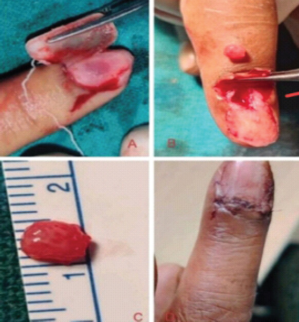
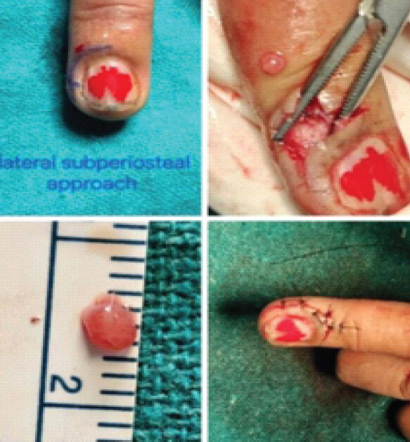
Surgical excision remains the gold standard of treatment, with approaches such as transungual, lateral subperiosteal, or direct nail bed incision depending on tumor location and surgeon preference [12,14,15,16]. The transungual approach offers direct visualization of subungual lesions and is associated with excellent symptom relief and minimal recurrence if the tumor is completely excised [15]. Alternatively, lateral subperiosteal dissection provides comparable outcomes with potentially better nail bed preservation [14,16].
Importantly, several clinical studies document dramatic reductions in pain postoperatively, as measured by the VAS. In a retrospective series of 23 patients, pre-operative mean VAS was 8.7, which declined to 0.8 following complete excision (P < 0.01) [17]. Similarly, a case series of four digital glomus tumors reported a mean VAS decrease from 7.75 to 1.0 after surgery (P = 0.0028) [18].
Glomus tumors of the hand are rare and misdiagnosed as there is multiple differential diagnosis of fingertip pain. There is diagnostic delay due to small size, atypical locations, and non-specific symptoms. Persistent localized digital pain, tenderness, and cold sensitivity may be overlooked despite the availability of simple bedside tests described in this study. The resulting diagnostic delay can lead to prolonged pain and unnecessary investigations or treatment and financial burden to medical facility or patients.
Therefore, this study was undertaken to evaluate the diagnostic value of simple clinical tests, early referral in concerned department and outcomes following surgical excision, with the aim of promoting early diagnosis and effective management, particularly in resource-limited settings. This study also contributes additional clinical data on glomus tumors from an Indian tertiary care center.
Objectives
This study aims to further delineate the clinical presentation, diagnostic strategies, make algorithm, and surgical outcomes of digital glomus tumors, contributing to improved recognition and management of the glomus tumor yet often overlooked yet highly treatable condition.
Materials and Methods
A retrospective review was conducted at the Department of Plastic Surgery, PGIMS, Rohtak, India. The study included seven patients with histopathologically confirmed glomus tumors of the hand diagnosed between 2021 and 2024. Inclusion criteria were clinical presentation consistent with glomus tumor, confirmation by histopathology, and availability of complete clinical records.
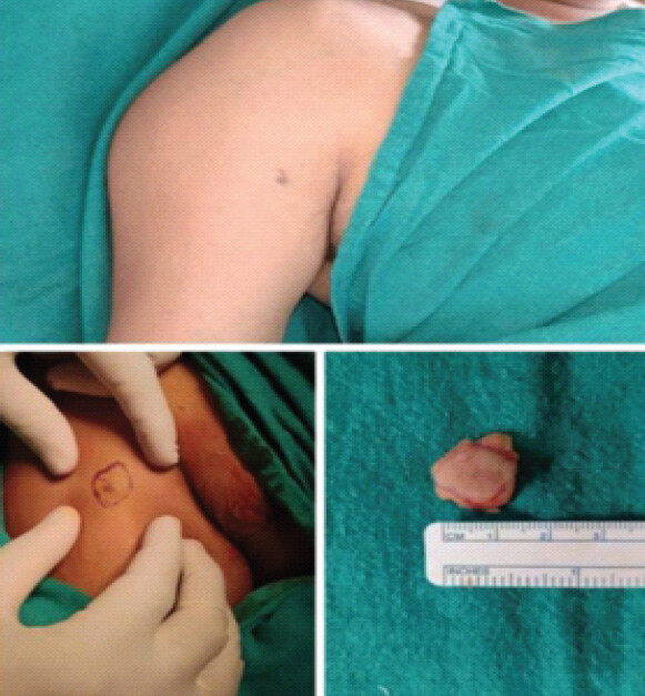
All patients underwent surgical excision with either a transungual (Fig. 1) or lateral subperiosteal approach (Fig. 2) and with wide local excision (Fig. 3), depending on tumor location. Perioperative and post-operative observations, complications, and follow-up data (minimum 7 months) were recorded. Pain was evaluated pre- and postoperatively using the VAS. Recurrence was monitored at regular outpatient visits.
Data were analyzed using descriptive statistics. Continuous variables were expressed as mean and range, while categorical variables were presented as frequencies and percentages. Pre-operative and post-operative VAS scores were compared to assess improvement in pain following surgery.
Results
A total of seven patients with histopathologically confirmed glomus tumors were included in the study. Among the seven patients, five were female and two were male, with a mean age of approximately 42 years (range: 32–51 years). The tumors were more commonly located on the right side in five out of seven cases. Anatomical distribution included subungual (n = 3), pulp of the finger (n = 1), lateral nail fold (n = 2), and lateral aspect of the arm (n = 1). Right side more commonly affect (5 cases) then left side (2 cases).
Clinically, all patients presented with pain as the primary symptom. Tenderness was reported in six patients, and five experienced cold sensitivity. The average duration of symptoms before diagnosis was approximately 17.5 months. On clinical examination, Love’s test was positive in all patients (100%), indicating high diagnostic significantly improved postoperatively to scores between 1 and 3. All excised lesions were confirmed as solid glomus tumors on histopathology.
All patients were followed up for 7 months, during which no tumor recurrence was observed. Postoperatively, all patients reported significant pain relief. The mean VAS score improved from 8.7 before surgery to 2.0 after surgery. No recurrences were observed during the follow-up period (Table 1).
Clinical profile and surgical outcome of all seven patients
| Patient ID | Age (years) | Sex | Location of tumor | Side | Symptoms | Duration (months) | Love test | Hildreth test | Cold sensitivity test | Pre-op VAS score | Tumor size (cm) | Surgical approach | Post-op complication | Follow-up (months) | Post-op VAS score | Recurrence |
|---|---|---|---|---|---|---|---|---|---|---|---|---|---|---|---|---|
| GT-01 | 32 | F | Index finger Subungual | Rt. | Pain, tenderness, cold sensitivity | 15 | Positive | Positive | Positive | 8 | 0.3 | Transungual | None | 7 | 2 | No |
| GT-02 | 45 | F | Ring finger Pulp of Finger | Lt. | Pain, cold sensitivity | 24 | Positive | Positive | Positive | 10 | 0.9 | Direct | None | 7 | 3 | No |
| GT-03 | 51 | F | Thumb, subungual | Rt. | Pain, tenderness, nail discoloration | 12 | Positive | Positive | Positive | 8 | 0.5 | Transungual | Nail Deformity | 7 | 1 | No |
| GT-04 | 39 | M | Index finger, subungal | Rt. | Pain, tenderness | 18 | Positive | Positive | Positive | 10 | 0.6 | Direct | None | 7 | 2 | No |
| GT-05 | 45 | F | Index finger Lateral Nail Fold | Lt. | Pain, tenderness, cold sensitivity | 24 | Positive | Negative | Positive | 9 | 0.4 | Direct | None | 7 | 3 | No |
| GT-06 | 34 | F | Thumb, lateral nail fold | Rt. | Pain, tenderness | 12 | Positive | Negative | Negative | 7 | 0.4 | Direct | None | 7 | 1 | No |
| GT-07 | 45 | M | Arm lateral, aspect | Rt. | Pain, tenderness, cold intolerance | 12 | Positive | Negative | Positive | 9 | 0.8 | Wide local excision | None | 7 | 1 | No |
F: Female, M: Male, OP: Operative, VAS: Visual analog scale, Rt: Right, Lt: Light
One patient (14.3%) developed post-operative nail deformity following transungual excision. No other complications or tumor recurrences were observed during the 7-month follow-up period.
Discussion
Glomus tumors are rare, benign neoplasms arising from the glomus body, typically located in the digits, particularly the subungual region as in our study, we have only one case which is at different site. In this case series, a strong female predominance (five out of seven cases) was observed, which is consistent with previously published data indicating that glomus tumors occur more frequently in women, especially in the fourth to fifth decades of life [4, 19, 20].
Pain was a universal symptom in this study, with most patients also exhibiting point tenderness and cold sensitivity – features that comprise the classical clinical triad. The Love’s pin test, a highly sensitive clinical test for glomus tumors, was positive in all patients in this series, confirming its diagnostic utility. However, the Hildreth’s test and cold sensitivity test, while supportive, were not positive in all patients, indicating some inter-patient variability in clinical signs [4, 21].
Surgical excision was curative in all cases, with substantial improvement in symptoms and no recurrence observed during follow-up. The transungual approach, although effective for subungual tumors, did result in nail deformity in one case a known risk of this technique [22]. All other tumors were approached directly, avoiding nail disruption. This reinforces the importance of selecting a surgical approach based on tumor location and size [23].
In terms of symptomatic relief, patients demonstrated significant improvement in pain scores. The mean VAS score improved from 8.7 preoperatively to 2.0 postoperatively, indicating excellent surgical outcomes. However, the mean duration of symptoms before diagnosis was notably long (17.5 months), reflecting a common delay in recognizing this condition (Table 2).
Comparison of present study with previously published studies on glomus tumors
| Study | Number of patients | Mean age (years) | Female (%) | Mean diagnostic delay | Diagnostic methods | Surgical approach | Recurrence | Key findings |
|---|---|---|---|---|---|---|---|---|
| Assmus and Dombert (2002) | 36 | 51-52 | 64% | 8 years | Clinical exam+MRI (selected cases) | Mostly transungual triangular incision | 1 case | Majority in distal phalanx; pain and cold intolerance common |
| Van Geertruyden et al. (1996) | 51 | 59 | 19% | 10 years | Clinical suspicion (90%), X-ray changes (36%) | Direct transungual excision | 2 cases | Blue discoloration 29%; nail deformity/pulp nodule 33% |
| Dhingra et al .(2022) | 4 | 39.2 | 75% | 5.75 years | Clinical tests (Love, Hildreth)+MRI | Transungual/lateral approach | None | VAS improved from 7.75 to 1 (P=0.0028) |
| Present study | 7 | 42 | 71% | 17.5 months | Clinical tests + MRI | Transungual (3) and direct excision (4) | None | VAS improved from 8.7 to 2.0; Love’s test positive in 100% |
MRI: Magnetic resonance imaging, VAS: Visual Analog Scale
International literature frequently reports diagnostic delays of 3–7 years due to the subtlety of symptoms and the rarity of the condition. However, in this series, the mean delay in diagnosis was 17.5 months, significantly shorter than reported globally. This may reflect greater clinical suspicion, improved diagnostic pathways, or better healthcare access at the treating center. This diagnostic delay has been consistently reported in literature, often leading to unnecessary treatments and investigations [18]. These findings offer valuable insight into how the recognition and management of glomus tumors may be evolving within the Indian healthcare system. It also suggests that regional differences in healthcare infrastructure and awareness can significantly impact time to diagnosis and, subsequently, patient outcomes.
Limitations
The sample size was relatively small, reflecting the rarity of glomus tumors and multiple differential diagnosis. In addition, the study was retrospective in nature and conducted at a single tertiary care center. The follow-up duration was limited, and longer follow-up would be required to fully evaluate recurrence rates. Despite these limitations, the study provides useful clinical insights into the diagnosis, early, and effective diagnosis due to early referral system and management of glomus tumors in a tertiary care setting.
Conclusion
Glomus tumors of the hand are uncommon but should be considered in patients presenting with persistent, localized digital pain, particularly when associated with tenderness and cold sensitivity. On the basis of this study, every localized fingertip pain should follow a Proposed Diagnostic Algorithm (Fig. 4) for glomus tumor and referred to proper department.
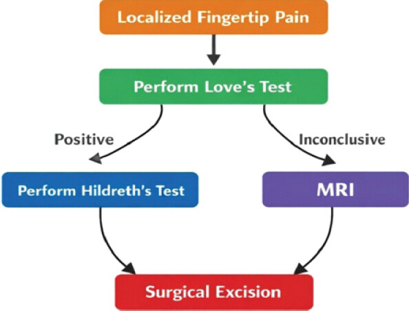
In this study, simple bedside clinical tests, especially Love’s test, demonstrated high diagnostic utility and may facilitate early recognition, while MRI provided useful pre-operative localization. Surgical excision resulted in substantial pain relief, with the mean VAS score improving from 8.7 to 2.0, no tumor recurrence during follow-up, and minimal post-operative morbidity and complications. These findings emphasize the importance of clinical suspicion and systematic evaluation to reduce diagnostic delay and enable effective treatment of glomus tumors as soon as possible to improve patient quality of life and prevent prolonged suffering, particularly in resource-limited settings. There is limited literature from Indian tertiary centers specifically analyzing glomus tumors. My study adds regional data and reflects the effectiveness of clinical diagnostic tools even without routine MRI use – relevant for cost-conscious setups.
Our study highlights the diagnostic value of simple clinical tests, particularly Love’s test, which showed high positivity in our series. The findings also demonstrate that accurate diagnosis can often be achieved through careful clinical evaluation, which is especially relevant in resource-limited settings where routine MRI may not always be feasible.
Clinical Message
“Glomus tumor should be considered in patients with persistent, severe, localized digital pain, particularly when Love’s test is positive. Early clinical recognition can reduce diagnostic delay and financial burden, while complete surgical excision provides definitive treatment and significant pain relief.”
Conflict of Interest:
Nil
Source of Support:
Nil
Consent:
The authors confirm that informed consent was obtained from the patient for publication of this article
How to Cite this Article
Sharma A, Kumar A, Singh K, Jain I, Sharma P, Singh E. Early Recognition of Glomus Tumor of Hand Using Clinical Tests: Experience from an Indian Tertiary Center. Journal of Orthopaedic Case Reports 2026 October;16(10): 339-344.
References
- Van Geertruyden J, Lorea P, Goldschmidt D, De Fontaine S, Schuind F, Kinnen L. Glomus tumours of the hand. A retrospective study of 51 cases. J Hand Surg Br 1996;21:257-60. [Google Scholar] | [PubMed]
- Mravic M, LaChaud G, Nguyen A, Scott MA, Dry SM, James AW. Clinical and histopathological diagnosis of glomus tumor: An institutional experience of 138 cases. Int J Surg Pathol 2015;23:181-8. [Google Scholar] | [PubMed]
- Wood W. On painful subcutaneous tumours. Edinb Med Surg J 1812;8:283-91. [Google Scholar] | [PubMed]
- Bhaskaranand K, Navadgi BC. Glomus tumour of the hand. J Hand Surg Br 2002;27:229-31. [Google Scholar] | [PubMed]
- Drapé JL, Idy-Peretti I, Goettmann S, Wolfram-Gabel R, Dion E, Grossin M. Subungual glomus tumors: Evaluation with MR imaging. Radiology 1995;195:507-15. [Google Scholar] | [PubMed]
- Singh D, Garg RS, Vikas, Garg Y, Arora V. Glomus tumor - a rarity; M. R. I- a big help in early diagnosis. J Orthop Case Rep 2016;6:38-9. [Google Scholar] | [PubMed]
- Morey VM, Garg B, Kotwal PP. Glomus tumours of the hand: Review of literature. J Clin Orthop Trauma 2016;7:286-91. [Google Scholar] | [PubMed]
- Seo JH, Lee HS, Kim SW, Jeong JJ, Choi YR. Subungual glomus cell proliferation in the toe: A case report. J Foot Ankle Surg 2014;53:628-30. [Google Scholar] | [PubMed]
- aisanSmilevitch D, Chaput B, Grolleau JL, Rongieres M, Mansat P. Improvement in quality of life after surgery for glomus tumors of the fingers. Chir Main 2014;33:330-5. [Google Scholar] | [PubMed]
- Muramatsu K, Ihara K, Hashimoto T, Tominaga Y, Taguchi T. Subungual glomus tumours: Diagnosis and microsurgical excision through a lateral subperiosteal approach. J Plast Reconstr Aesthet Surg 2014;67:373-6. [Google Scholar] | [PubMed]
- Biswas D, Sikdar S, Islam T. Outcomes of the transungual approach in 56 consecutive digital subungual glomus tumours. J Hand Surg Asian Pac Vol 2024;29:240-7. [Google Scholar] | [PubMed]
- Falcone MO, Asmar G, Chassat R. Subungual glomus tumor. Hand Surg Rehabil 2024;43S:101607. [Google Scholar] | [PubMed]
- Folpe AL, Fanburg-Smith JC, Miettinen M, Weiss SW. Atypical and malignant glomus tumors: Analysis of 52 cases, with a proposal for the reclassification of glomus tumors. Am J Surg Pathol 2001;25:1-12. [Google Scholar] | [PubMed]
- Mukherjee S, Bandyopadhyay G, Saha S, Choudhuri M. Cytodiagnosis of glomus tumor. J Cytol 2010;27:104-5. [Google Scholar] | [PubMed]
- Dhingra M, Niraula BB, Regmi A, Bansal S, Singh V, Phulware RH. Glomus tumour of hand--a commonly misdiagnosed pathology: A case series. J West AfrColl Surg 2022;12:39-45. [Google Scholar] | [PubMed]
- Schiefer TK, Parker WL, Anakwenze OA, Amadio PC, Inwards CY, Spinner RJ. Extradigital glomus tumors: A 20-year experience. Mayo Clin Proc 2006;81:1337-44. [Google Scholar] | [PubMed]
- Lee DW, Yang JH, Chang S, Won CH, Lee MW, Choi JH. Clinical and pathological characteristics of extradigital and digital glomus tumours: A retrospective comparative study. J EurAcadDermatolVenereol 2011;25:1392-7. [Google Scholar] | [PubMed]
- Carroll RE, Berman AT. Glomus tumors of the hand: Review of the literature and report on twenty-eight cases. J Bone Joint Surg Am 1972;54:691-703. [Google Scholar] | [PubMed]
- Singal A, Bisherwal K, Agrawal S, Bhat S, Diwakar P. Clinico-epidemiological profile and management outcome of subungual digital glomus tumor-Indian experience. DermatolTher 2022;35:e15745. [Google Scholar] | [PubMed]
- Fabro M, Fabro SR, Klitzke BB, De Araújo GL, Machado CA. Glomus tumor: Revitalizing concepts. Radiol Bras 2015;48:269-70. [Google Scholar] | [PubMed]
- Stewart DR, Sloan JL, Yao L, Mannes AJ, Moshyedi A, Lee CC. Diagnosis, management, and complications of glomus tumours of the digits in neurofibromatosis type 1. J Med Genet 2010;47:525-32. [Google Scholar] | [PubMed]
- Vinay K, Narang T, Roshini N, Baskaran N, Reddy A, Patel S. Clinico-epidemiological characteristics and long-term surgical outcomes in patients with glomus tumor: A retrospective study. Indian Dermatol Online J 2025;16:426-31. [Google Scholar] | [PubMed]
- Patel T, Meena V, Meena P. Hand and foot glomus tumors: Significance of MRI diagnosis followed by histopathological assessment. Cureus 2022;14:e30038. [Google Scholar] | [PubMed]
© 2026 Journal of Orthopaedic Case Reports - Published by Indian Orthopaedic Research Group
About the Authors
-

Abhishek Sharma
-

Akshay Kumar
-

Kuldeep Singh
-

Iti Jain
-

Preksha Sharma
-

Eashnoor Singh
Related Articles in Journal of Orthopaedic Case Reports
 September 1, 2026 The Tiny Tumor with Disproportionate Pain: Subungual Glomus Tumor of the Thumb Managed with Nail-Sparing Excision: A Case Report
 July 1, 2026 Extradigital Glomus Tumor of the Knee Mimicking Osteoarthritis: A Case Report
 October 1, 2026 Giant Cell Tumor of the Lateral Malleolus Treated by Tumor Excision and Reconstruction using A 180° Rotated Proximal Fibular Autograft – A Rare Case Report
 September 1, 2026 Management of Scapular Osteochondroma: An Uncommon Report of Two Cases