


Introduction
Total hip arthroplasty (THA) is one of the most successful orthopedic procedures for relieving pain, restoring joint function, and improving quality of life in patients with end-stage hip disorders. Despite excellent long-term implant survival, post-operative instability, impingement, and component malposition remain important causes of patient dissatisfaction and revision surgery. Increasing evidence suggests that these complications are influenced not only by implant positioning but also by the dynamic relationship between the lumbar spine, pelvis, and hip joint, collectively referred to as the spinopelvic complex [1,2].
The orientation of the acetabular component is not static but changes with alterations in pelvic position during standing, sitting, and other functional activities. Spinopelvic parameters, including pelvic tilt (PT), sacral slope (SS), pelvic incidence (PI), and lumbar lordosis (LL), influence functional acetabular orientation and consequently affect hip stability, range of motion, and the risk of prosthetic impingement or dislocation [1,3]. Recognition of these dynamic biomechanical interactions has shifted the focus from reliance on a universal acetabular “safe zone” toward individualized assessment of spinopelvic mobility during pre-operative planning and post-operative evaluation [4].
Patients undergoing THA frequently exhibit altered spinopelvic mechanics secondary to hip osteoarthritis, avascular necrosis, or degenerative changes of the lumbar spine. Restoration of hip motion following arthroplasty may modify pelvic orientation and spinal alignment, thereby influencing post-operative biomechanics and functional recovery [2,5]. Consequently, assessment of spinopelvic alignment in multiple functional positions has become increasingly important for understanding post-operative changes and optimizing implant positioning.
Although several recent studies have highlighted the clinical significance of the spinopelvic relationship in THA, prospective clinical data evaluating post-operative changes in spinopelvic parameters together with functional outcomes remain limited, particularly in the Indian population [1,4,6]. Therefore, the present study was undertaken to evaluate changes in spinopelvic alignment following primary THA and to determine their relationship with post-operative functional outcomes assessed using the Harris hip score (HHS).
Materials and Methods
Study design and setting
This prospective observational study was conducted in the Department of Orthopedics at People’s College of Medical Sciences and Research Centre (PCMS and RC), Bhopal, India. The study included consecutive patients who underwent primary THA during the study period. The study protocol was approved by the Institutional Ethics Committee (Approval No. PCMS/OD/PS/2024/738, Dated: March 26, 2024), and written informed consent was obtained from all participants before enrolment.
Sample size
As the primary objective of this study was to evaluate the correlation between spinopelvic parameters and post-operative functional outcome (HHS), the minimum required sample size was estimated using the formula for testing a Pearson correlation coefficient:
Where Z1−α/2 = 1.96 (two-tailed α = 0.05), Z1−β = 0.84 (80% power), and r is the anticipated correlation coefficient between a spinopelvic parameter and HHS. Based on prior literature reporting moderate associations (standardized coefficients of approximately 0.35) between pre-operative spinopelvic parameters and post-operative hip function after THA [7], an expected correlation of r = 0.35 was assumed. Thus, a minimum of approximately 57 patients was required to detect a correlation of this magnitude with 80% power. Given the observational, consecutive-enrollment design of this study, a census sampling approach was adopted; all eligible patients undergoing primary THA during the study period were enrolled, yielding a final sample of 80 patients.
Study variables
The study variables included demographic, anthropometric, radiographic, implant-related, and functional outcome measures. Demographic and anthropometric variables comprised age, sex, body weight, and body mass index (BMI). Spinopelvic parameters assessed on radiographic evaluation included PT, SS, PI, LL, and PFA. Implant-related variables recorded during surgery included the sizes of the acetabular and femoral components, as well as the intraoperative cup inclination and cup anteversion. Functional outcome was evaluated using the HHS.
Inclusion criteria
-
Patients aged 18 years or older
-
Either gender
-
Patients undergoing primary THA for any clinical indication.
Exclusion criteria
-
Previous lumbar spine surgery
-
Knee joint pathology affecting lower-limb alignment or function
-
Neuromuscular disorders involving the spine or lower limbs
-
Congenital deformities of the hip or spine.
Functional assessment
Functional outcome was evaluated using the HHS at predefined intervals to assess post-operative recovery and functional improvement over time. Assessments were performed preoperatively to establish baseline hip function, followed by post-operative evaluations on day 2 and day 14 to monitor early recovery. Subsequent follow-up assessments were conducted at 3 months and 6 months after surgery to determine intermediate- and short-term functional outcomes following THA.
Radiological assessment
Standardized lateral radiographs of the lumbosacral spine and pelvis were obtained both preoperatively and postoperatively (Fig. 1 and 2) to evaluate spinopelvic alignment in different functional positions. Radiographic assessments were performed with the patient in the standing, sitting, lateral decubitus, and lateral decubitus with 90° hip flexion positions. These standardized imaging positions facilitated comprehensive evaluation of dynamic changes in spinopelvic parameters associated with posture and hip movement.
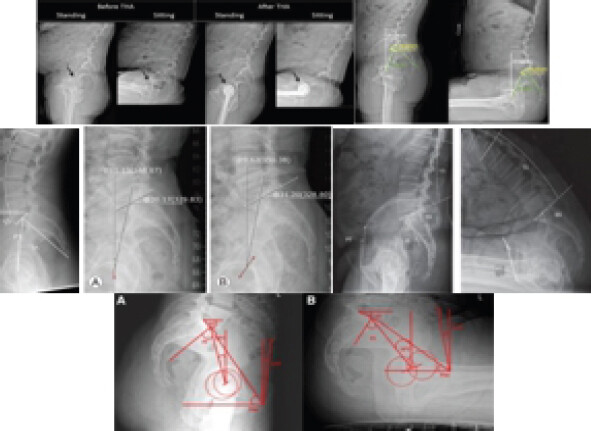
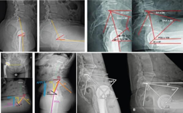
These positions were selected to evaluate dynamic spinopelvic mobility as described in previous literature. All radiographs were obtained in the lateral projection, and spinopelvic parameters (PT, SS, PI, LL, and PFA) were measured using standard radiographic landmarks.
Data collection procedure
After informed consent, each participant was registered under the study protocol. Demographic, clinical, operative, and radiological data were recorded in a standardized case record form. Radiographic measurements were performed preoperatively and at post-operative follow-up. Functional scores were documented at each predefined interval. The collected data were entered into Microsoft Excel and subsequently analyzed.
Statistical analysis
Data were analyzed using appropriate statistical software. Continuous variables were expressed as mean ± standard deviation, while categorical variables were presented as frequency and percentage. Pre-operative and post-operative spinopelvic parameters were compared using the paired Student’s t-test. Changes in HHS over time were analyzed using repeated-measures analysis of variance. Correlation between spinopelvic parameters and functional outcome was assessed using the Pearson correlation coefficient. P < 0.05 was considered statistically significant.
Results
A total of 80 patients undergoing primary THA were included in the study. The mean age of the study population was 48.33 ± 6.29 years. The highest proportion of patients belonged to the 41–50-year age group (35.0%), followed by the 51–60-year (25.0%) and 31–40-year (22.5%) age groups. Males constituted 60.0% of the study population, while females accounted for 40.0%. The mean BMI was 26.33 ± 2.29 kg/m2, with 45.0% of patients classified as overweight, 35.0% as having a normal BMI, and 20.0% as obese. All patients underwent primary THA for secondary osteoarthritis resulting from avascular necrosis of the femoral head (Table 1).
Baseline demographic and clinical characteristics of patients undergoing primary total hip arthroplasty (n=80)
| Variable | Category | Value (%) |
|---|---|---|
| Age (years) | 18–30 | 8 (10.0) |
| 31–40 | 18 (22.5) | |
| 41–50 | 28 (35.0) | |
| 51–60 | 20 (25.0) | |
| >60 | 6 (7.5) | |
| Mean±SD | 48.33±6.29 | |
| Gender | Male | 48 (60.0) |
| Female | 32 (40.0) | |
| Body mass index (kg/m2) | Normal (18.5–24.9) | 28 (35.0) |
| Overweight (25.0–29.9) | 36 (45.0) | |
| Obese (>30) | 16 (20.0) | |
| Mean±SD | 26.33±2.29 | |
| Primary diagnosis | Secondary osteoarthritis due to avascular necrosis | 80 (100) |
SD: Standard deviation
Comparison of spinopelvic parameters in the standing position before and after surgery demonstrated significant post-operative changes in most variables (Table 2). Mean PT decreased significantly from 18.63 ± 3.85° preoperatively to 14.20 ± 3.11° postoperatively (P < 0.0001). Conversely, SS increased from 36.54 ± 4.61° to 41.10 ± 4.26° (P < 0.0001), LL increased from 42.34 ± 5.12° to 46.80 ± 4.94° (P < 0.0001), and PFA increased from 181.36 ± 8.42° to 186.24 ± 7.95° (P = 0.0092). PI remained unchanged following surgery (52.80 ± 6.76° vs. 53.00 ± 6.53°; P = 0.87).
Comparison of standing spinopelvic parameters before and after total hip arthroplasty
| Parameter | Pre-operative (Mean±SD) | Post-operative (Mean±SD) | P-value |
|---|---|---|---|
| Pelvic tilt (°) | 18.63±3.85 | 14.20±3.11 | <0.0001 |
| Sacral slope (°) | 36.54±4.61 | 41.10±4.26 | <0.0001 |
| Pelvic incidence (°) | 52.80±6.76 | 53.00±6.53 | 0.87 |
| Lumbar lordosis (°) | 42.34±5.12 | 46.80±4.94 | <0.0001 |
| Pelvic femoral angle (°) | 181.36±8.42 | 186.24±7.95 | 0.0092 |
SD: Standard deviation
Significant post-operative changes in spinopelvic alignment were also observed in the sitting, lateral decubitus, and lateral decubitus with 90° hip flexion positions (Table 3). In the sitting position, PT decreased significantly, whereas SS and LL increased following surgery. PFA demonstrated a modest but statistically significant reduction. In the lateral decubitus position, post-operative reductions in PT were accompanied by significant increases in SS, LL, and PFA. Similarly, in the lateral decubitus position with 90° hip flexion, PT decreased significantly after surgery, while SS and LL increased. PFA showed a significant post-operative reduction in this position.
Comparison of spinopelvic parameters in different functional positions before and after total hip arthroplasty
| Position | Parameter | Pre-operative (Mean±SD) | Post-operative (Mean±SD) | P-value |
|---|---|---|---|---|
| Sitting | Pelvic tilt (°) | 26.34±5.29 | 22.13±4.75 | 0.0003 |
| Sacral slope (°) | 24.82±4.93 | 28.69±4.59 | 0.0012 | |
| Lumbar lordosis (°) | 30.51±6.10 | 34.97±5.64 | 0.0011 | |
| Pelvic femoral angle (°) | 123.44±10.35 | 118.27±11.64 | 0.036 | |
| Pelvic tilt (°) | 20.13±4.34 | 16.35±3.49 | 0.0006 | |
| Lateral decubitus | Sacral slope (°) | 35.32±5.02 | 38.46±4.66 | 0.009 |
| Lumbar lordosis (°) | 39.72±5.94 | 43.85±5.41 | 0.0017 | |
| Pelvic femoral angle (°) | 176.52±9.28 | 181.36±8.75 | 0.018 | |
| Lateral decubitus with 90° hip flexion | Pelvic tilt (°) | 32.47±6.21 | 27.34 ± 5.42 | <0.0001 |
| Sacral slope (°) | 18.49±5.53 | 22.24±4.84 | <0.0001 | |
| Lumbar lordosis (°) | 25.36±6.87 | 29.41±6.32 | 0.0075 | |
| Pelvic femoral angle (°) | 109.75±13.26 | 103.48±12.17 | 0.03 |
SD: Standard deviation
Functional outcome improved progressively throughout the follow-up period (Table 4). The mean HHS increased from 42.80 ± 6.50 preoperatively to 55.40 ± 5.80 at 2 days, 68.25 ± 6.10 at 14 days, 82.60 ± 5.40 at 3 months, and 90.35 ± 4.75 at 6 months after surgery. The overall improvement in HHS across the follow-up period was statistically significant (P < 0.0001).
Functional outcome assessed using the Harris hip score following total hip arthroplasty
| Follow-up interval | Harris hip score (Mean±SD) |
|---|---|
| Pre-operative | 42.80±6.50 |
| 2 days post-operative | 55.40±5.80 |
| 14 days post-operative | 68.25±6.10 |
| 3 months post-operative | 82.60±5.40 |
| 6 months post-operative | 90.35±4.75 |
| Overall P-value | <0.0001 |
SD: Standard deviation
Correlation analysis demonstrated significant associations between several spinopelvic parameters and post-operative functional outcome as measured by the HHS (Table 5). PT showed a significant negative correlation with functional outcome (r = −0.36, P = 0.010). In contrast, SS (r = 0.41, P = 0.003), PI (r = 0.39, P = 0.005), and PFA (r = 0.33, P = 0.018) demonstrated significant positive correlations with the HHS. LL exhibited a weak, non-significant correlation with functional outcome (r = 0.07, P = 0.629).
Correlation between spinopelvic parameters and functional outcome following total hip arthroplasty
| Spinopelvic parameter | Correlation coefficient (r) | P-value |
|---|---|---|
| Pelvic tilt (°) | –0.36 | 0.01 |
| Sacral slope (°) | 0.41 | 0.003 |
| Pelvic incidence (°) | 0.39 | 0.005 |
| Lumbar lordosis (°) | 0.07 | 0.629 |
| Pelvic femoral angle (°) | 0.33 | 0.018 |
Radiographic evaluation of acetabular component positioning showed that all implants were placed within the Lewinnek safe zone. Cup inclination was maintained within the recommended range of 30–50°, while cup anteversion remained between 5° and 25° in all patients, indicating appropriate acetabular component positioning.
Discussion
This prospective study demonstrated that primary THA produced significant improvements in spinopelvic alignment and functional outcome, with progressive gains in HHS from 42.80 ± 6.50 preoperatively to 90.35 ± 4.75 at 6 months, alongside significant post-operative reductions in PT and increases in SS, LL, and PFA, while PI remained stable.
The post-operative decrease in PT observed in the present cohort contrasts with the findings of Wagner et al., [8] who, in a prospective two-center study of 424 hips, reported a small but statistically significant increase in standing PT 1 year after THA, independent of surgical approach. This discrepancy likely reflects differences in pre-operative sagittal balance and underlying hip pathology; our cohort consisted exclusively of secondary osteoarthritis due to avascular necrosis rather than primary degenerative osteoarthritis, and patients with more pronounced pre-operative posterior compensation (elevated PT) tend to show a reduction toward normal values after restoration of hip flexion contracture, whereas those with lower baseline tilt may drift upward. This divergence underscores that the direction of PT change after THA is not uniform across populations and depends heavily on preoperative spinopelvic status.
The stability of PI across the surgical period in our cohort (52.80–53.00°, P = 0.87) is consistent with its established role as a fixed anatomical parameter unaffected by soft-tissue or joint-level intervention. This is corroborated by Ishikura et al. [9], who, in a retrospective cohort of 167 THA patients stratified by PI – LL mismatch, found that PI, LL, PT, and SS remained essentially stable from the pre-operative to post-operative period regardless of mismatch status, reinforcing that THA primarily modifies dynamic, compensatory parameters rather than the underlying pelvic morphology itself.
The significant negative correlation between PT and functional outcome observed here (r = −0.36, P = 0.010) aligns with recent evidence identifying elevated PT as a clinically meaningful predictor of inferior function after THA. Shafiei et al. [10], in a retrospective cohort of 825 hips, found that standing PT ≤ −10° was the only individual pre-operative spinopelvic risk factor significantly associated with poorer post-operative Hip Disability and Osteoarthritis Outcome Score/Joint Replacement (HOOS-JR) scores, and that patients with two or more spinopelvic risk factors had significantly lower functional scores than those without. Taken together with our findings, this supports incorporating PT assessment into routine pre-operative planning rather than treating it as an incidental radiographic measurement.
By contrast, LL showed only a weak, non-significant correlation with functional outcome in our cohort (r = 0.07, P = 0.629). This is consistent with the broader uncertainty in the literature regarding the relationship between individual spinopelvic parameters and patient-reported outcomes. Vatandoost et al. [11], in a systematic review and narrative synthesis of 51 studies, found that evidence specifically linking physical measures of spinopelvic alignment to patient-reported outcome measures after THA was extremely limited, comprising only six studies overall, with substantial heterogeneity in measurement approach and follow-up timing preventing meaningful synthesis. Our finding of a null LL correlation, set against significant correlations for PT, SS, and PI, is therefore consistent with a literature in which some spinopelvic parameters track functional recovery more reliably than others, and underscores the need for standardized parameter selection in future work.
Restoration of spinopelvic-related alignment appears to parallel functional gains more broadly, not only in the sagittal plane assessed here but also in the coronal plane. Lai et al. [12], studying 158 patients with coronal pelvic obliquity undergoing direct anterior approach THA, reported that surgical correction of pelvic obliquity was accompanied by significant improvement in HHS. Similarly, Ozawa et al. [13], in a cohort of 103 THA patients, found that post-operative HHS Activity subscores were significantly lower in patients with residual pelvic obliquity of 2° or more compared to those without. While our study did not assess coronal obliquity, these findings suggest that the association between spinopelvic malalignment and functional recovery after THA extends across anatomical planes, and that a more comprehensive multiplanar assessment may better explain the residual variance in functional outcome not accounted for by sagittal parameters alone.
Unmeasured factors relating to spinopelvic dynamic mobility and musculoskeletal frailty may also contribute to this residual variance. Okamoto et al. [14], in a study of 244 THA patients using propensity-matched analysis, found that both abnormal (stiff or hypermobile) spinopelvic mobility and coexisting osteosarcopenia were independently associated with lower likelihood of achieving a patient-acceptable symptom state after THA. Since the present study assessed only static radiographic parameters without dynamic SS change or musculoskeletal composition, future work incorporating these additional dimensions may help explain functional outcomes not captured by PT, SS, or PI alone.
Finally, all patients in this cohort achieved acetabular component positioning within the Lewinnek safe zone, which may partly explain the absence of dislocation or instability events despite variation in spinopelvic parameters. Nonetheless, the correlation data presented here, together with recent evidence on preoperative spinopelvic risk stratification [10], support the view that static safe-zone conformity alone may be insufficient to guarantee optimal function, and that individualized spinopelvic assessment should complement standard acetabular positioning targets in pre-operative planning for THA.
This study has several limitations. First, although the sample of 80 patients exceeded the minimum calculated sample size, it’s relatively small-size limits statistical power, particularly for detecting smaller associations and subgroup differences. The single-center design at a tertiary-care teaching hospital also limits external validity and generalizability. Despite consecutive recruitment, selection bias cannot be excluded, and the cohort may not represent the broader THA population. Second, the 6-month follow-up primarily reflects short- to intermediate-term recovery and cannot determine whether spinopelvic changes persist or affect long-term functional outcomes, implant survival, late instability, dislocation, or revision surgery. The absence of a non-THA control group also prevents definitive attribution of observed changes to THA rather than natural disease progression, post-operative recovery, or other temporal factors. Third, the cohort was relatively young (mean age, 48.33 years), and all patients underwent THA for secondary osteoarthritis associated with avascular necrosis of the femoral head. This relatively homogeneous population limits extrapolation to older patients undergoing THA for primary osteoarthritis or other indications, in whom age-related spinal degeneration and baseline spinopelvic characteristics may differ. Fourth, functional outcomes were assessed using the HHS alone. The absence of additional validated patient-reported measures, such as Western Ontario and McMaster Universities Osteoarthritis Index, HOOS/HOOS-JR, or EuroQol 5-Dimension, limits assessment of pain, symptoms, activity, and quality of life. Furthermore, Pearson correlations indicate associations rather than causation; therefore, the observed relationships between spinopelvic parameters and HHS should not be interpreted as evidence of a direct causal effect. Fifth, multivariable regression was not performed to account for potential confounders such as age, sex, BMI, baseline functional status, pre-operative deformity, disease severity, and comorbidities. Consequently, the reported unadjusted associations may partly reflect measured or unmeasured confounding. Sixth, although radiographs were obtained in multiple positions, dedicated quantification of spinopelvic mobility, such as the change in SS between standing and sitting, was not performed. Thus, positional alignment was assessed without fully characterizing spinopelvic stiffness or hypermobility. Detailed evaluation of coexisting lumbar pathology, sagittal imbalance, and degenerative spinal conditions was also lacking, and these factors may have influenced the findings. Seventh, interobserver and intraobserver reliability of radiographic measurements was not formally assessed. Therefore, measurement variability, particularly related to radiographic positioning and anatomical landmark identification, cannot be excluded. Finally, clinical instability, prosthetic dislocation, impingement, and other implant-related complications were not specifically evaluated as endpoints. Although all components were within the traditional Lewinnek safe zone and no instability or dislocation occurred during follow-up, these findings do not establish that spinopelvic optimization improved implant stability. The Lewinnek safe zone should also not be considered a definitive surrogate for functional cup orientation because acetabular orientation varies with pelvic position and individual spinopelvic mobility. Accordingly, the findings should be interpreted as demonstrating an association between post-operative spinopelvic alignment and functional outcome rather than proving causality or improved implant stability. Larger, multicenter prospective studies with longer follow-up, standardized assessment of spinal pathology and spinopelvic mobility, formal radiographic reliability testing, comprehensive patient-reported outcomes, and multivariable analysis are warranted to validate these findings and clarify their clinical implications.
Conclusion
Primary THA resulted in significant improvements in spinopelvic alignment and functional outcomes in patients with secondary osteoarthritis due to avascular necrosis of the femoral head. Significant post-operative reductions in PT, along with increases in SS, LL, and PFA across different functional positions, reflected restoration of spinopelvic biomechanics, while PI remained unchanged. Functional recovery, as assessed by the HHS, improved progressively throughout the follow-up period and demonstrated significant correlations with several spinopelvic parameters, particularly PT, SS, PI, and PFA. Furthermore, appropriate acetabular cup positioning within the Lewinnek safe zone was achieved in all patients. These findings underscore the importance of comprehensive spinopelvic assessment in patients undergoing THA, as optimization of spinopelvic alignment may contribute to improved post-operative function and favorable clinical outcomes.
Clinical Message
Assessment of spinopelvic alignment should be incorporated into the routine evaluation of patients undergoing THA, as post-operative changes in key spinopelvic parameters are associated with functional recovery. This study demonstrated that improvements in PT, SS, LL, and PFA were accompanied by significant gains in HHS, while PI remained unchanged. These findings support individualized pre-operative planning and post-operative radiographic assessment to optimize implant positioning and functional outcomes. Comprehensive evaluation of the spinopelvic relationship may contribute to improved patient satisfaction and reduce the risk of biomechanical complications following THA.
Conflict of Interest:
Nil
Source of Support:
Nil
Consent:
The authors confirm that informed consent was obtained from the patient for publication of this article
How to Cite this Article
Bhargava P, Rao H, Shah M, Dashore M, Mehrotra R, Mahmood ST. A Study of Spinopelvic Relationship and its Impact on Functional Outcome of Total Hip Arthroplasty. Journal of Orthopaedic Case Reports 2026 October;16(10): 345-352.
References
- Dennis DA, Shimmin AJ, Vigdorchik JM, Schwarzkopf R, Pierrepont JW, Huddleston JI 3rd. Total Hip Arthroplasty and the spinopelvic relationship: What's the latest! Instr Course Lect 2024;73:131-51. [Google Scholar] | [PubMed]
- Ram Sudhan S, Surendran S, Gopinath NP, Jijulal CU, Muhammed Fazil VV, Gopinathan P. Spinopelvic alignment and precise cup placement in total hip arthroplasty - A systematic review. J Orthop 2024;60:105-14. [Google Scholar] | [PubMed]
- Rice SJ, D'Abarno A, Luu HH. Robotic-assisted total hip arthroplasty and spinopelvic parameters: A review. Hip Pelvis 2024;36:87-100. [Google Scholar] | [PubMed]
- Grammatopoulos G, Innmann M, Phan P, Bodner R, Meermans G. Spinopelvic challenges in primary total hip arthroplasty. EFORT Open Rev 2023;8:298-312. [Google Scholar] | [PubMed]
- Tateiwa T, Ishida T, Kusakabe T, Masaoka T, Endo K, Shishido T. Hip disorders and spinopelvic alignment: A current literature review. J Joint Surg Res 2023;1:62-9. [Google Scholar] | [PubMed]
- Ike H, Choe H, Kobayashi N, Kumagai K, Inaba Y. A narrative review of spinopelvic alignment changes after total hip arthroplasty. J Clin Med 2026;15:2228. [Google Scholar] | [PubMed]
- Ochi H, Homma Y, Baba T, Nojiri H, Matsumoto M, Kaneko K. Sagittal spinopelvic alignment predicts hip function after total hip arthroplasty. Gait Posture 2017;52:293-300. [Google Scholar] | [PubMed]
- Wagner M, Verhaegen J, Vorimore C, Innmann M, Ottawa Arthroplasty GroupGrammatopoulos G. Changes of spino-pelvic characteristics post-THA are independent of surgical approach: A prospective study. Arch Orthop Trauma Surg 2025;145:165. [Google Scholar] | [PubMed]
- Ishikura H, Nishiwaki T, Kudo M, Minoji S, Tanaka T, Tanaka S. Influence of pelvic incidence-lumbar lordosis mismatch on surgical outcomes of total hip arthroplasty: A retrospective cohort study. Eur J Orthop Surg Traumatol 2025;35:252. [Google Scholar] | [PubMed]
- Shafiei SH, Plaskos C, Bromwich L, Baré JV, McMahon S, Shimmin A. Impact of preoperative spinopelvic risk factors on functional outcomes after total hip arthroplasty: A retrospective cohort study. Bone Joint J 2025;107-B:777-83. [Google Scholar] | [PubMed]
- Vatandoost S, Kowalski K, Lanting B, Ng KC, Soltanabadi S, Rushton A. Association between physical measures of spinopelvic alignment and physical functioning with patient reported outcome measures (PROMs) after total hip arthroplasty: Protocol for systematic review and meta-analysis. PLoS One 2024;19:e0304382. [Google Scholar] | [PubMed]
- Lai T, Shen K, Lan Y, Chen J, Feng E. Correlation analysis of clinical outcomes for patients with coronal pelvic obliquity after total hip arthroplasty in direct anterior approach. Orthop Surg 2025;17:1769-81. [Google Scholar] | [PubMed]
- Ozawa Y, Osawa Y, Takegami Y, Iida H, Takemoto G, Imagama S. Risk factors for residual pelvic obliquity one year after total hip arthroplasty. Eur J Orthop Surg Traumatol 2024;34:3319-27. [Google Scholar] | [PubMed]
- Okamoto Y, Wakama H, Saika T, Tani K, Otsuki S. Association of spinopelvic mobility and osteosarcopenia with total hip arthroplasty outcomes. J Exp Orthop 2025;12:e70395. [Google Scholar] | [PubMed]
© 2026 Journal of Orthopaedic Case Reports - Published by Indian Orthopaedic Research Group
About the Authors
-

Parv Bhargava
-

Harish Rao
-

Mrudul Shah
-

Mohit Dashore
-

Ravi Mehrotra
-

Syed Tariq Mahmood
Related Articles in Journal of Orthopaedic Case Reports
 October 1, 2026 Short-Term Clinicoradiological Outcomes of Primary Total Hip Arthroplasty With and Without Intraoperative Fluoroscopic Guidance: A Comparative Prospective Non-randomised Controlled (Quasi-experimental) Study
 September 1, 2026 The Role of Lumbar Sagittal Balance on Clinical Results after Posterior Interbody Fusion for Patients with Isthmic Spondylolisthesis: A Retrospective Study
 July 1, 2026 A Comparative Study of Functional Outcome Following Total Hip Replacement by Posterolateral and Anterolateral Approach
 June 1, 2026 Functional Outcomes of Total Hip Arthroplasty using Modified Harris Hip Score and Oxford Hip Score: A Prospective Study