


Introduction
Giant cell tumor (GCT) of bone is a locally aggressive primary bone neoplasm with benign histology, predominantly affecting young adults in the epiphyseal regions of long bones [1,2]. Despite its benign classification, GCT exhibits unpredictable behavior, including local recurrence and, rarely, pulmonary metastases [3]. Multicentric involvement is uncommon, occurring in <1% of cases, and may be synchronous or metachronous [4,5,6]. Metachronous lesions are typically identified within 3–5 years following treatment of the primary tumor (mean approximately 7 years), although rare cases with significantly prolonged intervals exceeding 10–20 years have been reported [6,7]. We present a rare case of metachronous multicentric GCT with a 16-year disease-free interval, successfully managed with zoledronic acid monotherapy.
Case Report
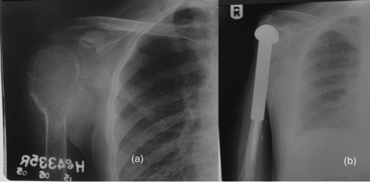
A 48-year-old man presented with progressively worsening left hip pain of several months’ duration. Sixteen years earlier, he had undergone wide resection of a giant cell tumor (GCT) involving the proximal right humerus, followed by endoprosthetic reconstruction (Fig. 1). Histopathological examination at that time confirmed a conventional GCT without evidence of malignant transformation. The patient remained asymptomatic and under regular surveillance for more than a decade.
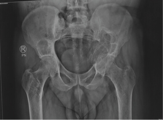
Plain radiographs and NCCT of the pelvis demonstrated a large expansile osteolytic lesion involving the left periacetabular region and adjacent iliac bone, with cortical breach and involvement of the proximal femur. Additional lytic lesions were present in the bilateral proximal femora and right iliac bone. The imaging findings raised the possibility of multicentric giant cell tumor (GCT), although metastatic disease and hyperparathyroidism-related brown tumors were also considered in the differential diagnosis (Fig. 2).
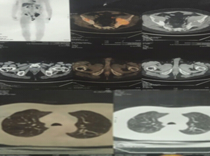
Whole-body positron emission tomography–computed tomography (PET-CT) demonstrated metabolically active lytic lesions involving the bilateral femoral neck and bilateral iliac bones with increased fluorodeoxyglucose uptake. No pulmonary metastases or additional skeletal lesions were identified, favoring a diagnosis of metachronous multicentric giant cell tumor rather than disseminated metastatic disease (Fig. 3).
Histopathological examination of the pelvic lesion revealed a highly cellular neoplasm composed of numerous uniformly distributed osteoclast-like multinucleated giant cells within a background of mononuclear stromal cells. The stromal cells were round to oval with focal spindle-cell morphology and displayed fine chromatin, inconspicuous nucleoli, and pale eosinophilic cytoplasm. Foci of hemorrhage were also present. Immunohistochemistry demonstrated strong positivity for H3F3A G34W, confirming the diagnosis of conventional giant cell tumor of bone (Fig. 4).
![Figure 4: Histopathological examination of the lesion. Hematoxylin and eosin-stained sections demonstrate hemorrhage and a cellular tumor composed of numerous osteoclast-like multinucleated giant cells (black arrows) within a background of mononuclear stromal cells (a, Hematoxylin and Eosin [H&E] ×100; b, H&E ×200). The stromal cells are round to oval and focally spindle-shaped with fine chromatin, inconspicuous nucleoli, and pale eosinophilic cytoplasm (c, H&E ×400). Immunohistochemistry shows diffuse nuclear positivity for H3F3A G34W, confirming the diagnosis of giant cell tumor of bone (d, ×400).](https://jocr.co.in/wp/wp-content/uploads/297FF4-converted-1.jpg)
As part of the diagnostic evaluation of multiple osteolytic lesions, serum calcium, phosphorus, alkaline phosphatase, and parathyroid hormone (PTH) levels were assessed and found to be within normal limits. These findings helped exclude metabolic conditions such as brown tumors secondary to hyperparathyroidism as potential differential diagnoses [8,9].
Given the multicentric involvement of the pelvis and proximal femur, the anatomically complex periarticular location of the lesions, and the significant morbidity associated with extensive surgical resection and reconstruction, a non-operative treatment strategy was considered most appropriate. The patient was treated with six intravenous infusions of zoledronic acid (4 mg/dose) at monthly intervals. The patient was supplemented with calcium and Vitamin D and was monitored clinically and radiologically during treatment. Therapy was well tolerated, with no treatment-related adverse events.
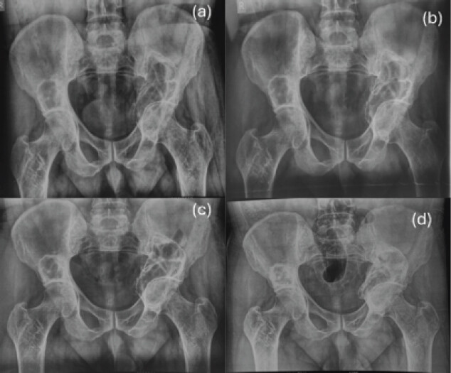
Clinical and radiographic follow-up was performed at 3-month intervals during the 1st year, every 6 months during the 2nd year, and annually thereafter. Serial annual pelvic radiographs demonstrated a sustained radiological response to therapy. The initially expansile osteolytic lesion showed progressive peripheral and intralesional sclerosis, gradual trabecular reconstitution, and stabilization of lesion margins over time. At final follow-up, marked sclerosis and osseous remodeling were evident without radiographic signs of progression (Fig. 5). Clinically, the patient experienced complete resolution of pain within 3 months of initiating treatment and returned to unrestricted functional activity. At the latest follow-up, 4 years after treatment initiation, there was no evidence of local progression, pulmonary metastasis, or development of new skeletal lesions.
Discussion
Multicentric GCT is a rare entity, representing less than 1% of all GCTs, and metachronous lesions are a subset of these cases [4,5,6]. Most metachronous lesions are diagnosed within 3–5 years of treatment of the index lesion, although intervals of over 10–20 years have been documented, highlighting the highly unpredictable natural history of this tumor [6,7].
Metachronous multicentric GCT is defined by the development of anatomically separate lesions with a disease-free interval between them. The usual sites involved are distal femur, proximal tibia, distal radius and proximal humerus, consistent with the common distribution of solitary GCT [1,5]. Pelvic involvement, as seen in our case, is less common and poses unique management challenges because of the proximity to the hip joint and the morbidity associated with extensive reconstruction.
The pathogenesis of metachronous disease is not fully understood. The suggested mechanisms include the activation of dormant neoplastic stromal cells, de novo formation of independent primary tumors, and less likely, occult metastatic spread [4,6]. The absence of pulmonary metastases in our patient and in most reported cases supports the idea of independent tumorigenesis instead of true metastatic disease.
Brown tumors of hyperparathyroidism are included in the differential diagnosis of multiple lytic lesions of bone and can closely resemble GCT radiologically and histologically [8]. Biochemical evaluation including serum calcium and PTH is an integral part of diagnostic work-up [9]. Normal PTH levels in our patient ruled out hyperparathyroidism and supported the diagnosis of true multicentric GCT.
Multicentric GCT should be treated on an individual basis. Surgical treatment is the standard for accessible lesions, but periarticular and pelvic lesions may require extensive procedures associated with significant morbidity and functional compromise. Bisphosphonates such as zoledronic acid inhibit osteoclast-mediated bone resorption, induce apoptosis of osteoclast-like giant cells, and inhibit proliferation of neoplastic stromal cells [10,11].
In addition to its antiresorptive effects, zoledronic acid has demonstrated a direct tumoricidal effect on GCT cells. Cheng et al. showed that nitrogen-containing bisphosphonates induce apoptosis in both osteoclast-like giant cells as well as neoplastic stromal cells, with zoledronic acid being the most potent [12]. Lau et al. also showed that zoledronic acid significantly reduced proliferation and induced apoptosis in most GCT stromal cell lines and downregulated expression of RANKL [13].
These experimental data are consistent with clinical data reported by Dubey et al. [14] demonstrating increased intralesional sclerosis, ultrastructural evidence of giant cell apoptosis and favorable clinical results in patients treated with zoledronic acid intravenously. Together, these studies provide a strong biological foundation for the durable radiological stabilization and resolution of symptoms seen in our patient. While denosumab is gaining traction for unresectable or recurrent GCT, zoledronic acid remains a cost-effective and widely available alternative, particularly in resource-poor settings. In the present case, pain relief was achieved completely with the use of zoledronic acid monotherapy, and radiological stabilization was maintained for 4 years without the need for surgery.
The 16-year disease-free interval in our patient places this case among the most delayed metachronous presentations reported in the literature and underscores the need for prolonged, potentially lifelong, surveillance in patients treated for GCT.
Conclusion
This case represents one of the most delayed presentations of metachronous multicentric giant cell tumor reported in the literature. It highlights the unpredictable behavior of GCT and the importance of long-term follow-up. In carefully selected patients with favorable tumor biology and preserved structural integrity, zoledronic acid may provide an effective non-surgical treatment option, supported by both experimental and clinical evidence of direct tumoricidal activity.
Clinical Message
Metachronous multicentric giant cell tumor can occur after a prolonged disease-free interval. Thorough metabolic evaluation is essential to exclude mimics such as hyperparathyroidism. In selected patients with preserved structural integrity and favorable tumor biology, zoledronic acid may provide effective disease control without surgical intervention.
Conflict of Interest:
Nil
Source of Support:
Nil
Consent:
The authors confirm that informed consent was obtained from the patient for publication of this article
How to Cite this Article
Sankhwar DK, Yadav P, Mridha AR, George J, Shankar V, Kumar VS. Delayed Metachronous Multicentric Giant Cell Tumor of Bone after a 16-Year Disease-Free Interval: A Case Report. Journal of Orthopaedic Case Reports 2026 October;16(10): 297-301.
References
- Campanacci M, Baldini N, Boriani S, Sudanese A. Giant-cell tumor of bone. J Bone Joint Surg Am 1987;69:106-14. [Google Scholar] | [PubMed]
- Athanasou NA. Giant cell tumor of bone. Orthop Clin North Am 2015;46:299-308. [Google Scholar] | [PubMed]
- Rock MG, Pritchard DJ, Unni KK. Metastases from histologically benign giant-cell tumor of bone. J Bone Joint Surg Am 1984;66:269-74. [Google Scholar] | [PubMed]
- Hoch B, Inwards C, Sundaram M, Rosenberg AE. Multicentric giant cell tumor of bone: Clinicopathologic analysis of 30 cases. Clin Orthop Relat Res 2006;450:231-6. [Google Scholar] | [PubMed]
- Errani C, Ruggieri P, Asenzio MA, Toscano A, Colangeli S, Rimondi E. Giant cell tumor of the extremity: A review of 349 cases from a single institution. Cancer Treat Rev 2010;36:1-7. [Google Scholar] | [PubMed]
- Bertoni F, Unni KK, Beabout JW, Ebersold MJ. Multicentric giant cell tumor of bone. Cancer 1988;62:2319-24. [Google Scholar] | [PubMed]
- Haskell A, Wodowoz O, Johnston JO. Metachronous multicentric giant cell tumor: A case report and literature review. Clin Orthop Relat Res 2003;412:162-8. [Google Scholar] | [PubMed]
- Keyser JS, Postma GN. Brown tumor of the mandible. Am J Otolaryngol 1996;17:407-10. [Google Scholar] | [PubMed]
- Silverberg SJ, Bilezikian JP. Evaluation and management of primary hyperparathyroidism. J Clin Endocrinol Metab 1996;81:2036-40. [Google Scholar] | [PubMed]
- Balke M, Campanacci L, Gebert C, Picci P, Gibbons M, Taylor R. Bisphosphonate treatment of aggressive primary, recurrent and metastatic giant cell tumour of bone. BMC Cancer 2010;10:462. [Google Scholar] | [PubMed]
- Tse LF, Wong KC, Kumta SM, Huang L, Chow TC, Griffith JF. Bisphosphonates reduce local recurrence in extremity giant cell tumor of bone: A case-control study. Bone 2008;42:68-73. [Google Scholar] | [PubMed]
- Cheng YY, Huang L, Lee KM, Xu JK, Zheng MH, Kumta SM. Bisphosphonates induce apoptosis of stromal tumor cells in giant cell tumor of bone. Calcif Tissue Int 2004;75:71-7. [Google Scholar] | [PubMed]
- Lau CP, Huang L, Wong KC, Kumta SM. Comparison of the anti-tumor effects of denosumab and zoledronic acid on the neoplastic stromal cells of giant cell tumor of bone. Connect Tissue Res 2013;54:439-49. [Google Scholar] | [PubMed]
- Dubey S, Rastogi S, Sampath V, Khan SA, Kumar A. Role of intravenous zoledronic acid in management of giant cell tumor of bone - a prospective, randomized, clinical, radiological and electron microscopic analysis. J Clin Orthop Trauma 2019;10:1021-6. [Google Scholar] | [PubMed]
© 2026 Journal of Orthopaedic Case Reports - Published by Indian Orthopaedic Research Group
About the Authors
-

Dilip Kumar Sankhwar
-

Puneet Yadav
-

Asit Ranjan Mridha
-

Jaiben George
-

Vivek Shankar
-

Venkatesan Sampath Kumar
Related Articles in Journal of Orthopaedic Case Reports
 June 1, 2026 Zoledronic Acid in the Management of Melorheostosis of Radius and Ulna – A Rare Case Report with Literature Review
 August 1, 2026 Bisphosphonate-Related Pathological Femoral Fracture in a Child with ACTG2-Related Intestinal Failure and Metabolic Bone Disease: A Case Report
 July 1, 2026 A Rare Case of Giant Cell tumor of Bone in Distal Tibia
 May 1, 2026 Combined Post-operative Denosumab and Oral Bisphosphonate Therapy Following Resection of Spinal Giant Cell Tumor: A Case Report