


Introduction
Articular cartilage is robust and elastic type of connective tissue that plays a crucial role in the physiological mobility of joints by providing improved cushioning and reducing friction on articulated surfaces [1]. Articular cartilage can be damaged due to trauma, sports or normal aging degenerative process. Once damaged, it cannot heal itself due to its avascular nature and the limited mitotic potential of chondrocytes [2]. At present, there is no universal technique for replacement of cartilage defect suitable for all patients. The method chosen depends on the defect’s grade, size, and localization, along with age and activity level of the patients [1]. Various surgical methods have been discussed to address cartilage defects including microfracture, osteochondral autograft transfer system, mosaicplasty, autologous chondrocyte implantation, among others [3]. Another method is to use collagen gel, which has become an attractive solution for chondral defects in recent times. Cell free collagen matrix is an example of absorbable acellular Type 1 collagen implant. When applied to the focal area of cartilage lesion, it forms a protective layer over injured area and provides a matrix for migration of stem cells and chondrocytes, thus it aims to restore articular cartilage [4].
Promising results have been reported using the acellular collagen matrix for the cartilage defects of knee, hip and talus [4,5,6,7,8,9,10]. With respect to knee joints, most of the authors have used it in small to medium chondral defects (up to 2 cm2) [4,7,8]. However, its use in large cartilage defects (>2 cm2) is not very well studied. Furthermore, there is paucity of literature showing the precise quality of repaired cartilage, as conventional magnetic resonance imaging (MRI) cannot quantitatively measure the quality of cartilage repair. The quantitative MRI (qMRI) T2 mapping, such as Cartigram, can quantitatively measure alterations in the water and collagen content of hyaline articular cartilage precisely and T2 value gives a quantitative assessment of quality of articular cartilage [11, 12]. Here, we have reported two cases with larger cartilage defect (up to 3.5 cm2) of the knee treated with acellular collagen matrix that showed excellent early healing depicted in qMRI T2 mapping with equally good early functional outcomes.
Case Report
We have reported two cases of isolated International Cartilage Repair Society Grade IV [13] articular cartilage defects of large size in the knee treated with acellular collagen matrix and showed the quality of repair using qMRI T2 mapping with early functional outcomes using Lysholm and International Knee Documentation Committee (IKDC) score. The surgical technique and post-operative rehabilitation were same for both the patients, which are described as follows.
Surgical procedure
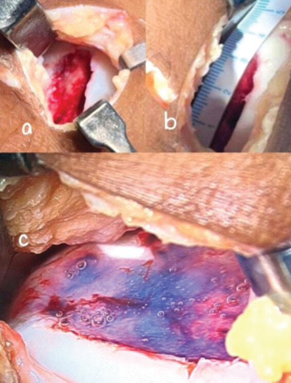
The surgical procedures were done under spinal anesthesia. Both the cases were operated by the same surgeon. With pneumatic tourniquet applied, first diagnostic arthroscopy was done and loose bodies were removed. Under arthroscopy, the defect was visualized and its size was carefully measured using a probe. In both cases, the defect was in medial femoral condyle. Following diagnostic arthroscopy, a mini-open arthrotomy was performed to address the defect. The cartilage defects were carefully debrided using a motorized shaver and curette. The size of the defects was documented. After debridement, it was irrigated and dried to ensure a clean and moisture-free surface. Cell free collagen matrix, previously stored at −20°C, was taken out of refrigeration and allowed to defrost at room temperature over 15–20 min. During this period, it transitioned from a solid white state to a transparent gel form, indicating readiness for application. The acellular collagen matrix was applied into the defect using pre-attached cannula until the defect was filled completely. Following application, the area was left undisturbed for gelification which was determined by the change of color from transparent to white. The limb was maintained in the same position throughout the setting phase. Once the collagen matrix had solidified, standard layered wound closure was performed followed by compression dressing application.
Post-operative rehabilitation protocol
postoperative rehabilitation protocol.
| Time | Exercises |
|---|---|
| 0–2 weeks | Immobilized in extension, ice, compression |
| Ankle pumps | |
| Non-weight-bearing | |
| Active straight leg raise without extensor lag | |
| Patellar mobilization | |
| 2–6 weeks | Knee flexion increased by 30° every 2 weeks |
| Non-weight-bearing | |
| Isometric quadriceps, , hamstring strengthening exercise, side lying hip abduction | |
| 6–8 weeks | Partial weight-bearing with brace and walker |
| Full knee range of motion achieved | |
| 8–12 weeks | Full weight-bearing allowed |
| Strengthening of quadriceps, hamstring | |
| Balancing exercises | |
| 12–24 weeks | Single leg strength exercises (single leg heel raise, single leg dead lift, single leg squat) |
| Gym based strengthening of quadriceps, VMO, hamstrings | |
| Strengthening of hip abductors and flexors. | |
| Things to avoid: Exercises into knee flexion >90°, cutting/pivoting, sports specific activity | |
| After 6 months | Return to normal activities |
VMO: Vastus medialis obliquus
Follow-up
Regular follow-ups were carried out at one month interval for 6 months. Clinical outcomes were evaluated based on Lysholm and IKDC scores at 3rd, 6th and 12th month. At 6 months, a Cartigram (qMRI T2 mapping) was done to check the healing of the cartilage repair.
Case report 1
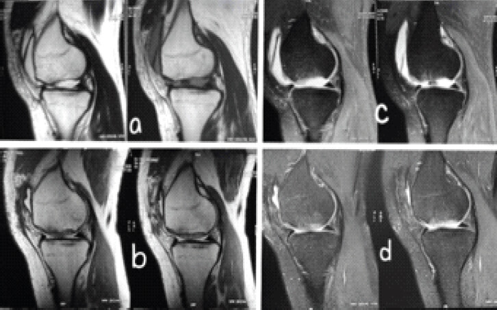
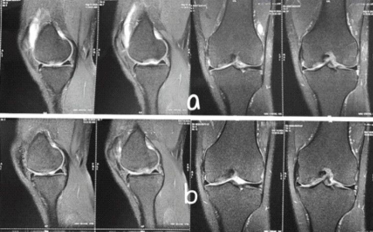
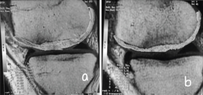
A 17-year-old male (non-athlete) presented with a two-month history of non-traumatic right knee pain and catching sensation, aggravated by weight-bearing activities. The pre-operative IKDC and Lysholm scores were 31.03 and 38, respectively. MRI of the knee revealed osteochondritis dissecans involving the medial femoral condyle, with a large osteochondral defect measuring approximately 3.2 × 2 cm and the presence of two sizable loose bodies (Fig. 1). He underwent arthroscopic removal of the loose bodies followed by mini-open cartilage repair using cell free collagen matrix as described above (Fig. 2). After debridement, the defect size was 3.5 × 2.5 cm. Post-operative IKDC score increased from 31.03 to 60.4 and 71.26 at 3rd and 6th months, respectively, whereas the Lysholm score increased from 38 to 69 and 85 at 3rd and 6th months, respectively. A follow-up Cartigram was performed at 6 months to evaluate the healing of the repair (Fig. 3). The T2 values at the site of cartilage repair of medial femoral condyle was 48–53, which is similar to the T2 values of adjacent normal cartilage (39–49), suggesting excellent healing.
Case report 2
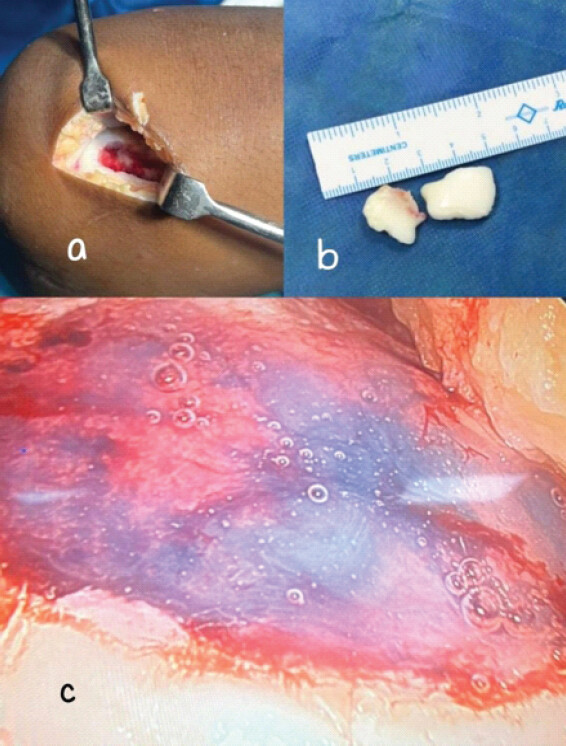
A 33-year-old man (non-athlete) presented with left knee pain for 3 months following fall from stairs. Pain was particularly on weight-bearing and knee movement. On clinical examination there was terminal loss of flexion due to pain. MRI was suggestive of large cartilage defect at the weight-bearing area of medial femoral condyle with two loose bodies (Fig. 4). The pre-operative IKDC and Lysholm score was 33.33 and 42, respectively. He also underwent diagnostic arthroscopy and loose body removal, followed by mini-open repair of cartilage defect using acellular collagen matrix (Fig. 5). After debridement, the defect size was 4 × 2 cm. Post-operative IKDC scores at 3rd, 6th, and 12th month were 62, 73.56, and 81.60, respectively, Lysholm scores at 3rd, 6th and 12th month were 71, 85 and 95. Follow-up Cartigram at 6 months suggested the T2 values at the site of cartilage repair site were 44–56, which is similar to the T2 values of adjacent normal cartilage (44–54), suggesting excellent healing (Fig. 6).
Discussion
Restoration of cartilage defects still remains a major obstacle in orthopedics. Extensive research has been done in the last decade to find suitable biomaterial which could be used. Cell free collagen matrix has become an attractive technique for restoring the cartilage defects. The basic principle of acellular collagen matrix is to fill the cartilage defect with matrix structure, which provides a scaffold for chondrocytes to migrate from the perilesional tissue, enabling attachment, proliferation and extracellular matrix production [7].
Schneider [7], in his study done in 2016 showed successful filling of all the cartilage defects of knee using cell free collagen matrix in thirteen patients with radiological correlation using conventional MRI sequence. The defects were of small and medium size (mean size <2.5 cm).
In another study, published in 2024, Simeonov found encouraging results by employing cell free collagen matrix to restore articular cartilage. He concluded that collagen matrix provides excellent surgical solution to younger patients with chondral defects <2 cm2 based on clinical outcomes. However, no follow-up MRI was done [4].
Similarly, in the study conducted by Syed et al. on cell free collagen matrix and treatment of cartilage defects in the knee, the mean chondral defect size was 11.82–17.82 mm. They have evaluated the outcomes using IKDC score and concluded that it could serve as a good treatment option for Grade IV chondral defects [8].
Schüttler et al. [6] and Efe et al. [5] in their respective studies showed clinical as well as MRI correlation for the repair of knee chondral defects of small size (<11 mm) using cell free collagen Type 1 matrix.
In the current literature, the cartilage defects of knee treated with acellular collagen matrix were of small to medium size (<2.5 cm). In our study, maximum dimension of both the defects was of more than 2.5 cm. In addition, there are not many studies on healing of knee articular cartilage defect and its MRI correlation. In fact, we were able to find only one study on knee cartilage defect repair using acellular collagen matrix, done by Schneider [7], where MRI studies were done in follow-up to show the repair quality. However, to the best of our knowledge, we were not able to find any English literature where Cartigram was done to assess the healing quality of cell free collagen matrix in cartilage injuries. T2 relaxation mapping with MRI, such as Cartigram, can help visualize and quantitatively evaluate the water content of cartilage. The changes in T2 values correlate with variations in water content and collagen structure and organization, as well as changes in hyaline cartilage composition and its depletion [11]. In addition, T2 mapping enables demonstration of treatment response based on changes in T2 values [14]. Here, we have assessed the healing of repaired cartilage defects using Cartigram at 6 months. In both the cases, the Cartigram showed excellent healing with T2 values of 48–53 in the first case and 44–56 in the second case, respectively, which were similar to T2 values of adjacent normal cartilage. The IKDC score in our study was 71.26 and 73.56 in the first and second cases, respectively, at 6 months. The mean IKDC scores at 6 months in other reference studies were 72.1 (Schneider), 66.25 (Syed et al.). We have applied cell free collagen matrix through mini-open arthrotomy instead of doing arthroscopically as the defect size was larger than usually described in literature.
Conclusion
This case report highlights the potential of cell free collagen matrix as an effective treatment for large Grade IV chondral defects of the knee, showing both excellent early functional recovery and promoting hyaline like cartilage regeneration as evidenced by qMRI Cartigram. To the best of our knowledge, this is among the first reports to document healing of large chondral defects of size >2.5 cm2 using cell free collagen matrix assessed by quantitative T2 mapping. Further prospective studies with larger cohorts and longer follow-ups are needed to establish its long-term efficacy.
Clinical Message
Cell free collagen matrix can be effectively used to treat large (2–4 cm) chondral defects with excellent early radiological and functional outcomes.
Conflict of Interest:
Nil
Source of Support:
Nil
Consent:
The authors confirm that informed consent was obtained from the patient for publication of this article
How to Cite this Article
Ajay, Parveen R, Mandal S, Mishra P, Mehmood Y, Ruhela A. Is Cell-Free Collagen Matrix Effective in the Treatment of Large Cartilage Defects of Knee? Early Functional and Quantitative Magnetic Resonance Imaging Cartigram Assessment in Two Patients. Journal of Orthopaedic Case Reports 2026 October;16(10): 57-62.
References
- Buryanov OA, Chornyi VS, Bazarov MO, Mohilnytskyy AО, Hutsailiuk VІ, Kusyak АP. Modern technologies for replacement of cartilage defects. Trauma 2024;25:45-53. [Google Scholar] | [PubMed]
- Steinert AF, Ghivizzani SC, Rethwilm A, Tuan RS, Evans CH, Nöth U. Major biological obstacles for persistent cell-based regeneration of articular cartilage. Arthritis Res Ther 2007;9:213. [Google Scholar] | [PubMed]
- Gracitelli GC, Moraes VY, Franciozi CE, Luzo MV, Belloti JC. Surgical interventions (microfracture, drilling, mosaicplasty, and allograft transplantation) for treating isolated cartilage defects of the knee in adults. Cochrane Database Syst Rev 2016;9:CD010675. [Google Scholar] | [PubMed]
- Simeonov E. Implantation of ChondroFiller Liquid® as a scaffold material for the treatment of chondral lesions of the knee joint. JIMAB 2024;30:5936-41. [Google Scholar] | [PubMed]
- Efe T, Theisen C, Fuchs-Winkelmann S, Stein T, Getgood A, Rominger MB. Cell-free collagen type I matrix for repair of cartilage defects-clinical and magnetic resonance imaging results. Knee Surg Sports Traumatol Arthrosc 2012;20:1915-22. [Google Scholar] | [PubMed]
- Schüttler KF, Götschenberg A, Klasan A, Stein T, Pehl A, Roessler PP. Cell-free cartilage repair in large defects of the knee: Increased failure rate 5 years after implantation of a collagen type I scaffold. Arch Orthop Trauma Surg 2019;139:99-106. [Google Scholar] | [PubMed]
- Schneider U. Controlled, randomized multicenter study to compare compatibility and safety of ChondoFiller liquid (cell free 2-component collagen gel) with microfracturing of patients with focal cartilage defects of the knee joint. Video J Orthop Surg 2016;1:1-8. [Google Scholar] | [PubMed]
- Syed RF, Thati S. Chondrofiller and treatment of cartilage defects in the knee. Acta Sci Orthop 2024;7:3-7. [Google Scholar] | [PubMed]
- Mazek J, Gnatowski M, Salas AP, O'Donnell JM, Domżalski M, Radzimowski J. Arthroscopic utilization of ChondroFiller gel for the treatment of hip articular cartilage defects: A cohort study with 12- to 60-month follow-up. J Hip Preserv Surg 2021;8:22-7. [Google Scholar] | [PubMed]
- Khan PS, Meleppuram JJ, Ahmed HN, Nizaj N, Nair AV, Ananthakrishnan R. Arthroscopic collagenous matrix therapy for osteochondral lesions of the talus. Arthrosc Tech 2025;14:103575. [Google Scholar] | [PubMed]
- Lee SH, Lee YH, Song HT, Suh JS. Quantitative T2 mapping of knee cartilage: Comparison between the synthetic MR imaging and the CPMG sequence. Magn Reson Med Sci 2018;17:344-9. [Google Scholar] | [PubMed]
- Safna MA, Sunil M, Kumar K. T2 mapping [Cartigram] in the evaluation of the articular cartilage in traumatic knee injury. MedPulse Int J Radiol 2019;9:59-62. [Google Scholar] | [PubMed]
- Van Den Borne MP, Raijmakers NJ, Vanlauwe J, Victor J, De Jong SN, Bellemans J. International cartilage repair society (ICRS) and Oswestry macroscopic cartilage evaluation scores validated for use in autologous chondrocyte implantation (ACI) and microfracture. Osteoarthritis Cartilage 2007;15:1397-402. [Google Scholar] | [PubMed]
- Mnezes NM, Gray ML, Hartke JR, Burstein D. T2 and T1rho MRI in articular cartilage systems. Magn Reson Med 2022;51:503-9. [Google Scholar] | [PubMed]
© 2026 Journal of Orthopaedic Case Reports - Published by Indian Orthopaedic Research Group
About the Authors
-

Ajay
-

Ruhi Parveen
-

Subhashis Mandal
-

Pallav Mishra
-

Yassir Mehmood
-

Ankit Ruhela
Related Articles in Journal of Orthopaedic Case Reports
 January 1, 2026 Single Stage Autologous Minced Cartilage Implantation for Chondral Defects of the Knee: A Case Series
 October 1, 2025 Hip Arthroscopy and Chondrofiller Application in Isolated Osteochondral Defect of the Femoral Head
 May 10, 2020 Acromioclavicular Joint Disk Tearing in Young Non-Arthritic Individuals: A Case Series
 October 1, 2025 Pedunculated Exostosis of the Pedicle of Lumbar Vertebra: A Rare Cause of Lumbar Pain and Swelling