


Introduction
Syndactyly is among the most common congenital malformations of the hand and is due to incomplete separation of the digital rays in the embryo [1]. It can be simple, which is only soft-tissue fusion, or complicated, which also involves the skeleton. Early surgical correction in childhood is advised to prevent functional impairment and progressive deformity during growth [2].
The main goals of syndactyly release and soft-tissue reconstruction are safely separating the digits, preserving the neurovascular structures and achieving a stable and acceptable-looking web space [3]. This can be done carefully through the redistribution of local soft tissues to avoid any complications post-surgery, like scar contracture or recurrence of webbing [4].
Surgical methods for syndactyly release have included zig-zag, dorsal rectangular flap and interdigitating flap, designed to redistribute skin and avoid linear scars. However, flap design is frequently dependent on surgical judgment rather than measurement, which may lead to asymmetric flap sizes and uneven tension in the reconstructed web space.
Adult manifestation of untreated syndactyly is rare and poses further reconstructive problems. Adjustments in soft-tissue elasticities along with adaptive anatomical changes from long-standing fusion may make surgical planning difficult [9]. In such cases, structured pre-operative planning is all the more necessary to obtain balanced tissue coverage and functional outcomes.
This technique takes on the flap planning for adult syndactyly release in a measurement-based flap planning way that highlights flap geometric design.
Case Report
A 23-year-old functionally active male presented with untreated congenital syndactyly of the middle and ring fingers. No surgical correction had been performed during childhood. He reported difficulty in independent movement of the involved digits during certain activities of daily living and also expressed concern regarding the cosmetic appearance of the hand.
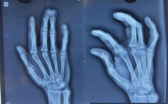
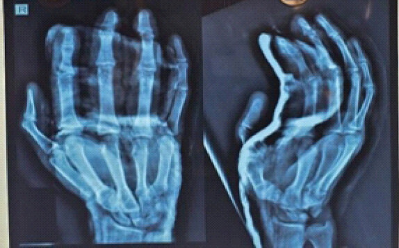
Clinical examination demonstrated syndactyly of the middle and ring fingers extending from the web space distally to the fingertips. Active and passive movements of the interphalangeal and metacarpophalangeal (MCP) joints were pain-free, although independent movement of the two involved digits was restricted by the syndactyly. The distal fusion was associated with confluent nail plates, with no apparent involvement of the nail folds. There was no clinical evidence of neurovascular compromise. The remaining digits and the contralateral hand were clinically normal. No associated upper-limb congenital anomaly or clinical feature suggestive of a syndromic association was identified. Plain radiographs demonstrated soft-tissue syndactyly of the middle and ring fingers with osseous fusion involving the distal portions of the distal phalanges, consistent with complex syndactyly (Fig. 1). The remaining phalanges were normally aligned, and the interphalangeal and MCP joint spaces were preserved. As the distal phalanges were fused, surgical separation required both soft-tissue release and osseous separation.
In view of the functional restriction and the patient’s desire for correction, surgical separation and reconstruction were planned. The delayed adult presentation posed an additional reconstructive challenge because long-standing fusion was expected to be associated with altered soft-tissue distribution and limited available skin following separation. Therefore, rather than relying solely on visual estimation of flap dimensions, a measurement-based pre-incision planning protocol was used to define the flap geometry and planned web-space reconstruction before surgical release.
Measured flap planning technique
Pre-operative anatomical marking
On the day prior to surgery, pre-operative marking was carried out while the patient was supine and the upper limb was kept in the anatomical position. The MCP and proximal interphalangeal (PIP) joint levels were identified and marked on both involved digits. The longitudinal distances between the MCP and PIP joint markings were measured. Another reference point was located at the junction of the distal one-third of the measured MCP-PIP distance in each digit. Therefore, the reference point was determined from the measured length of the proximal phalanx rather than from visual estimation.
Step 1: Identification of the mid-dorsal axis
A longitudinal line was drawn in the middle of the dorsal surface of the syndactylized digits halfway between the middle and ring fingers. This mid-dorsal axis was the main longitudinal reference for subsequent flap markings and allowed checking the flap configuration on either side for symmetry.
Step 2: Measurement of digital width
The transverse width of syndactylized digits was measured with a ruler at the level of the proximal phalanx. The measurement was at right angles to the mid-dorsal axis. The measured width was used as a reference for the spacing of successive zig-zag markings rather than choosing the flap dimensions by visual approximation alone.
Step 3: Geometric zig-zag flap marking
Alternating triangular flaps were then marked on the dorsal and volar surfaces. Successive flap limbs were planned at equal measured intervals, with corresponding flap lengths maintained equal on either side of the central reference axis. The markings were continued distally while maintaining the alternating configuration so that the opposing flaps could subsequently be interdigitated without creating a continuous longitudinal scar.
Step 4: Web commissure planning
The apex of the reconstructed web space was proposed to be located proximal to the level of the adjacent normal web spaces. Based on this predetermined point, the reconstruction of the web space was planned using a vertically orientated rectangular dorsal flap and a horizontally orientated rectangular volar flap. Their dimensions were measured before incision and included in the overall measured flap design.
Step 5: Verification of symmetry
Before surgery, the completed markings were reassessed using the central axis and previously marked anatomical landmarks. Corresponding flap dimensions were compared to ensure equal flap lengths and appropriate alignment. Any discrepancy in the markings was corrected before incision. This final verification constituted an integral component of the measured planning technique.
The upper limb was placed on an arm table, and a supraclavicular block was given on the day of surgery. The identified anatomical landmarks and the measured flap markings were maintained and utilized as the operative guide (Fig. 2, 3, 4, 5). The digits were separated by careful dissection, identifying and preserving the digital neurovascular bundles. The opposing flaps were interdigitated to provide balanced soft-tissue coverage and reconstruction of the web space.
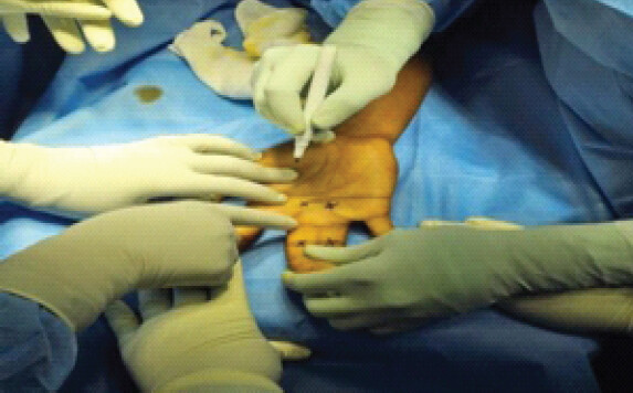
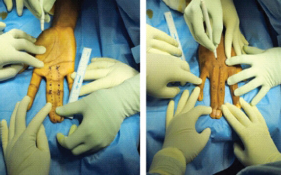
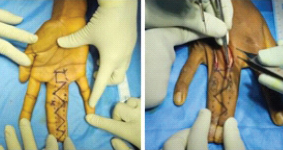
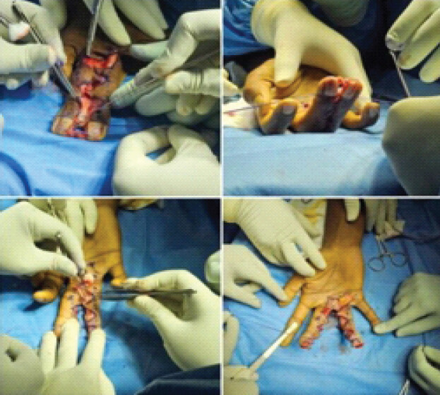
The distal portions of the fused phalanges were separated with a long curved osteotome under fluoroscopic guidance due to the distal osseous fusion. The nail beds were unusually well preserved. After the bony split, the position of the digits was confirmed fluoroscopically, flaps transposed as designed preoperatively, and the closure was completed without excessive tension. Post-operative radiographs revealed separation of the previously fused distal phalanges (Fig. 6).
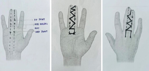
Structured measurement-based flap planning framework demonstrating symmetrical zig-zag geometry and web space planning before surgical digit separation.
Discussion
Syndactyly release and soft-tissue reconstructive surgery can be a demanding procedure that requires careful surgical planning to prevent complications such as web creep, scar contracture, and the asymmetry of digits [3,10].
Syndactyly release is commonly performed using zig-zag incisions, with interdigitating flaps that redistribute skin while helping prevent linear scars. These techniques have led to satisfactory outcomes if performed carefully. Flap planning, however, is often based on subjective estimation rather than objective measurement, which may vary flap geometry.
In this patient, the condition may be worsened due to decreased elasticity of the skin and a long history of structural adaptation in adults with syndactyly. Measuring-based flap designing affords a systematic protocol for creating symmetrical flaps. This also aids in achieving an even soft-tissue distribution post-separation.
Accurate mapping of the web commissure is critical because ineffectual web depth is a frequent contributor to unsatisfactory outcomes after syndactyly repair. Techniques that emphasize dorsal flap design and placement of the web apex have been associated with superior functional outcome with an esthetic touch.
The classical literature on syndactyly release has focused mainly on flap shape, web reconstruction, prevention of web creep, and long-term outcomes. Cronin [3] described the foundational zig-zag concept, while later authors discussed dorsal commissural flaps, interdigitating designs, simplified release methods, and causes of reconstruction failure. However, most descriptions do not formalize the pre-incision measurement process as a reproducible step. The present report differs by converting flap planning into a structured sequence: identifying the midline axis, measuring digital width, creating equal zig-zag intervals, planning the commissure, and verifying symmetry before incision. Table 1 portrays a comparative analysis of existing syndactyly techniques.
The principles of syndactyly reconstruction must be seen in relation to the wider field of congenital hand surgery. Modern reviews of congenital hand anomalies stress careful assessment of the anatomical pattern and individualised surgical planning [11,12]. Established principles of syndactyly reconstruction include appropriate timing of surgery, meticulous separation of digits, reconstruction of an adequate web commissure, preservation of neurovascular structures and provision of satisfactory soft tissue coverage [13–15]. These basic principles of reconstruction are also described in standard texts on congenital and operative hand surgery [16–18]. Moreover, classification of congenital upper-limb anomalies based on anatomical and developmental characteristics still is important for understanding the individual deformity and planning appropriate treatment [20].
Comparative analysis of existing syndactyly techniques.
| Author/Reference | Main contribution in literature | Limitation or gap in relation to your case | How your MFPT adds value |
|---|---|---|---|
| Cronin, 1956 [3] | Described the classic zig-zag principle for syndactyly repair to avoid linear scar contracture. | Focused on incision pattern, not on objective measurement of flap intervals. | MFPT retains zig-zag principles but adds measured spacing and symmetry verification. |
| Kozin, 2001 [2] | Provided broad principles of syndactyly classification, timing, surgical release, and complications. | Discusses principles, but does not provide a stepwise measurement-based marking protocol. | MFPT converts general planning principles into a reproducible operative marking sequence. |
| Tonkin, 2009 [4] | Analysed causes of failed syndactyly reconstruction, including web creep and poor soft-tissue planning. | Identifies failure patterns but does not propose a simple measurement-based preventive method. | MFPT addresses potential asymmetry and tension before incision, aiming to reduce preventable technical errors. |
| Manske and Halikis, 1990 [5] | Emphasised classification and surgical planning based on syndactyly type. | Classification helps decision-making but not exact intraoperative flap geometry. | MFPT supplements classification with practical geometric planning at the skin level. |
| Kay and McCombe, 2000 [6] | Discussed reconstructive principles and operative strategies in syndactyly release. | Technique planning remains largely experience-dependent. | MFPT provides a teachable framework, especially useful for trainees and delayed adult cases. |
| Yoon and Jones, 2019 [7] | Described interdigitating rectangular flaps for correction of syndactyly. | Focused on a flap design, not a universal measurement template for planning. | MFPT can be used as a planning adjunct before raising interdigitating flaps. |
| Sharma and Tuli, 2008 [8] | Presented a simplified technique for syndactyly release. | Simplification does not necessarily mean objective symmetry control. | MFPT adds measurable reproducibility without making the operation complex. |
| Niranjan and De Carpentier, 2005 [9] | Reported long-term outcomes following syndactyly correction. | Outcome-focused, with less emphasis on stepwise pre-incision planning. | MFPT focuses on the planning stage that may influence long-term scar behavior and web stability. |
| Buck-Gramcko, 1990 [10] | Described congenital hand malformations and reconstructive principles. | Broad congenital hand framework rather than a specific measured marking method. | MFPT narrows the concept to a precise operative planning method. |
| Ezaki, 2005 [19] | Reviewed syndactyly principles and operative management. | Conventional techniques are described, but measurement-based flap planning is not formalized. | MFPT formalizes what is often left to visual judgment. |
MFPT: Measurement-based flap planning technique
While this report represents a single case, the measured flap planning technique provides a structured approach to flap planning in syndactyly reconstruction. Use of anatomical landmarks, digital width measurement, planned flap symmetry, and pre-incision verification may reduce reliance on visual estimation only and provide a more consistent framework for surgical planning.
There are some limitations to this report. The technique was used in one adult patient with complex syndactyly, and no comparison was made with conventional flap planning. The length of follow-up limits our ability to assess late outcomes such as web creep, scar contracture, recurrence, and long-term functional improvement. Patient-reported and objective functional outcome measures were not prospectively collected with standardised measures. In this index case, although anatomical landmarks and digital width measurements were used in flap planning, the actual flap dimensions in numbers and angular relationships were not recorded prospectively. Clinically, post-operative flap symmetry was assessed, not on the basis of any predefined quantitative geometrical measurements. In addition, the procedure was performed by a single surgical team, and the interobserver and intersurgeon reproducibility has not been assessed. Future prospective studies should overcome these limitations by including predefined geometric parameters, standardised functional outcomes, independent assessment, multiple surgeons and longer follow-up.
What is new in the present report is not the introduction of a new flap pattern. The use of zig-zag incisions, dorsal commissural flaps, and interdigitating flap designs in syndactyly reconstruction is well established. The proposed change is in the structured process of planning for the incision, not in the use of the device. The technique of the measured flap design involves the identification of a central reference axis, measurement of the digital width with a ruler, equalization of the corresponding flap limbs, planned positioning of the apex of the web, and final verification of symmetry before incision. Thus, measurement-based flap planning technique is a planning adjunct to established syndactyly reconstruction, which is measurement-based, rather than a new reconstructive technique in its own right.
Conclusion
This case report illustrates the potential of integrating objective anatomical measurements and symmetry verification in pre-operative flap planning for adult complex syndactyly. Measured flap planning technique is best considered a planned adjunct to established syndactyly release methods rather than a new flap design or a technique of proven superiority. Further prospective evaluation with different syndactyly patterns, standardised functional outcomes, objective geometric assessment, independent evaluation, and longer follow-up is needed to establish its reproducibility and clinical utility.
Clinical Message
Adult complex syndactyly presents a unique reconstructive challenge due to long-standing tissue adaptation.
Planning the flap with a structured, measurement-based approach can reduce dependence on visual estimation and allow for symmetrical flap design and web-space reconstruction.
Conflict of Interest:
Nil
Source of Support:
Nil
Consent
The authors confirm that informed consent was obtained from the patient for publication of this article
How to Cite this Article
Rajadurai ORJW, Levi JJ, Kumar MS, Purushothaman K, Kanchan N, Moulvi SMM. Measured Flap Planning for Adult Complex Syndactyly Release: A Technical Case Report. Journal of Orthopaedic Case Reports 2026 October;16(10): 308-313.
References
- Braun TL, Trost JG, Pederson WC. Syndactyly release. Semin Plast Surg 2016;30:162-70. [Google Scholar] | [PubMed]
- Kozin SH. Syndactyly. J Am Acad Orthop Surg 2001;9:343-55. [Google Scholar] | [PubMed]
- Cronin TD. Syndactylism: A method of repair. Plast Reconstr Surg 1956;18:393-402. [Google Scholar] | [PubMed]
- Tonkin MA. Failure of syndactyly reconstruction. Hand Clin 2009;25:241-9. [Google Scholar] | [PubMed]
- Manske PR, Halikis MN. Surgical classification of syndactyly. Hand Clin 1990;6:557-64. [Google Scholar] | [PubMed]
- Kay SP, McCombe D. Syndactyly reconstruction. J Hand Surg Br 2000;25:505-10. [Google Scholar] | [PubMed]
- Yoon AP, Jones NF. Interdigitating rectangular flaps and dorsal pentagonal island flap for syndactyly release. J Hand Surg Am 2019;44:288-95. [Google Scholar] | [PubMed]
- Sharma RK, Tuli P. Syndactyly release: A simplified technique. Indian J Plast Surg 2008;41:199-204. [Google Scholar] | [PubMed]
- Niranjan NS, De Carpentier JP. Primary syndactyly correction: Long-term outcomes. J Plast Reconstr Aesthet Surg 2005;58:643-52. [Google Scholar] | [PubMed]
- Buck-Gramcko D. Congenital malformations of the hand. Hand Clin 1990;6:531-48. [Google Scholar] | [PubMed]
- Kozin SH, Zlotolow DA. Common pediatric congenital conditions of the hand. Plast Reconstr Surg. 2015;136(2):241e-257e. doi:10.1097/PRS.0000000000001469.. PMID: 26218399.. [Google Scholar] | [PubMed] | [CrossRef]
- Tonkin MA. Congenital hand anomalies. J Bone Joint Surg Br 2001;83:1-7. [Google Scholar] | [PubMed]
- Bae DS. Principles of syndactyly reconstruction. J Pediatr Orthop Soc North Am 2020;2:1-9. [Google Scholar] | [PubMed]
- Jones NF. Reconstruction of congenital hand anomalies. Plast Reconstr Surg 2011;128:241-50. [Google Scholar] | [PubMed]
- Kozin SH, Bishop AT. Syndactyly reconstruction techniques. Hand Clin 2007;23:1-13. [Google Scholar] | [PubMed]
- Flatt AE. The Care of Congenital Hand Anomalies. St Louis: Mosby; 1977. [Google Scholar] | [PubMed]
- Green DP, Hotchkiss RN, Pederson WC, Wolfe SW. Green's Operative Hand Surgery.. 7th ed. Philadelphia, PA: Elsevier; 2017. [Google Scholar] | [PubMed]
- Wolfe SW, Hotchkiss RN, Pederson WC, Kozin SH. Green's Operative Hand Surgery.. 6th ed. Philadelphia, PA: Elsevier; 2011. [Google Scholar] | [PubMed]
- Ezaki M. Syndactyly. Hand Clin 2005;21:631-41. [Google Scholar] | [PubMed]
- Manske PR, Oberg KC. Classification and developmental biology of congenital anomalies of the hand and upper extremity. J Bone Joint Surg Am. 2009;91(Suppl 4):3-18. doi:10.2106/JBJS. I.00008.. [Google Scholar] | [PubMed] | [CrossRef]
© 2026 Journal of Orthopaedic Case Reports - Published by Indian Orthopaedic Research Group
About the Authors
-

Jeff Walter Rajadurai OR
-

J Jebin Levi
-

M Sathesh Kumar
-

K Purushothaman
-

N Kanchan
-

S M M Moulvi
Related Articles in Journal of Orthopaedic Case Reports
 July 1, 2026 Adult Presentation of Congenital Macrodactyly of the Hand Managed with Debulking and Amputation of a Digit: Report of a Rare Case
 June 1, 2026 Staged Reconstruction for Dorsal Hand and Wrist Shear Injury: A Case Report
 March 1, 2026 The use of Hyaluronic Acid Matrix for Effective Skin Deficit Coverage in Pediatric Complex Syndactyly Reconstruction: A Case Report and Technique Review
 April 1, 2026 Incidental Intramedullary Lipoma of the Proximal Femur Detected Following Trauma: A Case Report