


Introduction
The humerus, the longest bone in the upper limb, extends from the shoulder to the elbow joint, supporting shoulder function and arm movement. Proximal humerus fractures account for 4–5% of all fractures, ranking third after hip and distal radius fractures [1]. These injuries predominantly affect the elderly due to osteoporosis, typically resulting from low-energy falls on an outstretched arm [2].
The Neer classification system, based on fragment displacement (>10 mm) and angulation (>45°), guides treatment decisions and outcome predictions [3]. More complex fracture patterns generally carry poorer prognoses and may require advanced imaging like computed tomography (CT) scans for accurate assessment [4].
Management options for displaced proximal humerus fractures include various fixation techniques. Open reduction and internal fixation with locking plates offers anatomical restoration but risks complications in osteoporotic bone, including screw cut-out, avascular necrosis, and impingement [5,6]. Intramedullary nailing often compromises rotator cuff integrity [7], while transcutaneous K-wire fixation is associated with pin migration, infection, and restricted shoulder motion [8].(Fig.1).
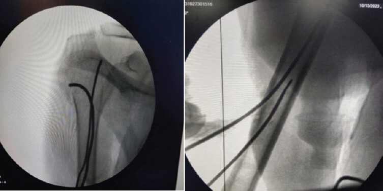
To minimize these complications, Takeuchi et al. developed a minimally invasive technique using three intramedullary J-nails [9]. This approach aims to achieve relative stability while preserving vascularity and soft tissue integrity. The J-nail technique offers potential advantages: Minimal soft tissue disruption, insertion below the deltoid attachment (avoiding shoulder and elbow joint impairment), and immediate post-operative pain relief with relatively stable fixation [9].
Despite these theoretical benefits, evidence regarding J-nail effectiveness for proximal humerus fractures in elderly patients remains limited. This study aims to evaluate functional outcomes following J-nail fixation in elderly patients with Neer 2-, 3-, and 4-part proximal humerus fractures. In addition, we will identify and characterize associated complications. By systematically assessing both benefits and limitations, this study seeks to determine whether J-nail fixation represents a viable alternative to conventional surgical approaches for this challenging fracture pattern in a vulnerable population.
Materials and Methods
Study design and setting
This is a single-center prospective, observational study conducted at the Department of Orthopaedics, Hamidia Hospital, Gandhi Medical College, Bhopal, from May 2023 to October 2024. The study protocol was approved by the Institutional Ethics Committee. A sample size of 30 patients was determined based on previous similar studies with a confidence interval of 95% and power of 80%.
Ethics approval
This study was approved by the Institutional Ethics Committee of Gandhi Medical College, Bhopal (Ethics Code: IEC Protocol No. 94/IEC/2023) on April 25, 2023. All participants provided written informed consent before enrollment in the study. The study was conducted in accordance with the ethical principles of the World Medical Association Declaration of Helsinki.
Study population
We enrolled 30 elderly patients (>60 years) with Neer type 2, 3, and 4 proximal humerus fractures who provided written informed consent. Patients unfit for anesthesia due to comorbidities or unwilling to participate were excluded.
Data collection
Detailed demographic data (age, sex, occupation), mechanism of injury, fracture characteristics, and medical history were recorded using a standardized case record form. Standard radiographs (anteroposterior and lateral views) and CT scans of the shoulder joint were performed for fracture classification according to Neer’s system [3]. Routine laboratory investigations, chest X-ray, electrocardiogram, and pre-anesthetic evaluation were completed. Baseline functional status was documented using the validated Constant and Murley [10] and Disabilities of the Arm, Shoulder, and Hand (DASH) [11] scores.
Surgical procedure
All procedures were performed by a single orthopedic surgeon under brachial block and general anesthesia. Patients were positioned supine with slight elevation of the injured shoulder. After standard surgical preparation and prophylactic antibiotics (ceftriaxone 1g IV), a minimally invasive approach was used.
A one-inch longitudinal incision was made on the lateral aspect of the upper arm just below the deltoid insertion. Following muscle splitting, entry holes were created at a 60° angle upward. The J-nail length was determined using K-wire measurements. TENS nails (2.4 mm) were customized intraoperatively with the proximal part bent at 30°. Two to three J-nails were inserted through separate entry points with dispersed nail heads to provide rotational stability as described by Takeuchi et al. [9].
Post-operative management
A standardized rehabilitation protocol was followed. The arm was supported with a sling for 1 week. Passive range of motion exercises began as tolerated, with active motion encouraged after 2–3 weeks based on pain and radiographic evidence of healing. External rotational movements were avoided until radiographic evidence of fracture callus formation.
Outcome assessment
Primary outcome measures included functional assessment using Constant and Murley [10] and DASH [11] scores. Secondary outcomes included radiographic evidence of fracture union, complications, and range of motion. These were evaluated at 2 weeks and at 3 and 6 months. Pre- and post-operative (6-month) outcomes were compared using paired t-tests with P < 0.05 considered statistically significant. Data were analyzed using EPI Info 7.0.
Results
Demographic and clinical characteristics
The study included 30 patients with a mean age of 67.3 ± 6.71 years. The majority (76.7%) were between 60 and 70 years. There was a slight male predominance (56.7% vs. 43.3%). The right side was affected in 56.7% of cases.
The most common mechanism of injury was fall (46.7%), followed by road traffic accidents (30.0%) and assaults (23.3%). According to the Neer classification, Type II fractures were most prevalent (63.3%), followed by Type III (26.7%) and Type IV (10.0%).
The mean duration between injury and surgery was 7.93 ± 1.8 days (range: 1–20 days). Nearly half of the patients (46.7%) had at least one comorbidity, including diabetes mellitus (16.7%), hypertension (16.7%), both conditions (6.7%), and tuberculosis (6.7%).
Functional outcomes
The Constant–Murley score showed significant improvement from a pre-operative mean of 12.60 ± 2.86 to 23.66 ± 4.55 at 2 weeks, 64.50 ± 20.82 at 3 months, and 82.96 ± 12.22 at 6 months post-operatively (P < 0.001). Similarly, the DASH score improved from 85.19 ± 4.92 at 2 weeks to 66.33 ± 8.41 at 3 months and 19.40 ± 10.20 at 6 months post-operatively (P < 0.001) (Table 1).
Functional outcomes based on Constant–Murley and DASH scores
| Constant–Murley score | Mean±standard deviation | F value | P-value |
|---|---|---|---|
| Pre-operative | 12.60±2.86 | 243.279 | <0.001* |
| 2-week postoperative | 23.66±4.55 | ||
| 3-month postoperative | 64.50±20.82 | ||
| 6-month postoperative | 82.96±12.22 | ||
| DASH Score | |||
| 2-week postoperative | 85.19±4.92 | 640.784 | <0.001* |
| 3-month postoperative | 66.33±8.41 | ||
| 6-month postoperative | 19.40±10.20 | ||
DASH: Disabilities of the arm, shoulder, and hand
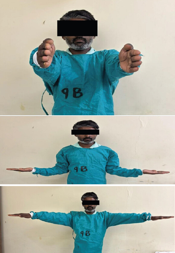
At the final follow-up, the mean range of motion was 127.33 ± 23.81° for abduction, 136.00 ± 22.07° for flexion, 36.00 ± 8.03° for extension, and 42.83 ± 11.79° for external rotation.
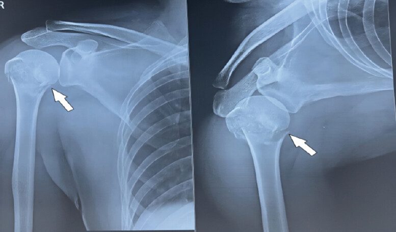
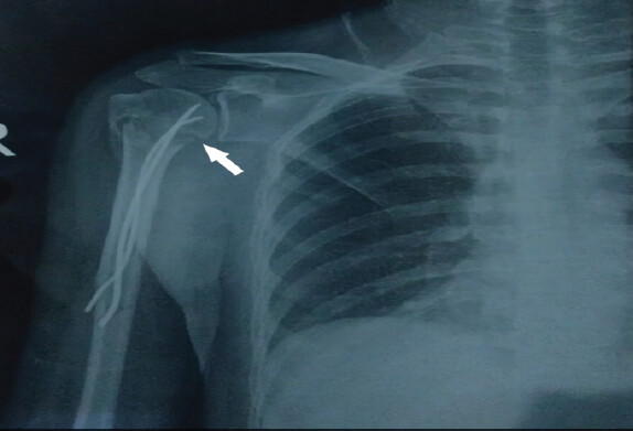
Based on the final DASH scores, 40.0% of patients achieved excellent results, 26.7% good, 16.7% fair, and 16.7% poor. Overall, 66.7% of patients had excellent or good outcomes (Table 1). (Fig. 2,3).
Radiological union and complications
Radiological union was achieved in all cases with a mean time of 12.8 ± 1.83 weeks. About 43.3% of fractures united within 10–12 weeks, while 56.7% united between 12 and 16 weeks.
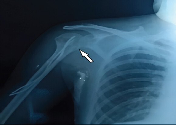
The overall complication rate was 30%. The most common complications were stiffness (13.3%), followed by malunion (10.0%), hardware migration (3.3%), and radial nerve injury (3.3%). Subgroup analysis revealed that complications were more frequent in Neer type 4 fractures (66.6%) compared to type 3 (37.5%) and type 2 (21.1%).(Fig.4).
Discussion
In this prospective study, the overall complication rate was 30%. The most common complication encountered was post-operative stiffness, reported in 4 patients (13.3%). This is consistent with stiffness being one of the leading problems after surgical management of proximal humerus fractures due to periarticular adhesions and delayed mobilization. Malunion was the second most frequent complication, observed in 3 patients (10.0%). Hardware migration and radial nerve palsy were less common, occurring in 1 patient each (3.3%).
Among the 19 patients with type II fractures, complications included 2 cases of stiffness, 1 case of radial nerve palsy, and 1 case of hardware migration, with no malunions reported. In contrast, type III fractures (n = 8) were associated with 2 cases of stiffness and 1 malunion, but no neurovascular injury or hardware-related problems. The most severe complication profile was observed in type IV fractures (n = 3), where 2 cases of malunion occurred, but no stiffness, nerve injury, or hardware migration was noted.
This distribution demonstrates that the nature and frequency of complications varied with fracture complexity. While type II fractures, which are relatively simpler, tended to develop stiffness and occasional hardware-related issues, the more complex type IV fractures were prone to malunion, likely reflecting greater initial displacement and technical difficulty in achieving and maintaining anatomical reduction. Type III fractures showed an intermediate pattern, combining stiffness with occasional malunion.
Clinically, this trend reinforces that fracture pattern strongly influences the post-operative complication profile. Simpler fractures (type II) may recover better functionally but still may have stiffness if early mobilization is delayed. In contrast, complex fractures (type IV) are more likely to heal with residual deformity, even with stable fixation, due to comminution and compromised bone stock. Recognition of these patterns can help in tailoring post-operative rehabilitation strategies and counseling patients regarding prognosis.
Contrary to established literature that typically reports female predominance in proximal humerus fractures, our series demonstrated a slight male preponderance (56.7%). Kannus et al. [12] attribute the usual female predominance to osteoporosis-related fragility fractures, whereas in our population, higher male exposure to road traffic accidents (30.0%) and assaults (23.3%) likely accounts for this variation. The mechanism of injury was predominantly low-energy falls (46.7%), consistent with Handoll et al.’s [13] meta-analysis identifying falls as the most common cause in the elderly. The right side was more frequently involved (56.7%), aligning with Court-Brown et al. [14] who noted increased vulnerability of the dominant upper limb.
In terms of fracture distribution, Neer type II fractures predominated (63.3%), differing from Chandak et al. [15] who primarily studied type III and IV fractures. Such variation in selection criteria must be considered when comparing results. Functional recovery in our study was encouraging, with DASH scores improving from 85.19 at 2 weeks postoperatively to 19.4 at 6 months (P < 0.001). Similarly, Constant–Murley scores improved from 12.60 preoperatively to 82.96 at 6 months, comparable to the mean score of 84.5 ± 5.99 reported by Chandak et al. [15] Range of motion recovery was also satisfactory, with mean abduction of 127.33°, flexion of 136.0°, extension of 36.0°, and external rotation of 42.83° at final follow-up. While these values are broadly comparable to Takeuchi et al., [9] external rotation was slightly restricted, possibly due to differences in rehabilitation adherence or fracture complexity. (Fig.5).
Radiological union was achieved in all patients with a mean time of 12.8 ± 1.83 weeks, consistent with Chandak et al. [15] who reported union within 12–14 weeks. This healing timeline reinforces the biological soundness of the J-nail construct. Functional outcomes were stratified by fracture type, showing excellent-to-good results in 66.7% overall, with better results in type II (68.4%) than type III (62.5%) and type IV (33.3%). This finding is in agreement with Neer’s original observation3 that more complex patterns carry a poorer prognosis.
The technical advantages of the J-nail, as highlighted by Takeuchi et al., [9] were also evident in our study: Minimal soft tissue dissection, preservation of humeral head vascularity, avoidance of rotator cuff violation common with intramedullary nailing [7], and facilitation of early mobilization. Compared to Proximal Humeral Internal Locking System plating, which risks screw cutout and soft tissue stripping [5,6], antegrade nailing that compromises the rotator cuff [7], and K-wire fixation associated with pin tract infections and muscle transfixation [16], the J-nail offers a balanced alternative with reduced morbidity. These advantages, along with the favorable complication profile, make it a promising minimally invasive fixation method for elderly patients with proximal humeral fractures.
Strengths and limitations
This study’s primary strength is its prospective evaluation of the J-nail technique as a “biological fixation” strategy, prioritizing soft-tissue and vascular preservation in a vulnerable elderly population. The use of standardized functional scores provides a robust assessment of recovery. However, limitations include a modest sample size (n = 30) and the absence of a control group, which limit comparative power. The 6-month follow-up, while sufficient for union, does not allow for assessment of long-term complications like avascular necrosis. Finally, the inclusion of heterogeneous Neer fracture types means results should be interpreted with attention to fracture complexity. Regarding the 30% complication rate, it is important to note that the majority of these events were minor and managed conservatively, representing a favorable safety profile compared to the severe hardware-related failures often associated with more invasive open reduction techniques.
Limitations
This study has several limitations that should be acknowledged. The 6-month follow-up period may be insufficient to detect long-term complications such as avascular necrosis [6] or post-traumatic osteoarthritis. The absence of a direct comparison group treated with alternative fixation methods (locking plates [5,6] or conservative management [17]) prevents definitive conclusions about the relative efficacy of the J-nail technique. Future research should address these limitations through larger multicenter randomized controlled trials with longer follow-up periods, comparison groups, comprehensive outcome measures including patient satisfaction and cost-effectiveness analyses, and systematic CT evaluation [4] for better assessment of complex fracture patterns and their correlation with outcomes.
Conclusion
The J-nail technique represents a viable, minimally invasive alternative for the management of proximal humerus fractures in elderly patients. Our study demonstrates that this approach achieves satisfactory functional outcomes with two-thirds of patients attaining excellent to good results at 6 months post-operatively. The technique provides adequate stability for fracture healing while minimizing surgical trauma and associated complications.
The procedure’s simplicity, shorter operative time, limited soft tissue disruption, and early mobilization potential make it particularly suitable for elderly patients with osteoporosis and medical comorbidities. As noted by Takeuchi et al., the J-nail technique achieves relative stability while maintaining vascularity and soft tissue integrity, which is crucial for healing in this vulnerable population.
The complication profile compares favorably with more invasive fixation methods described by Agudelo et al., though the effectiveness diminishes with increasing fracture complexity. Consistent with Neer’s classification implications, we found that 4-part fractures had poorer outcomes than 2- or 3-part fractures.
Based on our findings, we conclude that the J-nail technique offers a balanced approach between surgical intervention and biological respect, providing sufficient stability while maintaining the fracture hematoma and periosteal blood supply essential for healing in elderly patients.
Clinical Message
Minimally invasive J-nail fixation is an effective and biologically favorable treatment option for Neer type 2–4 proximal humerus fractures in elderly patients, providing reliable fracture union, good functional recovery, and early mobilization with minimal soft tissue disruption; however, careful case selection is essential as outcomes may be less optimal in more complex fracture patterns.
Conflict of Interest:
Nil
Source of Support:
Nil
Consent:
The authors confirm that informed consent was obtained from the patient for publication of this article
How to Cite this Article
Pathak A, Mishra S, Bansal A, Chouksey A, Martin O, Parmar S. Minimally Invasive J-Nail Fixation For Neer Type 2, 3, and 4 Proximal Humerus Fractures in Elderly Patients: A Prospective Clinical and Radiological Outcome Study. Journal of Orthopaedic Case Reports 2026 October;16(10): 448-454.
References
- Shi Y, Li F, Liang W, Liu J. Effect of different reduction intramedullary nails on spiral fracture of middle and lower tibia. Contrast Media Mol Imaging 2022;2022:4716978. [Google Scholar] | [PubMed]
- Olerud P, Ahrengart L, Ponzer S, Saving J, Tidermark J. Internal fixation versus nonoperative treatment of displaced 3-part proximal humeral fractures in elderly patients: A randomized controlled trial. J Shoulder Elbow Surg 2011;20:747-55. [Google Scholar] | [PubMed]
- Neer CS 2nd. Displaced proximal humeral fractures. II. Treatment of three-part and four-part displacement. J Bone Joint Surg Am 1970;52:1090-103. [Google Scholar] | [PubMed]
- Kilcoyne RF, Shuman WP, Matsen FA3rd, Morris M, Rockwood CA. The Neer classification of displaced proximal humeral fractures: Spectrum of findings on plain radiographs and CT scans. AJR Am J Roentgenol 1990;154:1029-33. [Google Scholar] | [PubMed]
- Agudelo J, Schürmann M, Stahel P, Helwig P, Morgan SJ, Zechel W. Analysis of efficacy and failure in proximal humerus fractures treated with locking plates. J Orthop Trauma 2007;21:676-81. [Google Scholar] | [PubMed]
- Gerber C, Werner CM, Vienne P. Internal fixation of complex fractures of the proximal humerus. J Bone Joint Surg Br 2004;86:848-55. [Google Scholar] | [PubMed]
- Bhandari M, Guyatt GH, Swiontkowski MF, Tornetta P, Sprague S, Schemitsch EH. Treatment of humerus fractures: Comparison of intramedullary nails with plates. J Bone Joint Surg Am 2006;88:943-54. [Google Scholar] | [PubMed]
- Soete PJ, Clayson PE, Costenoble VH. Transitory percutaneous pinning in fractures of the proximal humerus. J Shoulder Elbow Surg 1999;8:569-73. [Google Scholar] | [PubMed]
- Takeuchi R, Koshino T, Nakazawa A, Numazaki S, Sato R, Saito T. Minimally invasive fixation for unstable two-part proximal humeral fractures: Surgical techniques and clinical results using J-nails. J Orthop Trauma 2002;16:403-8. [Google Scholar] | [PubMed]
- Constant CR, Murley AH. A clinical method of functional assessment of the shoulder. Clin Orthop Relat Res 1987;214:160-4. [Google Scholar] | [PubMed]
- Hudak PL, Amadio PC, Bombardier C. Development of an upper extremity outcome measure: the DASH (disabilities of the arm, shoulder and hand) [corrected. The upper extremity collaborative group (UECG). Am J Ind Med 1996;29:602-8. [Google Scholar] | [PubMed]
- Kannus P, Palvanen M, Niemi S, Parkkari J, Järvinen M, Vuori I. Osteoporotic fractures of the proximal humerus in elderly finnish persons: Sharp increase in 1970-1998 and alarming projections for the new millennium. Acta Orthop Scand 2000;71:465-70. [Google Scholar] | [PubMed]
- Handoll HH, Elliott J, Thillemann TM, Aluko P, Brorson S. Interventions for treating proximal humeral fractures in adults. Cochrane Database Syst Rev 2022;6:CD000434. [Google Scholar] | [PubMed]
- Court-Brown CM, Garg A, McQueen MM. The epidemiology of proximal humeral fractures. Acta Orthop Scand 2001;72:365-71. [Google Scholar] | [PubMed]
- Chandak R, Sharma M, Jha AK. Review of clinical and radiological outcome of 3- and 4-part proximal humerus fracture managed with J nails in elderly osteoporotic individuals. Int J Res Orthop 2021;7:728. [Google Scholar] | [PubMed]
- Wachtl SW, Marti CB, Hoogewoud HM, Jakob RP, Gautier E. Treatment of proximal humerus fracture using multiple intramedullary flexible nails. Arch Orthop Trauma Surg 2000;120:171-5. [Google Scholar] | [PubMed]
- Goudie EB, MacDonald DJ, Robinson CM. Functional outcome after nonoperative treatment of a proximal humeral fracture in adults. J Bone Joint Surg Am 2022;104:123-38. [Google Scholar] | [PubMed]
© 2026 Journal of Orthopaedic Case Reports - Published by Indian Orthopaedic Research Group
About the Authors
-

Aditya Pathak
-

Santosh Mishra
-

Akhil Bansal
-

Anil Chouksey
-

Obeir Martin
-

Sunil Parmar
Related Articles in Journal of Orthopaedic Case Reports
 November 1, 2025 A Study on Functional Outcome of Hemiarthroplasty for Proximal Humeral Fractures – An Observational Study
 October 1, 2025 Outcomes of Proximal Humeral Fracture Fixation Using Minimally Invasive Plate Osteosynthesis: A Prospective Clinical and Radiographic Study
 October 1, 2026 Association between Coronal Mechanical Axis Correction and Early Functional Outcomes after Primary Total Knee Arthroplasty: A Retrospective Observational Study
 October 1, 2026 Functional Outcome of Operative Titanium Elastic Nailing System Versus Non-Operative Figure-of-Eight Bandage in Displaced Mid-Shaft Clavicle Fractures: A Randomized Controlled Study