


Introduction
Myositis ossificans (MO) is a benign, solitary, self-limiting, ossifying soft-tissue mass occurring within skeletal muscles [1]. It can be traumatic or atraumatic in origin. Its pathophysiology remains incompletely understood and typically occurs in young, active males [2]. Most frequently, patients present with pain and stiffness following an injury or several minor trauma. However, paresthesia, weakness, lymphedema, and venous thromboembolism have been reported due to displacement or compression of nearby neurovascular structures [3]. MO is most commonly diagnosed solely on plain radiographs. Nonetheless, computed tomography (CT) can provide a precise diagnosis when calcification patterns are radiographically undetectable [4]. Patients complaining of severe symptoms or those who failed non-operative treatment are candidates for surgical excision aiming for limitation of pain and restoration of function [2,5].
We present the case of a patient with left hand swelling who was found to have MO of the thenar eminence that was mistaken for an accessory trapezium fracture.
Case Report
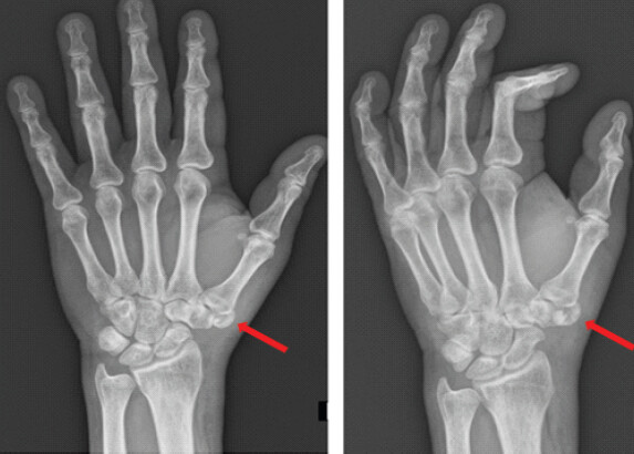
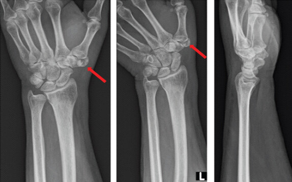
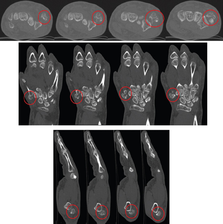
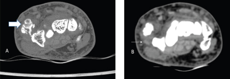
This is a case of a 66-year-old right-handed male patient, smoker, known to have hypertension, coronary artery disease, and dyslipidemia, taking Aspirin and Plavix. He presented to our clinic for atraumatic left hand swelling and erythema. He reports mechanical pain and limitation of movement of his left thumb. History goes back to 10 days before presentation, when he first started complaining of his symptoms after going to the gym. On presentation, physical exam revealed swelling and erythema of the left thenar eminence, with a painful and limited range of motion of his left thumb. Neurovascular examination was otherwise intact. Standard radiographs of his left hand (Fig. 1) and wrist (Fig. 2) were done, showing a suspicious accessory trapezium fracture. Imaging was then completed by a CT scan revealing the presence of a 14 × 11 × 11mm accessory trapezium noted on the dorsal and medial aspect of the base of the first metacarpal bone, showing fragmentation and cortical lucencies suggestive of a fracture (Fig. 3).
The patient was therefore treated by a short arm thumb spica cast for 6 weeks.
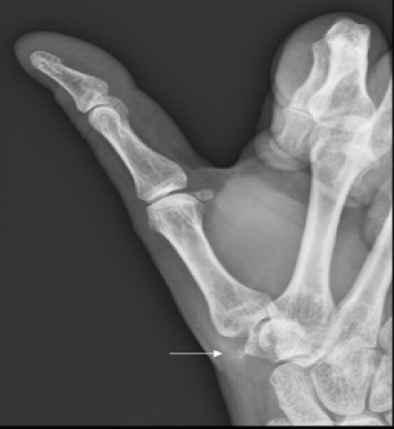
At 6 weeks follow up, the patient was pain-free and asymptomatic. A follow-up X-ray (Fig. 4) was done, showing resorption of the fractured accessory trapezium that was evident solely through remnants.
The resorption of the supposed accessory bone prompted us to take a second look on the CT scan, with a second radiologist concluding to a well-corticated bony structure with a mature bony matrix suggestive of MO (Fig. 5).
Discussion
Accessory ossicles of the wrist originate from an anomaly in fusion of the secondary ossification centers [6]. More than 20 accessory ossicles exist in the wrist with an incidence of 9.6%, with the most common being os triangulare and os ulnostyloideum. Trapezium secondarium, or accessory trapezium, has an incidence of 0.3% and is typically located between the bases of the first and second metacarpal bones at the superomedial aspect of the trapezium [7]. These ossicles are usually asymptomatic and are generally found as incidental radiologic findings. Nonetheless, they become symptomatic when they cause impingement (compressing or displacing adjacent structures), or upon trauma. They may also cause exercise-related pain [7,8,9]. There are only a handful of cases of accessory carpal bones ever reported. In fact, the very few cases of accessory ossicles of the wrist were found in association with other wrist abnormalities (carpal coalition, osteonecrosis…) [10,11]. Not only is there a scarcity of cases involving accessory ossicles of the wrist in the literature, but no cases of accessory ossicle fractures have been yet described.
In our patient, the accessory bone was in fact located at the dorsomedial aspect of the trapezium between the first and second metacarpals. However, the rarity of such cases, combined with the resorption of the accessory trapezium on follow-up X-rays, prompted us to consider differential diagnoses such as MO.
MO is a localized and self-limiting form of heterotopic ossification involving muscles. It most commonly affects the quadriceps, gluteus, and the brachialis muscles [12]. The hand is an uncommon site for MO; only a handful of cases have yet to be reported [5,13,14,15,16]. Radiographic diagnosis is made when these lesions appear with a densely calcified peripheral rim (zoning phenomenon) with a lucent center on plain radiographs and CT scan [2]. Magnetic resonance imaging may be the best modality to evaluate soft-tissue masses; however, MO appears as a heterogeneous mass with extensive surrounding muscle edema [17]. Non-operative treatment is advised and includes brief immobilization and cryotherapy. Surgical excision is indicated in the presence of severe pain and refractory cases [2].
Our patient presented for severe pain and swelling of the left thenar eminence following possible repetitive trauma (gym activities). Imaging showed hallmarks of MO features (zoning phenomenon with a lucent center) detected on CT scan in a rather uncommon location. Moreover, his symptoms were alleviated by immobilization and rest. The resorption of the calcification on follow-up radiographs highlighted the self-limiting feature of MO and its favorable prognosis.
Therefore, after an extensive review of the literature, we reported a case of a MO of the thenar eminence of the left hand which was originally mistaken for a fracture of an accessory trapezium. With this in mind, physicians are encouraged to consider MO as a possible diagnosis for soft-tissue swelling of the hand.
Conclusion
MO of the hand is rare and is, in most cases, asymptomatic. However, it can cause pain, stiffness, and compression symptoms. We reported a rare finding of a MO presenting in the Thenar eminence of a 66-year-old male patient that was treated conservatively, leading to its resorption after 6 weeks of follow-up.
Clinical Message
In patients presenting with acute hand pain and soft-tissue swelling, especially when imaging findings are atypical or evolve over time, clinicians should consider myositis ossificans in the differential diagnosis. Serial imaging and reassessment can prevent misdiagnosis as a fracture and help ensure appropriate, conservative management.
Conflict of Interest:
Nil
Source of Support:
Nil
Consent:
The authors confirm that informed consent was obtained from the patient for publication of this article
How to Cite this Article
Ghanem W, Jabbour F, AbdelNour H, Ezzeddine H, Faddoul S, Moucharafieh R. Myositis Ossificans of the Thenar Eminence mimicking a Fracture of an Accessory Trapezium: A Case Report. Journal of Orthopaedic Case Reports 2026 October;16(10): 86-89.
References
- Kransdorf MJ, Meis JM, Jelinek JS. Myositis ossificans: MR appearance with radiologic-pathologic correlation. AJR Am J Roentgenol 1991;157:1243-8. [Google Scholar] | [PubMed]
- Walczak BE, Johnson CN, Howe BM. Myositis ossificans. J Am Acad Orthop Surg 2015;23:612-22. [Google Scholar] | [PubMed]
- Mavrogenis AF, Soucacos PN, Papagelopoulos PJ. Heterotopic ossification revisited. Orthopedics 2011;34:177. [Google Scholar] | [PubMed]
- Shehab D, Elgazzar AH, Collier BD. Heterotopic ossification. J Nucl Med 2002;43:346-53. [Google Scholar] | [PubMed]
- Al-Qattan MM, Al-Fahdil L, Al-Shammari HM, Joarder AI. Management of myositis ossificans of the hand: A case report and a review of the literature. J Hand Surg Am 2017;42:576.e1-4. [Google Scholar] | [PubMed]
- Oberg KC, Feenstra JM, Manske PR, Tonkin MA. Developmental biology and classification of congenital anomalies of the hand and upper extremity. J Hand Surg Am 2010;35:2066-76. [Google Scholar] | [PubMed]
- Gursoy M, Coban I, Mete BD, Bulut T. The incidence of accessory ossicles of the wrist: A radiographic study. J Wrist Surg 2021;10:458-64. [Google Scholar] | [PubMed]
- Mespreuve M, Bosmans F, Waked K, Vanhoenacker FM. Hand and wrist: A kaleidoscopic view of accessory ossicles, variants, coalitions, and others. Semin Musculoskelet Radiol 2019;23:511-22. [Google Scholar] | [PubMed]
- Netter FH. Atlas of Human Anatomy 7th ed. Philadelphia, PA:Elsevier 2018. [Google Scholar] | [PubMed]
- Boya H, Ozcan O, Araç S, Tandoğan R. Incomplete scapholunate and trapeziotrapezoid coalitions with an accessory carpal bone. J Orthop Sci 2005;10:99-102. [Google Scholar] | [PubMed]
- Lane LB, Gould ES, Stein PD, Coffey E. Unilateral osteonecrosis in a patient with bilateral os centrale carpi. J Hand Surg Am 1990;15:751-4. [Google Scholar] | [PubMed]
- Weiss SW, Goldblum JR. Enzinger and Weiss's Soft Tissue Tumors 4th ed. St Louis, MO:Mosby 2001:1389-405. [Google Scholar] | [PubMed]
- Akahane T, Mori N, Nakatsuchi Y. Myositis ossificans occupying the thenar region: A case report. J Med Case Rep 2015;9:105. [Google Scholar] | [PubMed]
- Goto H, Hatori M, Kokubun S, Makino M. Myositis ossificans in the tip of the thumb: A case report. Tohoku J Exp Med 1998;184:67-72. [Google Scholar] | [PubMed]
- Kusuma S, Lourie GM, Lins RE. Myositis ossificans of the hand. J Hand Surg Br 1999;24:128-30. [Google Scholar] | [PubMed]
- De Smet L, Degreef I. Myositis ossificans of the hand in a child: Case report. J Pediatr Orthop B 2012;21:539-41. [Google Scholar] | [PubMed]
- Ehara S, Nakasato T, Tamakawa Y, Yamataka H, Murakami H, Abe M. MRI of myositis ossificans circumscripta. Clin Imaging 1991;15:130-4. [Google Scholar] | [PubMed]
© 2026 Journal of Orthopaedic Case Reports - Published by Indian Orthopaedic Research Group
About the Authors
-

Wendy Ghanem
-

Fouad Jabbour
-

Hicham AbdelNour
-

Hady Ezzeddine
-

Sami Faddoul
-

Ramzi Moucharafieh
Related Articles in Journal of Orthopaedic Case Reports
 October 1, 2026 Myositis Ossificans Affecting the Rotator Cuff Muscles and Proximal Humerus: A Rare Clinical Entity
 May 1, 2026 Surgical Management of Painful Os Vesalianum: A Case Report and Literature Review
 June 1, 2025 Rare Case of Subscapularis Myositis Ossificans with Intra-articular Extension
 October 1, 2024 Eating Is Living: A Case Report of Post-Traumatic Myositis Ossificans in Elbow