


Introduction
Back degloving injuries are a rare but severe form of trauma characterized by the separation of skin and subcutaneous tissue from the underlying fascia, muscle, or bone. These injuries typically result from high-energy mechanisms, such as motor vehicle accidents or industrial accidents, in which shear forces cause avulsion of the skin from deeper structures. Based on severity and mechanism of injury, these wounds may present as either open or closed, each carrying distinct risk profiles [1]. Closed back degloving injuries, also known as Morel-Lavallée lesions, are more difficult to identify, as the overlying dermis remains intact. Delayed diagnosis precludes early intervention and fluid drainage, permitting the accumulation of blood and necrotic fat within the degloved potential space [2,3]. Conversely, open degloving injuries present with visible wounds, facilitating prompt diagnosis and necessitating more aggressive management, including operative intervention. The primary concern with open injuries is the risk of infection leading to sepsis and impaired wound healing [4].
Due to the high morbidity and mortality associated with these injuries, multidisciplinary surgical intervention is essential to optimize outcomes. Initial management of open degloving wounds consists of surgical debridement and excision of non-viable tissue to reduce infectious risk [1,5]. When a sufficient area of dermis remains intact and viable, it may be utilized immediately as an autograft in conjunction with the avulsed skin [6]. This is followed by advanced wound care, such as negative pressure wound therapy (NPWT), to promote granulation tissue formation and prepare the wound bed for definitive closure [2]. In most cases, anatomic reconstruction involves the use of dermal regeneration templates and split-thickness autografts once an adequate baseline of wound healing has been established [6,7].
Case Report
Initial presentation
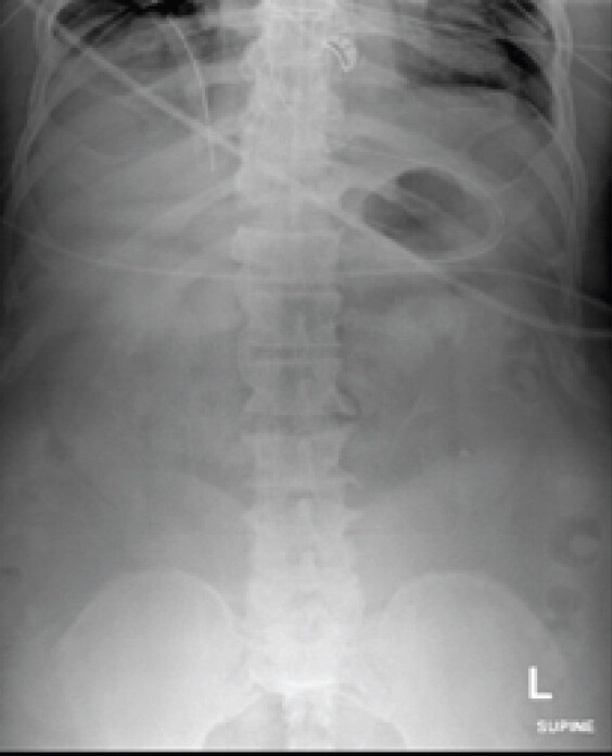
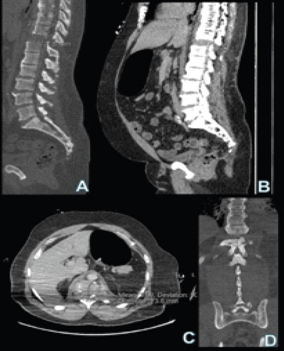
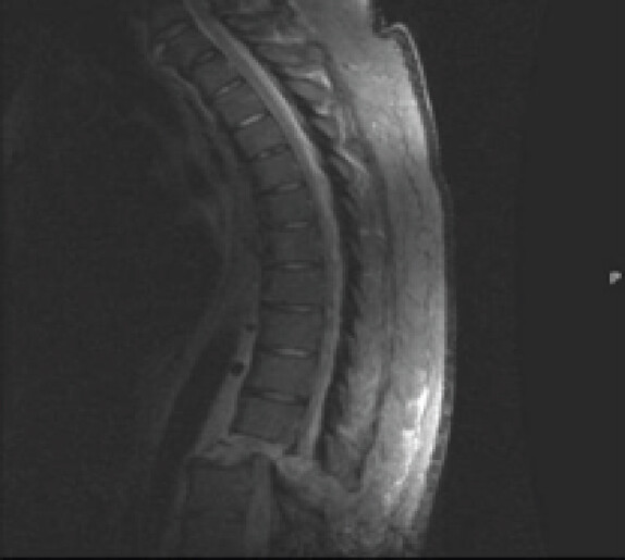
We present a 61-year-old male – with a medical history of diabetes mellitus, hypercholesterolemia, hypertension, venous stasis, iron deficiency anemia, and prior cerebral infarction – who presented after a motorcycle-versus-car collision. He was unresponsive at the scene with a Glasgow Coma Scale of 9. He presented with an absent left lower extremity pulse. The patient sustained extensive polytraumatic injuries across multiple organ systems. Neurological and spinal injuries included bilateral subarachnoid hemorrhages, C7–T1 widening, a T11–T12 distraction injury (2 cm) with associated retrolisthesis (8 mm), a closed burst fracture of T12, and right vertebral artery dissection. Orthopedic injuries consisted of an open comminuted bicondylar distal femur fracture of the left lower extremity, a lateral patellar facet fracture, a right distal radius fracture, and a left posterior T12 rib fracture. Vascular injury included left popliteal artery occlusion. Additional injuries consisted of right pneumothorax, left hemothorax, a nasal bone fracture, and open back wounds. Clostridioides difficile colitis was acquired during the hospitalization. Workup included computed tomography of the thoracic and lumbar spine, magnetic resonance imaging (MRI), and serial radiographs, which collectively confirmed complex vertebral injuries and progressive soft-tissue involvement (Fig. 1, 2, 3).
Hospital course
Following admission, the patient underwent an extensive series of multidisciplinary operative interventions spanning several months. On August 21, 2024, the vascular surgery team performed an above-knee arterial bypass from the popliteal to posterior tibial artery, along with a four-quadrant fasciotomy of the left lower extremity to address vascular compromise. Concurrently, the orthopedic surgery team applied a left knee spanning external fixator for provisional stabilization.
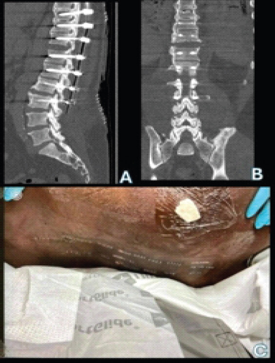
On admission day 8, the orthopedic spine team performed open reduction of the T11–T12 fracture-dislocation with percutaneous instrumented fusion from T9 to L3 (Fig. 4). Fasciotomy wounds of the left lower extremity were subsequently closed by the general surgery team on admission day 21. Two days later, the orthopedic trauma team removed the external fixator and performed open reduction and internal fixation of the left open intra-articular bicondylar distal femur fracture.
The patient’s course was complicated by progressive wound breakdown and purulence along the thoracolumbar spine, necessitating the first irrigation and debridement (I&D) on admission day 27 by the orthopedic spine team (Fig. 5). The patient eventually required NPWT due to the infection of his thoracolumbar incisions. Wound smear and cultures tested positive for Pseudomonas aeruginosa and Enterobacter cloacae complex, as well as returning Candida auris. He was started on meropenem, levofloxacin, and micafungin. Repeat I&D procedures were performed on admission days 44 and 47, the latter including a wound vacuum-assisted closure (VAC) exchange (Fig. 6). Due to persistent infection, the orthopedic spine and plastic surgery teams jointly performed removal of the infected spinal instrumentation with re-instrumentation from T9 to L2 on admission day 54, along with an additional wound VAC exchange (Fig. 7). Spinal debridement and wound VAC changes were continued on admission days 56 and 58, the latter including paraspinal muscle debridement performed by the orthopedic surgery team (Fig. 8). A final I&D with wound VAC change was performed on admission day 63 (Fig. 9). Concurrent to these serial I&D procedures, the tissue/fluid cultures also began to grow Candida Auris. Blood culture and cerebrospinal fluid cultures were negative at this point.
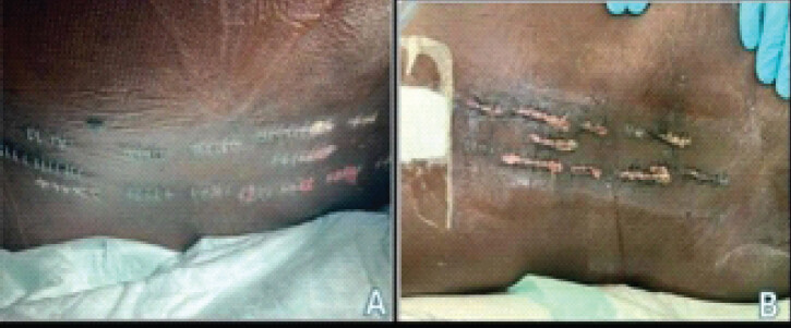
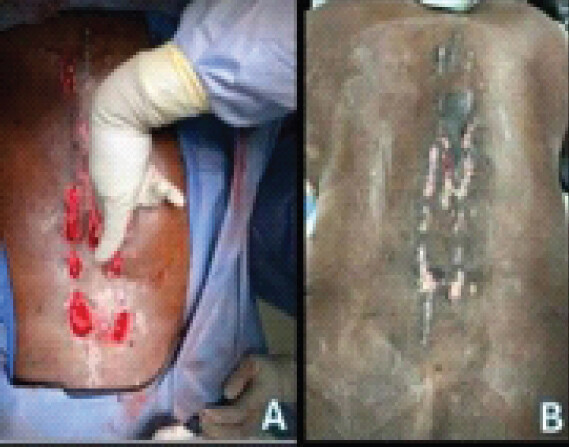
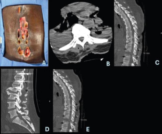
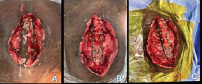
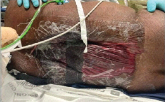
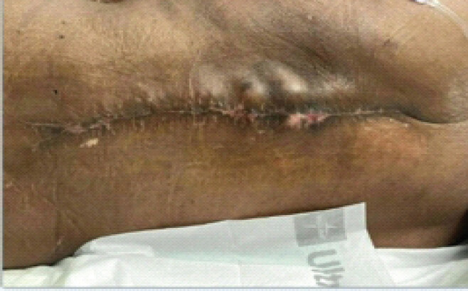
On admission day 65, the plastic and reconstructive surgery team performed complex primary closure of the posterior spinal wound (Fig. 10). This was complicated by a post-operative hematoma requiring surgical evacuation on admission day 69.
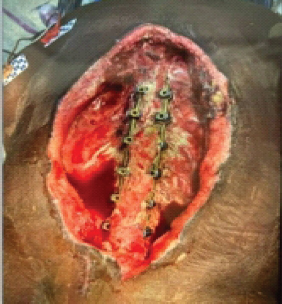
Throughout the hospitalization, the patient received broad-spectrum antibiotic and antifungal therapy, serial NPWT therapy, tracheostomy placement, and comprehensive supportive care. The overall hospital course was prolonged by recurrent wound breakdown, infection, and sepsis, ultimately requiring hardware removal, spinal re-instrumentation, and multistage wound reconstruction involving orthopedic spine, plastic surgery, trauma surgery, and vascular surgery teams. As of admission day 143, the patient’s wound is shown as imaged (Fig. 11). Blood and skin cultures were negative post-closure.
Readmission and follow-up period
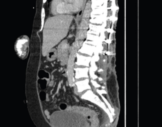
Following discharge, the patient was readmitted in March 2025 with sacrococcygeal osteomyelitis and a spinal epidural abscess, underscoring the persistence of infectious complications (Fig. 12). He also had a positive blood culture for multiple Gram-negative rods. The patient’s course was further complicated by multiple discharges and readmissions. Complications included pleural effusions necessitating bronchoscopies, recurrent respiratory infections, chest tube placement, and further hardware removal with repeat irrigation procedures. Despite ongoing multidisciplinary management, the patient demonstrated progressive clinical deterioration over the subsequent 5–6 months. The cumulative burden of chronic infection, spinal instability, and systemic decline precluded meaningful functional recovery. Ultimately, given the refractory nature of his injuries and complications, goals of care were redirected, and the patient was transitioned to hospice care.
Discussion
Back degloving injuries involving spinal hardware represent a uniquely complex management challenge. These injuries frequently result from high-energy trauma and may present with both open and closed degloving patterns [8,9]. In the present case, the patient initially sustained open wounds; however, a Morel-Lavallée lesion subsequently developed, introducing a closed internal degloving component that further complicated an already prolonged recovery. This progression was likely attributable to more than 1 month of exposed necrotic tissue and spinal hardware prior to the availability of sufficient viable tissue for flap coverage. A similar mechanism was observed with Reid et al. (2019) [9].
Morel-Lavallée lesions are post-traumatic closed soft-tissue injuries caused by shearing forces that separate the subcutaneous fat from the underlying fascia, disrupting local capillaries and lymphatics and creating a potential space in which blood, lymph, and necrotic fat accumulate. These lesions may go unrecognized initially, particularly in polytrauma patients in whom life-threatening injuries are prioritized. If not addressed in a timely manner, they can become encapsulated, are prone to infection, and may prove resistant to conservative therapy. Hudson et al. described such delayed presentations as contour deformities frequently requiring surgical intervention due to fibrous pseudocapsule formation and persistent fluid collections [2].
In the setting of spinal trauma, where instrumentation integrity and soft-tissue coverage are critical, the presence of a Morel-Lavallée lesion substantially increases the risk of wound breakdown and infection [10]. In this patient, recurrent wound dehiscence, purulent drainage, and eventual hardware exposure were likely compounded by the evolving lesion. NPWT and staged debridement were essential in controlling infection and preparing the wound bed for delayed closure. Nevertheless, this case illustrates that even with aggressive management, chronicity and recurrent infection can necessitate hardware removal, prolonged antibiotic therapy, and extended wound care.
These findings are consistent with prior literature that highlights the diagnostic and management complexity of Morel-Lavallée lesions. Hudson et al. emphasized conservative surgical approaches in chronic lesions, whereas Dini et al. and Sakai et al. demonstrated the utility of VAC therapy and dermal regeneration matrices in the management of degloving injuries [2,5,6]. Hakim et al. further underscore the importance of early recognition and aggressive surgical management in improving outcomes in complex trauma [1].
Morel-Lavallée lesions should be suspected in polytrauma patients with persistent or evolving soft-tissue swelling near high-friction interfaces, and early MRI or ultrasound is recommended to facilitate diagnosis prior to pseudocapsule formation. Delayed diagnosis may ultimately require surgical excision, prolonged VAC therapy, and hardware revision. Multidisciplinary coordination among plastic surgery, infectious disease, and rehabilitation services is essential to optimize patient outcomes. These injuries are associated with significant morbidity and mortality, and should be managed with a high level of interdisciplinary care.
Conclusion
This case demonstrates that closed degloving injuries can arise as a delayed and insidious complication of high-energy spinal trauma, even when the initial presentation is dominated by open wounds and polytraumatic injuries. In this patient, a Morel-Lavallée lesion developed over the instrumented thoracolumbar spine, precipitating an elongated course of recurrent wound breakdown, infection, hardware compromise, and ultimately multistage reconstruction. The report underscores that an intact dermis may conceal progressive deep soft-tissue necrosis, and that failure to recognize this early permits the accumulation of blood and necrotic fat within a potential space that is highly susceptible to infection. For the orthopedic surgeon managing spinal trauma, where instrumentation integrity depends on durable soft-tissue coverage, a high index of suspicion for evolving closed degloving injury is essential. Early advanced imaging, timely drainage, staged debridement with NPWT, and coordinated multidisciplinary care offer the best opportunity to control infection, preserve spinal stability, and avert the cascade of complications illustrated here.
Clinical Message
Morel-Lavallée lesions should be anticipated as a possibility in polytrauma spine surgery patients, particularly when there is evolving soft-tissue swelling over instrumentation. Because intact skin can mask extensive necrosis, early recognition via MRI or ultrasound, prompt drainage, and staged multidisciplinary management are critical to preventing wound breakdown and infection.
Conflict of Interest:
Nil
Source of Support:
Nil
Consent:
The authors confirm that informed consent was obtained from the patient for publication of this article
How to Cite this Article
Morar H, Ngwa F, Patel D, Pazionis T. Degloving in Disguise: Managing a Delayed Spinal Morel-Lavallée Lesion after Thoracic Fracture-Dislocation. Journal of Orthopaedic Case Reports 2026 October;16(10): 90-96.
References
- Hakim S, Ahmed K, El-Menyar A, Jabbour G, Peralta R, Nabir S . Patterns and management of degloving injuries: A single national level 1 trauma center experience. World J Emerg Surg 2016;11:35. [Google Scholar] | [PubMed]
- Hudson DA, Knottenbelt JD, Krige JE. Closed degloving injuries: Results following conservative surgery. Plast Reconstr Surg 1992;89:853-5. [Google Scholar] | [PubMed]
- Hudson DA. Missed closed degloving injuries: Late presentation as a contour deformity. Plast Reconstr Surg 1996;98:334-7. [Google Scholar] | [PubMed]
- Veena PW, Babu R, Venkatesh MS, Udayashankar C, Deepak KL. Degloving injuries of the abdominal wall. J Wound Care 2013;22:562-8. [Google Scholar] | [PubMed]
- Greenhill D, Haydel C, Rehman S. Management of the morel-lavallée lesion. Orthop Clin North Am 2016;47:115-25. [Google Scholar] | [PubMed]
- Dini M, Quercioli F, Mori A, Romano GF, Lee AQ, Agostini T. Vacuum-assisted closure, dermal regeneration template and degloved cryopreserved skin as useful tools in subtotal degloving of the lower limb. Injury 2012;43:957-9. [Google Scholar] | [PubMed]
- Sakai G, Suzuki T, Hishikawa T, Shirai Y, Kurozumi T, Shindo M. Primary reattachment of avulsed skin flaps with negative pressure wound therapy in degloving injuries of the lower extremity. Injury 2017;48:137-41. [Google Scholar] | [PubMed]
- Dodwad SN, Niedermeier SR, Yu E, Ferguson TA, Klineberg EO, Khan SN. The Morel-Lavallée lesion revisited: Management in spinopelvic dissociation. Spine J 2015;15:e45-51. [Google Scholar] | [PubMed]
- Reid DB, Daniels AH, Haque MU, Palumbo MA. Successful treatment of morel-lavallée lesion of the back with transcutaneous transmyofascial bolstered progressive tension suturing. Orthopedics 2019;42:e399-401. [Google Scholar] | [PubMed]
- Kanakaris NK, Ciriello V, Stavrou PZ, West RM, Giannoudis PV. Deep infection following reconstruction of pelvic fractures: Prevalence, characteristics, and predisposing risk factors. Eur J Trauma Emerg Surg 2022;48:3701-9. [Google Scholar] | [PubMed]
© 2026 Journal of Orthopaedic Case Reports - Published by Indian Orthopaedic Research Group
About the Authors
-

Henry Morar
-

Funiba Ngwa
-

Dhara Patel
-

Theresa Pazionis
Related Articles in Journal of Orthopaedic Case Reports
 October 1, 2026 Bilateral Cedell Fractures of the Talus Associated with Ankle Fractures in a Polytrauma Patient: A Rare Injury Managed with Damage Control Orthopedics and Staged Fixation
 October 1, 2026 Single-Stage Fixation of Bilateral Open Medial Hoffa Fractures Associated with Bilateral Patella Fracture and Bilateral Distal Radius Fractures: A Case Report
 September 1, 2026 Extensive Post-Traumatic Heterotopic Ossification with Multi-joint Involvement: A Case Report
 May 1, 2026 Segmental Achilles Tendon Loss With Skin Defect in an Open Calcaneal Fracture Managed Without Tendon Reconstruction: A Case Report