

Some moments in spine surgery are very unsettling, such as when a pedicle gives way. The pedicle is the narrow, cortical-walled corridor through which we deliver three-column fixation, and its integrity is precisely what makes a pedicle screw the reliable anchor that it is. When that corridor is compromised – because the drill drifts, the tap is used too aggressively, or the bone was never strong enough to begin with – the consequences run from the trivial to the catastrophic: A lost screw, an unstable construct, or a neurological injury. Yet the vocabulary we use to describe these events remains oddly imprecise. “Pedicle breach, miss/lateral cutout” is spoken of constantly; “pedicle break” and “pedicle burst” are met just as often across the operating table but are rarely named or defined (Fig. 1) [1].
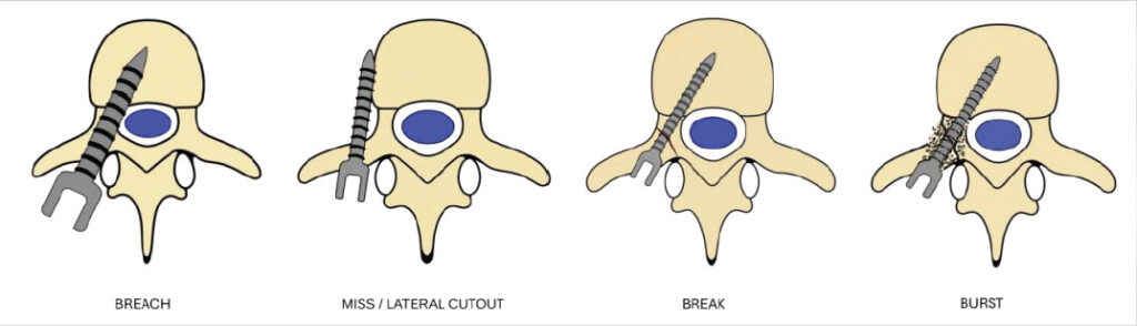
A breach is the disruption of a single cortical wall while the pedicle itself remains essentially intact; the screw may partially exit, but enough cortical bone survives to provide purchase, and when such a breach is minor (under 2 mm) and asymptomatic, it may not always demand revision [1]. A miss/lateral cutout is a complete absence of the pedicle laterally or just brushing the lateral wall. A break is categorically different – a complete fracture through all four walls that splits the pedicle in two and destroys the structural integrity on which screw purchase depends. It may be iatrogenic, following aggressive tapping, an oversized screw, or a misdirected trajectory, or it may be part of trauma [2]. A burst is the extreme edge of this spectrum: A comminuted, multicortical fracture in which the pedicle is fragmented to the point that little intact bone remains to accept a screw at all, whether as part of a thoracolumbar burst-type injury or in profoundly osteoporotic bone that simply cannot tolerate instrumentation [2,3]. The distinction is not academic. Management, salvage options, and the risk to neural elements vary sharply across these points.
A common reason these injuries go unnamed is that our classification systems are focused on 1 subgroup. For screw position, we have the Gertzbein–Robbins grading, which sorts a screw from grade A (entirely within the pedicle), through B (breach under 2 mm), C (2–4 mm), and D (4–6 mm), to E (>6 mm or wholly outside), with A and B generally accepted. C to E carry a rising risk of neurological compromise that may warrant revision depending on direction and the patient’s clinical state (Fig. 2) [1].
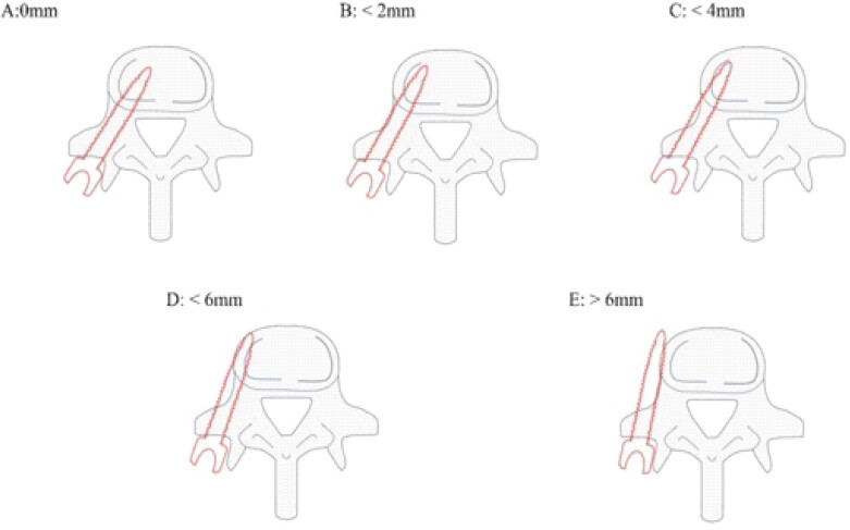
However, we have nothing comparable for the fractured pedicle itself. There is no widely adopted pedicle-only classification analogous to Gertzbein–Robbins; pedicle fractures are instead described by degree – partial cortical breach, complete split, or comminuted burst – and included in broader thoracolumbar systems such as the AO classification when trauma is the cause [2,4]. The AO system recognizes pedicle involvement within its account of posterior-element injury but does not dedicate a pedicle-specific grade [2]. The result is a strange asymmetry: We grade the position of the screw with precision, yet describe the failure of the bone around it only in loose, narrative terms.
Understanding how the pedicle giveaways makes the spectrum easier to anticipate. In elective instrumentation, failure is most often secondary to our own faults. An incorrect entry point, an inappropriate medial-lateral or cranio-caudal trajectory, over-reaming, excessive tapping, or a screw oversized relative to the pedicle diameter each erode cortical bone incrementally – first a breach, then a miss/lateral cutout, then a complete break, and finally comminution if we persist [1,5]. Repeated screw exchanges and forceful insertion into sclerotic or osteoporotic bone only accelerate the progression [3,6].
The bone we begin with matters just as much. A small, congenitally dysplastic pedicle, osteopenia or frank osteoporosis, a previously operated posterior element, chronic infection, or lytic metastatic disease all lower the threshold at which a screw becomes a fracture [3,6]. Moreover, in the trauma setting, the injury may precede surgical intervention entirely: Axial loading, burst-type vertebral fractures, and shearing forces through the posterior elements can break or comminute the pedicle at the moment of impact, long before any instrument is introduced [2].
This is why intraoperative recognition – reading the injury as it happens – is a skill worth developing deliberately, because the clinical picture guides us before the imaging does. A sudden loss of tactile resistance, an abrupt change in the direction of the probe, an unexpected medial or lateral drift, or a screw that toggles rather than seats are all signs that the corridor has been violated [5]. With experience, you tend to stop at the early signs of giveaway rather than proceeding to the extreme end of the spectrum.
Imaging then confirms and quantifies what the hands suspected. Fluoroscopy in anteroposterior, oblique, and lateral projections will catch gross violations. Still, computed tomography (CT) – intraoperative where available, post-operative otherwise – remains the gold standard for measuring a breach and planning what to do about it [1,5]. Applying Gertzbein–Robbins grading to that CT converts a vague impression into a decision: Accept, redirect, or revise.
Naming the injury, however, is only half the task. Once we know whether we are dealing with a breach, lateral miss/cutout, a break, or a burst, the harder question follows – how to salvage fixation without compounding the damage – and here the single most useful principle we can offer is that the salvage must be matched to the grade of injury, neither over-treating a trivial breach nor under-treating a destroyed pedicle. Much of that proportionality is decided before the incision. A pre-operative CT that measures pedicle dimensions informs screw diameter and trajectory. Flags in advance which patients will need augmentation – fenestrated screws, polymethylmethacrylate (PMMA), hydroxyapatite (HA) coated screw, or an extended construct – converts intraoperative surprise into a rehearsed plan, particularly in the osteoporotic or small-pedicled patient [1,6]. Where navigation or robotic assistance is available, it can reduce breach rates and permit real-time trajectory adjustment, though neither replaces tactile feedback or surgeon judgment, which remain indispensable [1,5].
The Breach
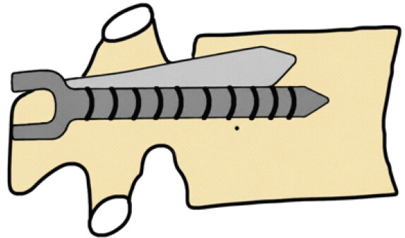
For the breach – a single violated wall with the pedicle otherwise intact – the response is usually the least invasive. If enough cortex remains, the screw can simply be redirected within the same pedicle along a modified trajectory, angling away from the breached wall to regain purchase without a second violation [1,5]. Screw size is the other lever: In good bone a marginally larger screw may recapture purchase by re-engaging cortical bone, whereas in fragile or osteoporotic bone a smaller-diameter screw, or a cement-augmented fenestrated screw, reduces the risk of extending the breach while improving pullout strength [5,6]. The exception that should never be rationalized away is the medial breach approaching neural elements, or a large breach (Gertzbein–Robbins C to E) with inadequate purchase – here early revision, not persistence, is the correct instinct (Fig. 3) [1, 5].
The Miss/Lateral Cutout
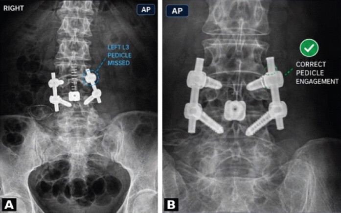
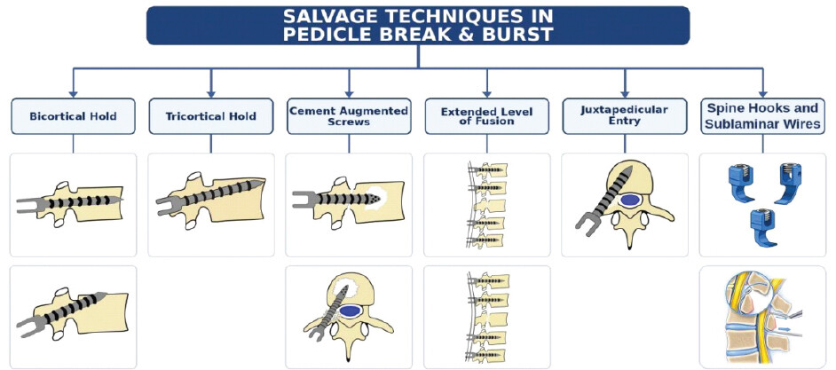
For a miss/lateral cutout – the lateral wall is breached; otherwise, the pedicle in toto is intact, so salvage strategies are less invasive. If the lateral wall is missed and all the walls of the pedicle are intact, careful redirection with a better entry point will help achieve a good hold of the pedicle without the need for other augmentation techniques. If there is lateral cutout, redirection of the screw with a smaller size, in itself, could just be enough, but sometimes augmentation with bone cement or bicortical or tricortical hold can also help. In a longer construct, skipping a level is also an alternative without risking any biomechanical stability of the construct (Fig. 4).
The Break
The break asks more of us. When the pedicle is fractured but not wholly comminuted, an extrapedicular – juxtapedicular, or “in-out-in” – trajectory allows the screw to start lateral to the ruined pedicle and travel into the vertebral body, engaging substantial vertebral-body cortex while bypassing the damage [7]. In selected cases a smaller size screw with an anterolateral or anterosuperior trajectory can secure bicortical fixation through the anterior cortex of the vertebral body, a maneuver that meaningfully augments pullout strength and is especially valuable in osteoporotic bone [7,8]. Fenestrated screws with PMMA augmentation or HA coated screw offer another route to purchase in an osteoporotic or marginally fractured pedicle, often sparing the patient a more extensive revision [3,9,10]. Moreover, where access is already open, and bone quality is reasonable, morselized or local graft can be laid into the pedicle defect and a larger screw placed once the graft consolidates – a strategy reserved, realistically, for planned staged revision rather than the urgent intraoperative moment [6].
The Burst
The burst forces a change of plan altogether, because a comminuted pedicle is, by definition, not reconstructable in situ. The most dependable response is to extend the construct by one or more levels cranially or caudally, distributing load across more screws and unloading the compromised segment [3]. When the pedicle is lost entirely, alternative anchors – laminar or transverse-process hooks, sublaminar wires, laminar screws, or spinous-process screws – allow us to bypass it completely [3]. Cement augmentation of screws at adjacent levels, whether through fenestrated screws or perivertebral delivery, can compensate for the purchase surrendered at the destroyed level [3,6]. Moreover, even where the pedicle itself is gone, an anterosuperior trajectory into the superior and anterior cortex of the vertebral body can sometimes still find cortical hold. If in a large construct, one of the pedicle gets burst, it can be skipped if it doesnot correspond to proximal or distal most screw. Sometimes a smaller diameter screw with superior or anterior cortex hold can prove to be good bailout option. (Fig. 5) [6, 8].
Principles That Run Through All Three
A few principles cut across every scenario. The first is to chase cortical bone: Whenever the pedicle is compromised, redirecting the screw to engage the superior endplate or anterior vertebral-body cortex buys far better purchase than cancellous bone alone [6,11]. The juxtapedicular technique earns its place particularly when the pedicle is too narrow, fractured, or osteoporotic to trust [7]. Cement augmentation, for all its power, demands discipline – controlled PMMA delivery through fenestrated screws can transform pullout strength, but careless technique invites cement leakage and pulmonary embolism [3]. Bicortical fixation, performed cautiously under fluoroscopic guidance, similarly rewards the patient with poor bone quality [6]. Above all, resist the temptation to re-drill a shattered pedicle repeatedly; each attempt tends to worsen the injury, and the wiser move is to change level, trajectory, or fixation modality early rather than late. Longer construct can be forgiving for a single pedicle violation and that level can be skipped if no salvage is possible.
When to Stage or Convert to Open Revision
Some situations call for restraint of a different kind – the decision to stage or convert to open revision. Any malpositioned screw with radiological or clinical evidence of neural impingement, such as a medial breach accompanied by a motor or sensory deficit, should be revised promptly and decompressed as needed; Gertzbein–Robbins grades C to E with neurological symptoms generally warrant it [1]. Equally, when in-situ salvage is likely to fail – severe comminution, active infection, or tumor infiltration – it is better to commit to an open revision with extended constructs and grafting than to defend a construct that cannot hold [3,7].
Reduced to its essentials, the intraoperative logic is straightforward. A minor breach or lateral miss with good purchase is redirected, or accepted if it is Gertzbein–Robbins A or B and asymptomatic. A fractured pedicle or lateral cutout is managed with an in-out or juxtapedicular trajectory or bicortical fixation, with cement augmentation where the bone is osteoporotic. A burst pedicle prompts us to extend fixation and reach for alternative anchors or cement-augmented screws at adjacent levels [5,7].
The value of naming breach, miss/lateral cutout, break, and burst is not taxonomic tidiness for its own sake. It is that a shared vocabulary gives us a shared, graded response – a way of meeting each failure with a technique proportionate to it, preserving construct stability while keeping the neural elements safe. That, in the end, is what separates a salvaged pedicle from a compounded one.
Clinical Message
Pedicle failure should not be treated as a single entity; distinguishing between breach, lateral miss/cutout, break, and burst helps guide appropriate salvage. Early intraoperative recognition and careful assessment of screw position and pedicle integrity allow timely redirection, alternative trajectories, augmentation, or construct extension. The principle is to use the least invasive salvage that provides adequate fixation while avoiding further pedicle damage and neurological risk.
Conflict of Interest:
Nil
Source of Support:
Nil
Consent:
The authors confirm that informed consent was obtained from the patient for publication of this article
How to Cite this Article
Hadgaonkar S, Pardeshi JK, Jadhav D, Shyam A. Not All Pedicle Violations Are Equal: Defining Breach, Miss, Break, and Burst and Their Salvage Strategies. Journal of Orthopaedic Case Reports 2026 October;16(10): 01-05.
References
- Gertzbein SD, Robbins SE. Accuracy of pedicular screw placement in vivo. Spine (Phila Pa 1976) 1990;15:11-4. [Google Scholar] | [PubMed]
- Vaccaro A, Oner C, Kepler C, Dvorak M, Schnake K, Bellabarba C. AOSpine Thoracolumbar Spine Injury Classification System: Fracture Description, Neurological Status, and Key Modifiers. Spine 2013;38(23):2028-2037. doi.org/10.1097/BRS.0b013e3182a8a381. [Google Scholar] | [PubMed] | [CrossRef]
- Krappinger D, Kastenberger TJ, Schmid R. Die Behandlung osteoporotischer Wirbelkörperfrakturen mit augmentierter Instrumentation [Augmented posterior instrumentation for the treatment of osteoporotic vertebral body fractures. Oper Orthop Traumatol 2012;24:4-12. [Google Scholar] | [PubMed]
- Gadoin D, Dagher M, Flouzat Lachaniette CH, Bouthors C, Yoon BC, Dubory A. Incidence of vertebral pedicle fracture occurrence in thoracic and lumbar spine trauma. A CT-scan anatomical assessment. Neurochirurgie 2026;72:101833. [Google Scholar] | [PubMed]
- Librianto D, Saleh I, Fachrisal, Utami WS, Hutami WD. Breach rate analysis of pedicle screw instrumentation using free-hand technique in the surgical correction of adolescent idiopathic scoliosis. J Orthop Case Rep 2021;11:38-44. [Google Scholar] | [PubMed]
- Tandon V, Kalidindi KK, Pacha S, Bhat MR. A prospective study on the feasibility, safety, and efficacy of a modified technique to augment the strength of pedicle screw in osteoporotic spine fixation. Asian Spine J 2020;14:357-63. [Google Scholar] | [PubMed]
- Kurland DB, Lendhey M, Delavari N, Winfield J, Mahoney JM, Becske T. Percutaneous juxtapedicular cement salvage of failed spinal instrumentation? Institutional experience and cadaveric biomechanical study. Oper Neurosurg 2024;26:38-45. [Google Scholar] | [PubMed]
- Murata A, Tsutsui S, Yamamoto E, Kozaki T, Nakanishi R, Yamada H. A bicortical pedicle screw in the cephalad trajectory is the best option for the fixation of an osteoporotic vertebra: A finite element study. Spine Surg Relat Res 2024;8:510-7. [Google Scholar] | [PubMed]
- Boucas P, Mamdouhi T, Rizzo SE, Megas A. Cement augmentation of pedicle screw instrumentation: A literature review. Asian Spine J 2023;17:939-48. [Google Scholar] | [PubMed]
- Song Z, Zhou Q, Jin X, Zhang J. Cement-augmented pedicle screw for thoracolumbar degenerative diseases with osteoporosis: A systematic review and meta-analysis. J Orthop Surg Res 2023;18:631. [Google Scholar] | [PubMed]
- Matsukawa K, Yato Y. Lumbar pedicle screw fixation with cortical bone trajectory: A review from anatomical and biomechanical standpoints. Spine Surg Relat Res 2017;1:164-73. [Google Scholar] | [PubMed]
© 2026 Journal of Orthopaedic Case Reports - Published by Indian Orthopaedic Research Group
About the Authors
-

Shailesh Hadgaonkar
-

Jaspal Khushalsingh Pardeshi
-

Diptesh Jadhav
-

Ashok Shyam
Related Articles in Journal of Orthopaedic Case Reports
 January 10, 2022 An Isolated Displaced Coracoid Fracture Treated with Open Reduction Internal Fixation with 4 mm Cannulated Cancellous Screw
 August 1, 2026 Lesser Trochanter Osteochondroma as an Etiology for Ischiofemoral Impingement: A case report
 September 1, 2026 Glomus Tumor of the 4th Toe: A Case Report
 November 23, 2017 Endoscopic Calcaneoplasty for Haglund’s Excision Using Two Lateral Portals