


Introduction
Total hip arthroplasty (THA) is one of the most common orthopaedic procedures performed in the US in the adult population [1]. This procedure is much less common in the pediatric and adolescent populations, although these numbers have been increasing over the past 20 years in part due to increased longevity of implants and use of more durable materials in THA [2]. This improvement in longevity is particularly important in younger patients because there is a need for increased survivorship of THA components given that this patient population is generally more active and will live longer than geriatric patients in which the vast majority of THAs are performed. The long-term implications of performing a THA in such a young patient must be considered, understanding that most patients will likely outlive their implants, necessitating revision surgery.
Consideration must be given to implant and bearing surface selection due to the potential for unique or distorted anatomy as well as the small stature of pediatric patients. Variations in components/bearing surfaces and types of fixation can be used, including ceramic-on-polyethylene, ceramic-on-ceramic, metal-on-polyethylene, metal-on-metal, as well as cemented versus cementless (press-fit) fixation. Modern highly cross-linked polyethylene has an excellent wear profile, improved from earlier polyethylene designs, making this choice desirable in young patients [2, 3, 4].
In the pediatric population, there are numerous pathologies that ultimately lead to the need to perform a THA. One of the most common causes of hip pathology resulting in THA is avascular necrosis (AVN) secondary to slipped capital femoral epiphysis [2,5]. Other causes include trauma, congenital deformities, AVN from corticosteroid use, and, less commonly, juvenile idiopathic arthritis (JIA). With advancements in disease-modifying antirheumatic drugs, THA performed for JIA is seen much less frequently than prior to the 2000s [2]. While attempts are made to salvage the joint and correct deformity in the very young patient, many of the patients who are diagnosed with advanced arthrosis may ultimately require THA.
Surgical approach is another consideration for the surgeon and patient when performing THA. It is often determined by the individual deformity, prior surgeries, presence of hardware, and whether or not a specific procedure, such as an extended trochanteric osteotomy, is planned. Prior literature has demonstrated no difference in post-operative pain, limp, complications requiring revision surgery, or return to activities when comparing surgical approaches [6].
The purpose of this case series was to describe indications for and evaluate the outcomes of THA in a series of five adolescent patients <18 years of age, as well as to discuss surgical approaches and specific components used.
Case Report
This study was approved by our Institutional Review Board.
Case 1
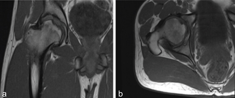
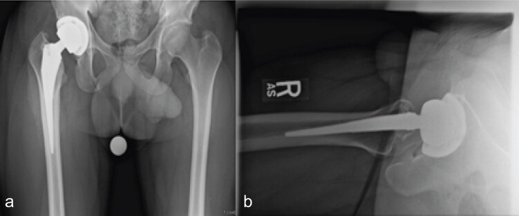
The patient was a 17-year-old male who presented to the pediatric orthopaedic clinic with increasing right hip pain over a 1-year period. The patient had a past medical history of growth hormone deficiency and was treated with growth factor replacements and oral steroids. The patient was active in multiple sports including football; however, there was no known preceding trauma. Before presentation, he had attempted conservative management for right hip pain, including over-the-counter anti-inflammatory medication and physical therapy without relief of symptoms. Imaging at that time showed concern for AVN of the right hip with associated collapse of the femoral head. In addition, imaging showed congenital coxa breva and coxa vara. Magnetic resonance imaging was completed and confirmed osteonecrosis of the right femoral head (Fig. 1). The patient was referred to adult reconstruction for THA. The patient also had genetic testing to rule out underlying mucopolysaccharidosis, which was found to be negative. The patient underwent right direct anterior THA. Intraoperatively, a 123° neck angle arthroplasty was used to accommodate his short varus neck to restore leg length and offset. He recovered from surgery without complications. His preceding right hip pain completely resolved; he returned to normal activities and has not been limited by arthroplasty. The patient follows up annually in clinic with serial radiographs, which demonstrate appropriate alignment of the THA without any signs of hardware failure at 5-year follow-up (Fig. 2).
Case 2
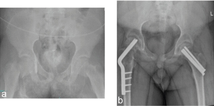
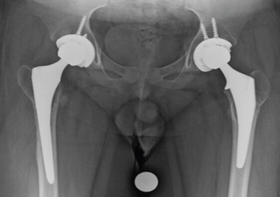
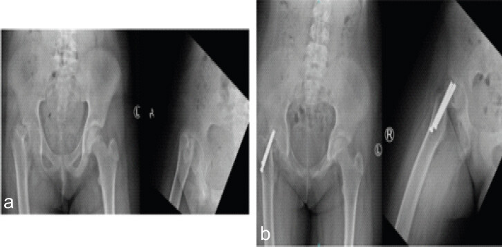
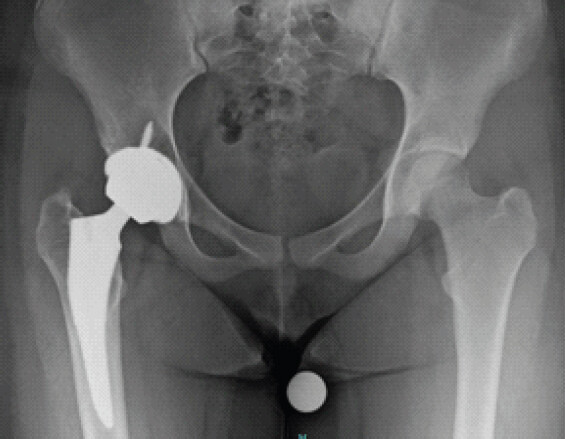
The patient was an 11-year-old male who presented after an all-terrain vehicle accident. The patient sustained multiple injuries, including bilateral femoral neck fractures, left forearm fracture, and non-operative spine fractures (Fig. 3). He underwent right femoral neck open reduction and internal fixation and left femoral neck closed reduction and percutaneous pinning. Initially, the patient did well post-operatively, progressing to weight-bearing with a walker at 2 months. Five months post-operatively, imaging showed concern for non-union of the right femoral neck. A computed tomography (CT) scan was completed at that time, which confirmed the non-union. Laboratory tests for non-union showed no abnormalities in inflammatory markers, Vitamin D, or complete blood count. He underwent revision surgery for femoral neck non-union with a right proximal femur valgus osteotomy. Intraoperative cultures were obtained and found to be negative. Three months post-operatively, the patient began experiencing increased right hip pain (Fig. 3). Appropriate investigation into an infectious etiology was completed, including advanced imaging and interventional radiology-guided hip aspiration. The underlying cause of the pain appeared to favor AVN over infection. The patient was referred to adult reconstruction for right total hip replacement. He underwent removal of hardware and conversion to right THA with robotic assistance, completed through a posterior approach given the need for removal of hardware. Intraoperatively, the blade plate was found to have perforated the femoral head. The patient’s pre-operative right hip pain resolved, and he progressed appropriately post-operatively. Approximately 2 months after the right THA was performed, he began to develop increasing pain in the left hip. His pain progressed until he was ambulating with the use of a walker and wheelchair. A CT scan demonstrated AVN of the left femoral head, and the decision was made to proceed with removal of hardware from the left hip and conversion to THA (Fig. 4). This procedure completely resolved his left hip pain, and he was able to return to normal ambulation without aids 2 years out from surgery.
Case 3
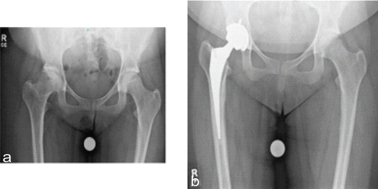
The patient was an 11-year-old female who presented to the pediatric orthopaedic clinic with worsening right knee pain and limp for 2 months after a fall. The patient was intermittently using crutches due to pain. Imaging at that visit showed a severe right slipped capital femoral epiphysis (Fig. 5). She underwent open reduction and pinning of her right femur given the severity of the slip. In addition, the patient had endocrine laboratories completed, which were negative. Post-operatively, she maintained partial weight-bearing with resolution of pain. At 6 weeks post-operatively, routine imaging was completed, which showed femoral head collapse and signs of AVN (Fig. 5). The patient was referred to adult reconstruction for right THA due to progressive pain and limitation of activity. The procedure was completed using a ceramic-on-ceramic bearing surface via a direct anterior approach (Fig. 6). The patient’s pre-operative pain completely resolved. At 5 years post-operatively, the patient continues to have no pain, but she noted occasional squeaking from the bearing surface. She was able to return to activities such as running without limitations.
Case 4
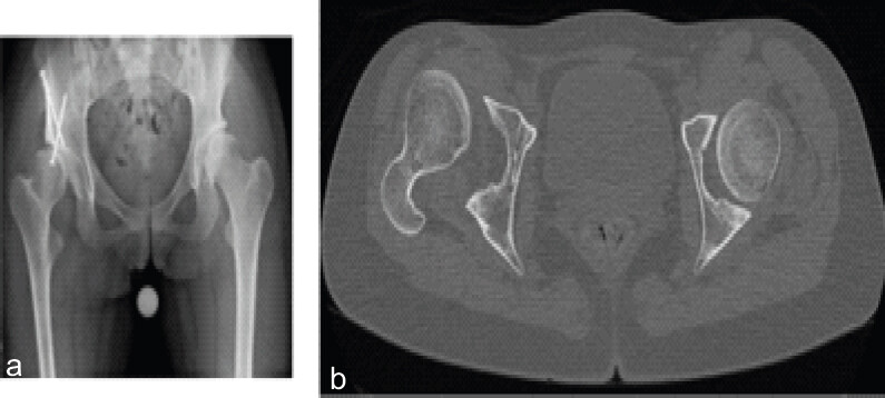
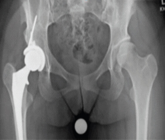
The 12-year-old female patient initially presented to the pediatric orthopaedic clinic for right hip pain that was ongoing for 1 year despite conservative management. On examination, she was found to have a cavovarus foot deformity and equinus contracture consistent with Charcot-Marie-Tooth disease, for which the patient had a positive family history. Additional imaging at initial presentation showed right hip dysplasia with a lateral subluxation of the femoral head. She subsequently underwent bilateral gastrocnemius lengthening and right hip periacetabular osteotomy in a staged fashion. Surgery initially resolved pre-operative symptoms. However, approximately 6 months post-operatively, the patient noted a change in her right leg rotational alignment and a limp with ambulation. Imaging at that time showed lateral subluxation of the femoral head consistent with a failed periacetabular osteotomy and anterior dislocation of the femoral head (Fig. 7). The patient was referred to adult reconstruction for THA due to progressive pain and limitation of activity. She underwent a right robotically assisted THA and removal of three acetabular screws through her prior direct anterior incision (Fig. 8). The THA completely resolved her pre-operative hip pain, and the patient returned to activities without limitations 3 years out from surgery.
Case 5
The patient was a 16-year-old female with a history of chromosomal abnormality and autism who presented to the pediatric orthopaedic clinic with right hip pain that had been ongoing for many years. Multiple years prior, she had been followed by a pediatric orthopaedic surgeon who diagnosed her with Perthes disease. She underwent two hip containment procedures at a young age. On initial presentation in clinic at age 13, she had radiographic signs of subchondral sclerosis and flattening of the femoral head. Despite radiographic findings, the patient’s symptoms were initially controlled with over-the-counter pain medications, and her symptoms were not impeding activity. At subsequent clinic visits, her pain began to increase with progression of radiographic findings and AVN with femoral head collapse (Fig. 9). The patient was referred to adult reconstruction for THA. She underwent right THA via an anterior approach. The patient had complete resolution of her pre-operative symptoms, and she returned to normal activities without limitations. At 3 years post-operatively, she continues to follow up in our clinic without any concerns or activity limitations (Fig. 9).
Discussion
At a mean follow-up of 3.6 years, no revisions or complications were noted in this population, and no evidence of radiographic lucency, subsidence of components, or hardware failure was seen. Clinically, all patients demonstrated improvement in pain and range of motion, and returned to normal function, including participation in sports and school. All patients in this series ambulated without assistance or limp at most recent clinic follow-up. Patient demographics, surgical approach, and bearing selection are shown in Table 1.
Patient demographics, hip pathology and surgical history, and surgical approach and component information. * Approach, bearing surface, fixation, and acetabular cup sizes were the same in bilateral hips.
| Case | Age (yrs) | Sex | BMI | Skeletal Maturity | Pathology | Prior Surgeries | Retained Hardware (Y/N) | Follow-up *yrs) | Surgical Approach | Bearing Surface | Cemented vs cementless | Acetabular cup size |
|---|---|---|---|---|---|---|---|---|---|---|---|---|
| 1 | 17 | M | 23.49 | Mature | Femoral head AVN (steroid-induced, growth hormone deficiency) | 0 | N | 4 | Direct Anterior | Ceramic-on-Polyethylene | Cementless | 52 |
| 2 | 11 | M | 37.13 | Immature | Failed ORIF with nonunion, AVN | 1 (prior ORIF) | Y | 1 | Posterior* | Ceramic-on-Polyethylene* | Cementless* | 50* |
| 3 | 11 | F | 24.22 | Immature | Femoral head collapse (AVN from SCFE) | 1 (open reduction/pinning) | Y | 3 | Direct Anterior | Ceramic-on-Ceramic | Cementless | 52 |
| 4 | 12 | F | 20.08 | Immature | CMT with hip dysplasia s/p failed PAO | 1 (PAO) | Y | 1 | Direct Anterior | Ceramic-on-Polyethylene | Cementless | 46 |
| 5 | 16 | F | 25.39 | Mature | Chromosomal abnormality, LCP, femoral head AVN | 2 (containment procedures) | N | 2 | Direct Anterior | Ceramic-on-Polyethylene | Cementless | 46 |
AVN = Avascular necrosis; ORIF = open reduction internal fixation, SCFE = slipped capital femoral epiphysis, CMT = Charcot Marie Tooth, LCP = Legg-Calve-Perthes, PAO = Periacetabular osteotomy
In this population, outcomes in general seem to be favorable regardless of surgical approach, with a dislocation rate of approximately <1% in a systematic review of over 2300 hips [6]. Cementless fixation is preferred in modern THA in adolescent patients, with press-fit implants now being the favored choice. Historically, THA in this population had varying degrees of success due to osteolysis and failure of fixation, and longevity of the construct as well as clinical outcomes have been inconsistent [7]. Modern ceramic-on-polyethylene and ceramic-on-ceramic bearings paired with cementless fixation give the potential for long-term survivorship in these young patients.
Timing for THA in this population is important, with the goal being to wait as long as medically safe while balancing patient symptoms such as pain and decreased ability to perform activities of daily living. The classic thinking regarding skeletal maturity and closure of the triradiate cartilage introduces the idea of adequate support for acetabular components with THA. Some of the patients in this case series had open triradiate cartilage at the time of surgery. A recent study by Rainer et al. discusses excellent results with a significant improvement in clinical outcomes, good implant survivorship, and low complication rates in patients with open triradiate cartilage undergoing THA. These results may indicate that awaiting closure of the acetabular triradiate cartilage may not be necessary if hip disease is advanced enough to cause significant pain or impact function [8].
Overall, THA is a reliable procedure for improving pain and function in patients with advanced structural hip disease, and modern components have seen increased utilization in the adolescent population [9]. Though this population presents unique challenges regarding durability of implants, literature after the year 2000 demonstrates improvements in clinical function with excellent radiologic outcomes up to 8 years post-operatively, with significant improvement in mobility and alleviation of pain [7,9,10,11]. The most recent studies to date confirm excellent 5–10 year survivorship with low revision rates and significant improvements in patient-reported outcome measures [12,13].
Conclusion
THA in young patients specifically remains an excellent treatment option given improvements in modern components (use of highly cross-linked polyethylene bearing surface) and a low rate of revision. Although additional planning, including templating, is often necessary for a successful surgical outcome, THA in the pediatric and adolescent populations has demonstrated excellent outcomes with cementless implant designs. However, long-term follow-up is still needed to further elucidate the full effects of more modern bearing surfaces and implant designs in this population.
Clinical Message
Total hip arthroplasty can offer life-changing benefits for adolescents in need. Although they are the minority, surgeons should feel comfortable offering surgery to adolescents with appropriate indications like those discussed in this paper.
Conflict of Interest:
Nil
Source of Support:
Nil
Consent:
The authors confirm that informed consent was obtained from the patient for publication of this article
How to Cite this Article
Ernst JC, Patton CN, Frye BM. Total Hip Arthroplasty in the Adolescent Population: A Case Series Journal of Orthopaedic Case Reports 2026 October;16(10): 493-499.
References
- Skopec L, Berenson RA, Simon B, Papanicolas I. Variation in processes of care for total hip arthroplasty across high-income countries. Health Aff Sch 2024;2:qxae043. [Google Scholar] | [PubMed]
- Kahlenberg CA, Gibbons JA, Jannat-Khah DP, Goodman SM, Mandl LA, Sculco PK. Use of total hip arthroplasty in patients under 21 years old: A US population analysis. J Arthroplasty 2021;36:3928-33.e1. [Google Scholar] | [PubMed]
- Atrey A, Ward SE, Khoshbin A, Hussain N, Bogoch E, Schemitsch EH. Ten-year follow-up study of three alternative bearing surfaces used in total hip arthroplasty in young patients: A prospective randomised controlled trial. Bone Joint J 2017;99-B:1590-5. [Google Scholar] | [PubMed]
- Metcalfe D, Peterson N, Wilkinson JM, Perry DC. Temporal trends and survivorship of total hip arthroplasty in very young patients: A study using the national joint registry data set. Bone Joint J 2018;100:1320-9. [Google Scholar] | [PubMed]
- Whitmarsh-Brown MA, Christ AB, Lin AJ, Siddiqui AA, Herman RY, Allison DC. Modernizing our understanding of total hip arthroplasty in the pediatric and young adult patient: A single-center experience. J Pediatr Orthop 2023;43:e290-8. [Google Scholar] | [PubMed]
- Te Velde JP, Buijs GS, Schafroth MU, Saouti R, Kerkhoffs GM, Kievit AJ. Total hip arthroplasty in teenagers: A systematic literature review. J Pediatr Orthop 2024;44:e115-23. [Google Scholar] | [PubMed]
- Polkowski GG, Callaghan JJ, Mont MA, Clohisy JC. Total hip arthroplasty in the very young patient. J Am Acad Orthop Surg 2012;20:487-97. [Google Scholar] | [PubMed]
- Rainer W, Shirley MB, Trousdale RT, Shaughnessy WJ. The open triradiate cartilage: How young is too young for total hip arthroplasty? J Pediatr Orthop 2021;41:e793-9. [Google Scholar] | [PubMed]
- Peagler CL Jr, Dobek AJ, Tabaie S. Trends in the use of total hip arthroplasty in the pediatric population: A review of the literature. Cureus 2023;15:e43978. [Google Scholar] | [PubMed]
- Van De Velde SK, Loh B, Donnan L. Total hip arthroplasty in patients 16 years of age or younger. J Child Orthop 2017;11:428-33. [Google Scholar] | [PubMed]
- Kamath AF, Sheth NP, Hosalkar HH, Babatunde OM, Lee GC, Nelson CL. Modern total hip arthroplasty in patients younger than 21 years. J Arthroplasty 2012;27:402-8. [Google Scholar] | [PubMed]
- Hong JX, Dravid A, Sankar WN, Sheth NP. Pediatric patients undergoing total hip arthroplasty: A single-center experience at average 5.3-year follow-up. J Arthroplasty 2025;40:S229-35. [Google Scholar] | [PubMed]
- Stein AM, Gaumé M, Demoures T, Mary P, Vialle R. Total hip arthroplasty in children with osteoarthritis: A retrospective cohort study. Arch Pediatr 2026;33:105471. [Google Scholar] | [PubMed]
© 2026 Journal of Orthopaedic Case Reports - Published by Indian Orthopaedic Research Group
About the Authors
-

Jennafir C Ernst
-

Caitlyn N Patton
-

Benjamin M Frye
Related Articles in Journal of Orthopaedic Case Reports
 October 1, 2026 Total Hip Arthroplasty for Advanced Femoral Head Osteonecrosis in a 24-Year-Old Competitive Wrestler: A Case Report
 July 1, 2026 A Comparative Study of Functional Outcome Following Total Hip Replacement by Posterolateral and Anterolateral Approach
 July 1, 2026 Managing Complications of Total Hip Arthroplasty for Avascular Necrosis Hip Performed Via Anterior Approach
 June 1, 2026 Functional Outcomes of Total Hip Arthroplasty using Modified Harris Hip Score and Oxford Hip Score: A Prospective Study