


Introduction
Legg-Calvé-Perthes disease (LCPD) is described as idiopathic avascular necrosis of the femoral head. Several etiologies have been proposed, including a mutation of type II collagen and thrombophilia causing necrosis of the femoral head [1]. LCPD has an incidence of approximately 1 in 10,000 children, affecting boys 5 times as much as girls [2]. Children generally present at age 5–8 years with mild pain, a limp or decreased range of motion of the hip. Staging has been classified based on radiographic imaging (early sclerosis, fragmentation, re-ossification, and healing) [1]. The lateral pillar classification has been accepted as a prognostic indicator, with <50% of the height of the lateral pillar of the femoral head indicative of poor outcomes [3]. Progression of the condition depends largely on the degree of femoral head deformation. Mild deformity may remain asymptomatic while more severe deformity may cause metaphyseal and acetabular changes, leading to pre-mature arthritis before the age of 50 [4].
Case Report
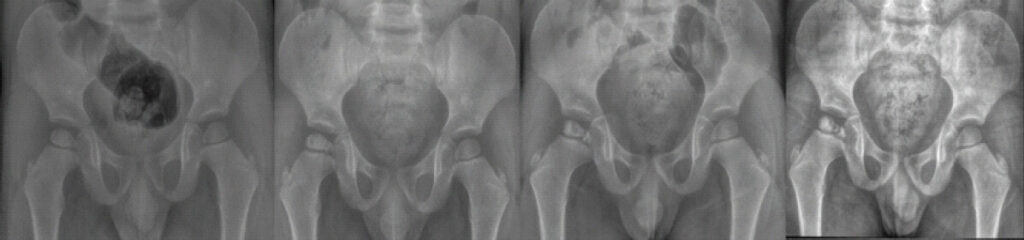
Twin A and Twin B are monozygotic males from a mother who received normal prenatal care and had no history of smoking. Each twin was raised in the same household. Pregnancy was complicated by pre-term pre-mature rupture of membranes at 35 weeks. Each twin was born through cesarean section at 35 weeks gestation. Twin A was born first, with no notable complications at birth. Twin B spent approximately 6 h on continuous positive airway pressure for decreased aeration at birth; however, there were no further complications at birth. Twin A presented to our office at 3 years 4 months of age with an antalgic gait for approximately 2 weeks in comparison to his brother, Twin B. Radiography at that time revealed no obvious abnormality to the bilateral hips. Lab work at that time, including inflammatory markers (erythrocyte sedimentation rate and C-reactive protein), complete blood count and comprehensive metabolic panel, which revealed an elevated alkaline phosphatase at 279 units/L, and no other significant abnormalities. Twin A followed up in our office 2 weeks later with improvement of symptoms. Given his improvement and no obvious diagnosis at that time, the decision was made to continue to observe Twin A. At the 2-month follow-up, his symptoms had almost completely resolved. Radiography of his hips and pelvis revealed slight lucency of the right lateral femoral head at that time. He was then seen again at 6 months from symptom onset, where radiography revealed fragmentation of the right femoral head, lateral pillar Class A, and the diagnosis of LCPD was determined most likely. During his past follow-up at 1 year after initial presentation, he was found to be ambulating well without limitations. His radiography remained at lateral pillar Class A and was found to be consistent with the re-ossification stage (Fig. 1).
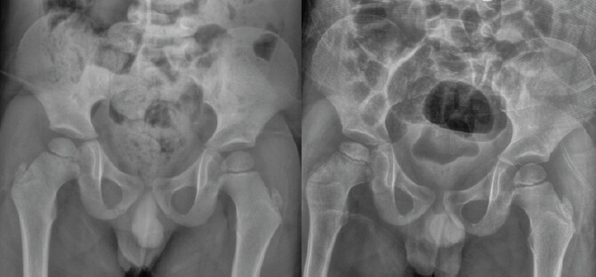
During the 1-year follow-up of Twin A, Twin B presented with approximately 2 weeks of antalgic gait and mild pain localized to the right hip. Radiography of the hips and pelvis revealed fragmentation of the medial aspect of the femoral head with slight height loss of the lateral femoral head, indicative of early LCPD. Twin B at 3-months follow-up continued to have a mild antalgic gait with minimal interval improvement. Given the fair prognosis of Twin A thus far, the decision was made to continue to observe Twin B in a similar fashion (Fig. 2).
Discussion
LCPD remains a condition of uncertain etiology, with environmental factors, such as second-hand smoke exposure and low socioeconomic status having been suggested to increase a patient’s risk [5]. In recent years, research has aimed to identify genetic components associated with the development of LCPD. Given its rarity, documenting familial cases of LCPD, especially among siblings, is essential to expanding the data needed for larger studies on possible genetic contributions to its etiology. We present a case of identical twins developing LCPD confirmed by radiographic imaging.
Multiple genetic factors have been suggested to play a role in the development of LCPD. In the present literature, the most prominent genetic factor is the association of LCPD with hereditary thrombophilia. In a meta-analysis by Woratanarat et al., the presence of Factor V Leiden was shown to increase the risk of LCPD [6]. Other studies have reached similar conclusions. In a study of LCPD in Mexican patients, a significant difference in prothrombin time as well as Factor V and Factor IX levels were seen in those with LCPD [7]. In a study on 169 patients at two centers in the Netherlands, Factor V Leiden, prothrombin C20210A mutations, protein C deficiency and elevated Factor VIII were all significantly associated with LCPD, further suggesting genetic aberrations in coagulation increase a patient’s risk of LCPD [8].
Interestingly, mutations in the type II collagen gene have also shown an association with LCPD. In a study of a Chinese family with known LCPD, a heterozygous mutation in exon 29 of COL2A1 was identified in family members specifically affected [9]. Similar familial studies have noted mutations at different sites of the COL2A1 gene in families with multiple family members affected by LCPD [10,11]. Interestingly, one case report also suggested an association of LCPD with osteogenesis imperfect [12].
Although genetic factors have been shown to increase the risk of developing LCPD, research specifically examining LCPD in twins remains limited. The largest twin study, involving 81 twin pairs from the Danish Twin Registry, suggested a low likelihood of a distinct genetic contribution, as the concordance rate among monozygotic twins was low [13]. However, only 10 monozygotic twin pairs were included, limiting the strength of this conclusion. In contrast, multiple case reports have documented monozygotic twins both developing LCPD, supporting the possibility of a specific genetic component [14–16].
Conclusion
Given the potential influence of genetic factors in the development of LCPD, investigating familial associations remains essential. However, present literature offers relatively limited insight into the relationship between monozygotic twins and LCPD. Although constrained by the absence of genetic testing, our case contributes to the growing body of evidence suggesting a genetic component in the disorder’s pathogenesis. Further research exploring the genetic mechanisms underlying LCPD is therefore warranted.
Clinical Message
There is a potential genetic contribution to LCPD pathogenesis. Recognition of a genetic component may warrant increased clinical vigilance and early screening in at-risk siblings to facilitate timely diagnosis and management.
Conflict of Interest:
Nil
Source of Support:
Nil
Consent
The authors confirm that informed consent was obtained from the patient for publication of this article
How to Cite this Article
Lane G, Christy G, Baker D, Raju S, Erkilinc M. Identical Twins with Legg-Calve-Perthes Disease: A Report of Two Cases. J Orthop Case Rep. 2026 November;16(11): 197-200. doi: 10.13107/jocr.2026.v16.i11.8438.
References
- Kim HK. Legg-Calvé-Perthes disease. J Am Acad Orthop Surg 2010;18:676-86. [Google Scholar] | [PubMed]
- Joseph B, Shah H, Perry DC. Epidemiology, natural evolution, pathogenesis, clinical spectrum, and management of Legg-Calvé-Perthes. J Child Orthop 2023;17:385-403. [Google Scholar] | [PubMed]
- Herring JA, Kim HT, Browne R. Legg-Calve-Perthes disease. Part I: Classification of radiographs with use of the modified lateral pillar and Stulberg classifications. J Bone Joint Surg Am 2004;86:2103-20. [Google Scholar] | [PubMed]
- Joseph B. Natural history of early onset and late-onset Legg-Calve-Perthes disease. J Pediatr Orthop 2011;31 Suppl 2:S152-5. [Google Scholar] | [PubMed]
- Bahmanyar S, Montgomery SM, Weiss RJ, Ekbom A. Maternal smoking during pregnancy, other prenatal and perinatal factors, and the risk of Legg-Calvé-Perthes disease. Pediatrics 2008;122:e459-64. [Google Scholar] | [PubMed]
- Woratanarat P, Thaveeratitharm C, Woratanarat T, Angsanuntsukh C, Attia J, Thakkinstian A. Meta-analysis of hypercoagulability genetic polymorphisms in Perthes disease. J Orthop Res 2014;32:1-7. [Google Scholar] | [PubMed]
- Rodríguez-Olivas AO, Reyes-Maldonado E, Casas-Ávila L, Galicia-Alvarado MA, Rosales-Cruz E, Zavala-Hernández C. Association of IL-23R rs1569922 and other probable frequent etiological factors with Legg-Calvé-Perthes disease in Mexican patients. Genes (Basel) 2025;16:1126. [Google Scholar] | [PubMed]
- Vosmaer A, Pereira RR, Koenderman JS, Rosendaal FR, Cannegieter SC. Coagulation abnormalities in Legg-Calvé-Perthes disease. J Bone Joint Surg Am 2010;92:121-8. [Google Scholar] | [PubMed]
- Li N, Yu J, Cao X, Wu QY, Li WW, Li TF. A novel p. Gly630Ser mutation of COL2A1 in a Chinese family with presentations of Legg-Calvé-Perthes disease or avascular necrosis of the femoral head. PLoS One 2014;9:e100505. [Google Scholar] | [PubMed]
- Miyamoto Y, Matsuda T, Kitoh H, Haga N, Ohashi H, Nishimura G. A recurrent mutation in type II collagen gene causes Legg-Calvé-Perthes disease in a Japanese family. Hum Genet 2007;121:625-9. [Google Scholar] | [PubMed]
- Su P, Li R, Liu S, Zhou Y, Wang X, Patil N. Age at onset-dependent presentations of premature hip osteoarthritis, avascular necrosis of the femoral head, or Legg-Calvé-Perthes disease in a single family, consequent upon a p. Gly1170Ser mutation of COL2A1. Arthritis Rheum 2008;58:1701-6. [Google Scholar] | [PubMed]
- Hong P, Zhao X, Liu R, Rai S, Song Y, Xu R. Perthes disease in a child with Osteogenesis imperfecta from a rare genetic variant: A case report. Front Genet 2022;13:920950 [Google Scholar] | [PubMed]
- Metcalfe D, Van Dijck S, Parsons N, Christensen K, Perry DC. A twin study of perthes disease. Pediatrics 2016;137:e20153542. [Google Scholar] | [PubMed]
- Hrubá M, Bárta P, Džupa V, Krbec M. Monozygotic twins with Legg-Calvé-Perthes disease and with non-identical lumbosacral malformation: A case report and literature review. Acta Chir Orthop Traumatol Cech 2023;90:353-7. [Google Scholar] | [PubMed]
- Dunn A. Coxa plana in monozygotic male twins. J Bone Joint Surg Am 1960;42-A:178-83. [Google Scholar] | [PubMed]
- Giannestras N. Legg-perthes disease in twins. J Bone Joint Surg Am 1954;36-A:149-52. [Google Scholar] | [PubMed]
© 2026 Journal of Orthopaedic Case Reports - Published by Indian Orthopaedic Research Group
About the Authors
-

Gabriel Lane
-

Gavin Christy
Related Articles in Journal of Orthopaedic Case Reports
 November 1, 2026 Slipped Capital Femoral Epiphysis Associated with Congenital Coxa Vara: A Case Report
 September 1, 2026 Low Kocher–Caird Scores High Clinical Suspicion: A Diagnostic Dilemma in Pediatric Hip Septic arthritis
 August 1, 2026 Surgical Excision of a Rare Ventral Scapular Osteochondroma using the Prone “Chicken-Wing” Position: A Case Report
 August 1, 2026 Serendipitous Healing of a Congenital Pseudoarthrosis of the Clavicle Following Acute Injury: A Case Report