


Introduction
Total knee arthroplasties (TKAs) are becoming increasingly prevalent, resulting in a rise in the frequency of revision TKAs. Revision TKAs are more challenging than primary ones due to the impact of greater bone loss on implant stability. Factors contributing to bone loss during revision TKAs include inflammation, bacterial infections, stress shielding, osteolysis due to wear and loosening of the original implant, periprosthetic fractures, damage incurred while removing the primary implant, osteonecrosis, bone cysts, and previous fractures of the tibial plateau or femoral condyles [1]. One contributing factor to aseptic loosening that leads to prosthetic failure is pigmented villonodular synovitis (PVNS) [2]. Originally characterized by Chassaignac in 1852 and subsequently detailed by Jaffe et al., in 1941, PVNS is identified as a range of benign neoplastic disorders distinguished by the presence of macrophages filled with hemosiderin, multinucleated giant cells, lipid-rich histiocytes, and inflammatory cell infiltrates within an enlarged synovium. The knee joint is most frequently affected, accounting for as much as 80% of cases, followed by the hip, ankle, and shoulder. It primarily impacts adults in their third to fifth decades of life. Recognized as a benign proliferation of cells, it is categorized into two forms: Localized-type (L-PVNS) and diffuse type (D-PVNS). These variants exhibit differences in their clinical and radiographic features but share similarities in histology, showing histiocyte proliferation and hemosiderin accumulation within the synovial tissue. Due to its gradual appearance and symptom similarities with other joint conditions such as rheumatoid arthritis, juvenile idiopathic arthritis, and synovial cysts, PVNS is often misdiagnosed or diagnosed at a later stage. The typical treatment for this condition involves surgically removing the affected synovium. Even with the implementation of rigorous resection methods, the rates of recurrence are still notable, varying between 18% and 46%, depending on the particular type and extent of the surgery. Additional treatment options are frequently investigated for managing cases of recurring or incompletely resectable PVNS [3]. Future revisions should consider new strategies for improving fixation and preserving native bone stock. In revision TKAs, the use of highly porous metal cones and sleeves has proven to be an effective technique for enhancing fixation [1]. At present, titanium metal sleeves are used in revision TKA for addressing significant contained Anderson Orthopedic Research Institute (AORI) type IIA, IIB, or III bone defects. While these sleeves serve a similar purpose to cones, they are designed specifically for each implant, integrated with the stem, and placed as a single unit alongside the entire prosthetic device. Metaphyseal sleeves might provide an additional benefit by acting as a supportive structure for reconstruction when the metaphysis is compromised [4,5,6,7,8,9,10,11,12,13,14,15,16,17,18,19,20].
This report discusses a case involving a 52-year-old man who underwent revision TKA after a right TKA performed 5 years ago with implant loosening and PVNS.
Case Report
A 52-year-old man came with intense pain and swelling in his right knee, along with difficulty walking that required assistance for the past week. He exhibited a varus deformity in his right knee and was ambulating with a lateral thrust while using support. Five years ago, he underwent a right TKA at another facility. He was a known diabetic with a hemoglobin A1c level of 6.5%, and his complete blood counts (CBC), erythrocyte sedimentation rate (ESR), and C-reactive protein (CRP) levels were all within normal ranges. Imaging studies showed subsidence of the tibial component, a 12° varus deformity on the Hip-Knee-Ankle (HKA) axis, and loosening of the implant from the earlier operated TKA on the right side (Fig. 1), with an AORI classification type 3 bone defects in both the femur and tibia.
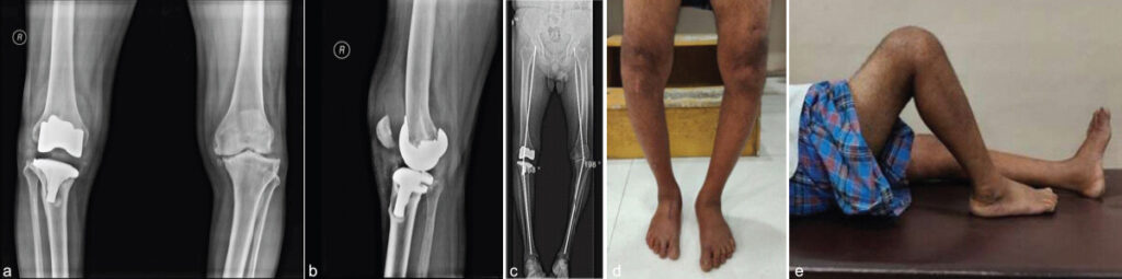
The effusion in the right knee was aspirated, revealing a reddish tinge, likely due to trauma, though it did not exhibit the classic appearance of being bloody or chocolate colored, which is typical for PVNS.
The patient failed to bring any of his earlier reports, including X-rays and discharge summaries. The rationale for the initial surgery and the intraoperative finding was unclear. We acknowledge that the original condition may have been PVNS, which was not identified at that time.
The treatment plan was
-
Debridement + implant removal + extensive synovectomy + antibiotic-loaded cement spacer in the first stage. This was chosen because infection could not be excluded with absolute certainty in a painful, loose TKA with extensive synovial inflammation; it was not undertaken because PVNS requires staged revision
-
Revision TKA in the second stage after histopathological confirmation of PVNS and clinical reassessment for infection.
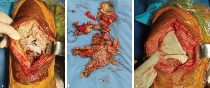
An anterior midline incision was made over the right knee after the patient was induced under spinal and epidural anesthesia. A medial parapatellar arthrotomy was performed, revealing inflamed synovial tissue on exposure. The tissue did not appear bloody or chocolate colored, which is typically associated with PVNS. The excised synovial tissue was sent for histopathological evaluation. Loosened implants were identified and removed with an osteotome and hammer, being careful to preserve as much bone stock as possible for the planned revision surgery. Given the aggressive synovial inflammation and the potential for an infectious cause, the decision was made to place a cement spacer. A synovectomy and thorough debridement of the remaining bone stock and surrounding tissues were completed.
A 5 mL and 10 mL syringe was utilized to prepare the intramedullary section of the cement spacer, which was mixed with gentamicin and vancomycin. The cement was manually shaped for the femoral condyles and the tibial plateau surrounding the inserted intramedullary section of the spacer (Fig. 2). The antibiotic-loaded spacer was used as a precautionary measure because the painful loose TKA and extensive synovial inflammation raised concern for an occult infective process despite normal inflammatory markers and a negative pre-operative aspiration culture. Once the cement had set, a pain relief solution was injected into the quadriceps, and the knee was reduced. The deformity was partially corrected using the cement spacer. Watertight closure of the arthrotomy and the wound was done. After the surgery, the patient was placed on anticoagulants, prophylactic antibiotics, and supplements for duration of 1 month, and was instructed to bear weight with assistance. After the initial stage of surgery, the patient experienced a significant reduction in pain. Sutures were removed on the 16th day after surgery.
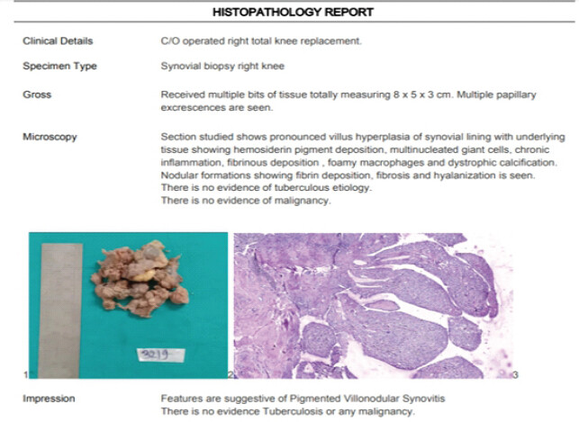
The histopathological analysis of the synovial tissue confirmed the diagnosis of PVNS (Fig. 3). The pathologist ruled out polyethylene debris-induced synovitis using established histopathological criteria. Specifically, polarized light microscopy showed no birefringent polyethylene particles, and no foreign body type granulomatous reaction typical of wear debris related pathology was identified. Instead, the synovium demonstrated a disease specific pattern of diffuse villous and nodular hyperplasia with prominent hemosiderin deposition, foamy macrophages, multinucleated giant cells, fibrin deposition, and fibrosis, which is diagnostic of PVNS.
The patient returned for a follow-up appointment after a month, during which his CBC, ESR, and CRP were retested and found to be within normal limits. He was scheduled for the second stage of revision TKA. The challenges for this surgery were to preserve bone stock, restore flexion and extension balance, preserve joint line, maintain coronal and sagittal plane alignment, maintain optimal ligament stability, maintain adequate bone implant interface reconstruction, and decide the amount of constriction needed.
The patient was prepared for surgery under spinal and epidural anesthesia. An incision was made along the anterior midline of the right knee. A medial parapatellar arthrotomy was performed, with no signs of infectious or inflamed tissues observed during exposure. The knee was subluxated, and the cement spacer was removed utilizing a drill, osteotome, and hammer while preserving as much of the bone stock as possible. A thorough debridement was carried out once more before proceeding with the revision TKA. It was decided to utilize metaphyseal sleeves, a tibial tray rotating platform, TC-3, a femoral stem, a tibial stem, and femoral adapters in the procedure. After thorough preparation, trial implants were inserted, the knee was reduced, and the stability, patellar tracking, range of motion, and ligament integrity were assessed and found to be satisfactory. The final implants were then placed, and following reduction, stability and integrity were evaluated, confirming satisfactory results. A watertight closure of the arthrotomy and the wound was completed. In the post-operative phase, the patient was prescribed anticoagulants, prophylactic antibiotics, and supplements for 1 month, along with instructions for weight-bearing with assistance.
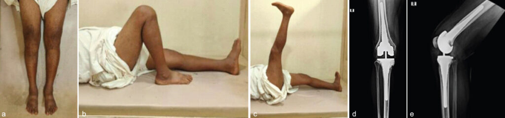
The deformity in the patient’s right knee was successfully corrected through revision right TKA, enabling him to walk without pain after the surgery. There were no complications at the surgical site, and no signs indicating a recurrence of PVNS were present. The SF-12 score, Hospital for Special Surgery Knee Score, Knee Society Score (KSS), and Oxford Knee Score for his right knee, assessed 1½ years post-surgery, demonstrated considerable improvement compared to his pre-operative values. His pre-operative and post-operative knee scores are presented in (Table 1), along with follow-up images in (Fig. 4). The HKA axis of his lower extremity has been corrected to neutral (180°).
Pre-operative and 1.5 years follow-up post-operative scores of 52-year-old male treated with the right revision total knee arthroplasty
| Score | Pre-operative score | Interpretation | Post-operative score | Interpretation |
|---|---|---|---|---|
| Short form score (SF-12) | ||||
| Physical component summary | 23.99938 | Below average | 56.57706 | Above average |
| Mental component summary | 19.06444 | Below average | 60.75781 | Above average |
| Hospital for special surgery knee score | 13 | Poor | 96 | Excellent |
| Knee society score | 6 | Poor | 90 | Excellent |
| Oxford knee score | 12 | Poor | 48 | Excellent |
Discussion
The failure of TKA can result in significant bone loss around the implant, which may compromise the stability and function of the ligaments. Careful surgical planning and precise identification of defects are essential for the successful execution of revision TKA.
PVNS is known to cause implant loosening through a mix of mechanical stress and bone degeneration due to the growth of abnormal synovial tissue. The PVNS tissue can damage the bone surrounding the implant, weaken the supporting structures, and ultimately result in aseptic loosening. This phenomenon is often observed in the tibial component, where the abnormal tissue can proliferate beneath the implant [21].
In cases of PVNS involving significant joint areas, extensive removal of the synovium is often necessary, which frequently compromises portions of the capsule and ligament structure. In these situations, implants that provide a higher degree of constraint are needed to maintain proper balance. Semi-constrained options, and even hinged systems, have been employed in such cases [2].
The Malefijt et al. [22] group enhanced the understanding of periprosthetic bone defects in the knee by presenting the differentiation between contained and uncontained defects, a concept that remains relevant today. Presently, the AORI classification proposed by Engh and Parks [23] is the most frequently cited system in the literature, representing a considerable improvement over earlier classifications by integrating aspects such as defect size, location, and soft-tissue involvement.
Hamlin et al. [24] documented four failures among 18 primary arthroplasties with PVNS. Three of these failures were attributed to aseptic loosening, which were addressed through revision arthroplasty, while one failure resulted from two recurrences of PVNS that required an above knee amputation.
Houdek et al. [25] documented that complications led to revisions in 10 out of 48 primary arthroplasties involving PVNS. Recurrence occurred in six patients, who underwent synovectomy and revision arthroplasty in four cases, local excision in one case, and transfemoral amputation in one case. In total, 25 patients faced at least one complication, with the most common being a decreased range of motion in the knee. The average KSS and functional score showed considerable improvement post-surgery (P < 0.001).
Chung and Park [21] reported a comparable case of TKA revision due to loosening of the tibial component in a single patient diagnosed with PVNS. A radiolucent lesion was identified beneath the tibial component during the patient’s last follow-up assessment before revision surgery. During the revision surgery, there was localized growth of synovial tissue with significant pigmentation observed around the anteromedial area of the tibial component. The tibial component was replaced after excising the abnormal tissues. The polyethylene’s articulating surface remained intact. A 5-mm thick medial metal block and an extension stem were utilized in the revision procedure. Histopathological examination of the abnormal tissue showed characteristic features of PVNS.
Certain principles must always be followed when dealing with bone loss during revision TKA. Extreme care must be taken to preserve as much host bone as possible while removing the implant. The treatment strategy will vary based on the remaining bone structure. It is essential to restore the joint line to achieve a stable and well-functioning knee. It is important to use stemmed components to divert stress from the joint line when addressing any periarticular concerns that involve more than a simple prosthetic enhancement. Although this topic has been examined in many studies and reviews, incorporating a stem in TKA, whether for primary or revision surgeries, can improve mechanical stability but may also result in stress shielding along the length of the stem. Both experimental and computational studies suggest that the addition of a stem reduces proximal tibial strain [11,13].
Radiotherapy is an important treatment for PVNS, improving local control rates in both adjuvant therapy and after incomplete resections. Nevertheless, the primary goal should always be to attain the most complete resection possible. For D-PVNS, it is recommended to deliver a total dose of 36–40 Gy using conventional fractionation (5 × 1.8–2.0 Gy/week), while for L-PVNS, lower total doses ranging from 20 to 36 Gy are sufficient. Employing computed tomography-based 3D radiation planning is advisable because the target area in joints must encompass the entire synovial space [26].
It is important to emphasize that a two-stage revision is not routinely indicated for PVNS-associated TKA failure. In the present case, the staged approach was selected because the patient had a painful and grossly loose TKA with extensive synovial inflammation and substantial AORI type III bone loss, while occult periprosthetic joint infection could not be excluded with complete certainty at the time of the first operation. The antibiotic-loaded cement spacer was therefore used as a precautionary measure while obtaining definitive histopathological and microbiological assessment. The subsequent diagnosis of PVNS should not be interpreted as an indication for staged revision in PVNS itself. In patients with confirmed PVNS and no clinical or laboratory suspicion of infection, thorough synovectomy combined with appropriate revision arthroplasty may be performed in a single stage when technically feasible.
Conclusion
A precise and timely diagnosis of PVNS of the knee is essential along with thorough pre-operative imaging and complete surgical excision to reduce the likelihood of recurrence and maintain joint function.
Successfully managing bone defects during TKA revisions is vital for obtaining favorable and lasting results. There are multiple management approaches available, each presenting distinct advantages and disadvantages, and there is no universal remedy for bone failure that applies to every case. Therefore, the decision-making process and the choice of method used should be customized to the patient; still, the procedure should focus on restoring bone stock in individuals, facilitating possible future revisions.
Clinical Message
This report is expected to encourage surgeons to consider pigmented villonodular synovitis (PVNS) in the differential diagnosis of patients who present with painful swelling after TKA and to recognize PVNS as a possible contributor to implant loosening. Importantly, PVNS by itself does not require a two-stage TKA; the staged approach in this case was selected because of the uncertainty surrounding occult infection and the severity of the failed TKA with extensive bone loss.
Conflict of Interest:
Nil
Source of Support:
Nil
Consent:
The authors confirm that informed consent was obtained from the patient for publication of this article
How to Cite this Article
Satvilkar MZNM, Malve SP, Vishwakarma UK. Revision Total Knee Arthroplasty in a Case of Operated Right Total Knee Arthroplasty with Implant Loosening and Pigmented Villonodular Synovitis. Journal of Orthopaedic Case Reports 2026 October;16(10): 45-51.
References
- Shafaghi R, Rodriguez O, Schemitsch EH, Zalzal P, Waldman SD, Papini M. A review of materials for managing bone loss in revision total knee arthroplasty. Mater Sci Eng C Mater Biol Appl 2019;104:109941. [Google Scholar] | [PubMed]
- Panciera A, Colangelo A, Di Martino A, Ferri R, Bulzacki Bogucki BD, Cecchin D. Total knee arthroplasty in pigmented villonodular synovitis osteoarthritis: A systematic review of literature. Musculoskelet Surg 2024;108:145-52. [Google Scholar] | [PubMed]
- Fazio A, Mazzella GG, Greco T, Comisi C, Fulchignoni C, Maccauro G. Diagnosis and treatment options in pigmented villonodular synovitis of the knee: A narrative review. J Clin Med 2025;14:5857. [Google Scholar] | [PubMed]
- Huten D, Pasquier G, Lambotte JC. Techniques for filling tibiofemoral bone defects during revision total knee arthroplasty. Orthop Traumatol Surg Res 2021;107:102776. [Google Scholar] | [PubMed]
- Bieganowski T, Buchalter DB, Singh V, Mercuri JJ, Aggarwal VK, Rozell JC. Bone loss in aseptic revision total knee arthroplasty: Management and outcomes. Knee Surg Relat Res 2022;34:30. [Google Scholar] | [PubMed]
- Jabbal M, Simpson AH, Walmsley P. Mechanisms of bone loss in revision total knee arthroplasty and current treatment options. Orthop Rev (Pavia) 2023;15:75359. [Google Scholar] | [PubMed]
- Hongvilai S, Tanavalee A. Review article: Management of bone loss in revision knee arthroplasty. J Med Assoc Thai 2012;95 Suppl 10:S230-7. [Google Scholar] | [PubMed]
- Bush JL, Wilson JB, Vail TP. Management of Bone Loss in Revision Total Knee Arthroplasty. Clin Orthop Relat Res 2006;452:186-92. [Google Scholar] | [PubMed]
- Aggarwal AK, Baburaj V. Managing bone defects in primary total knee arthroplasty: Options and current trends. Musculoskelet Surg 2021;105:31-8. [Google Scholar] | [PubMed]
- Lee YS, Chen AF. Managing bone loss in revision total knee arthroplasty. Ann Joint 2016;1:17-7. [Google Scholar] | [PubMed]
- Xie S, Conlisk N, Hamilton D, Scott C, Burnett R, Pankaj P. Metaphyseal cones in revision total knee arthroplasty: The role of stems. Bone Joint Res 2020;9:162-72. [Google Scholar] | [PubMed]
- Mozella AD, Barros Cobra HA. Bone Defects in Revision Total Knee Arthroplasty. Rev Bras Ortop 2020;56:138-46. [Google Scholar] | [PubMed]
- Rodríguez-Merchán EC, Gómez-Cardero P, Encinas-Ullán CA. Management of bone loss in revision total knee arthroplasty: Therapeutic options and results. EFORT Open Rev 2021;6:1073-86. [Google Scholar] | [PubMed]
- Ahsan P, Ezaz MS, Jahan I, Asma NS, Anjoom M. Total knee replacement in a young patient with valgus knee osteoarthritis: A case report. J Orthop Case Rep 2024;14:48-53. [Google Scholar] | [PubMed]
- Angerame MR, Jennings JM, Holst DC, Dennis DA. Management of bone defects in revision total knee arthroplasty with use of a stepped, porous-coated metaphyseal sleeve. JBJS Essent Surg Tech 2019;9:e14. [Google Scholar] | [PubMed]
- Khalifa AA, Said E, Refae H, Abdelaal AM. Knee Bony defects reconstruction in revision total knee arthroplasty for aseptic loosening. Orthop Res Online J 2019;5:1-7. [Google Scholar] | [PubMed]
- Lei PF, Hu RY, Hu YH. Bone defects in revision total knee arthroplasty and management. Orthop Surg 2019;11:15-24. [Google Scholar] | [PubMed]
- Marongiu G, Capone A, Verona M, Piovan G, Zorzi C, Kendoff D. The management of bone defects in revision knee arthroplasty: The role of porous metal cones and 3D-printed cones. Prosthesis 2023;5:1171-83. [Google Scholar] | [PubMed]
- Qiu YY, Yan CH, Chiu KY, Ng FY. Review article: Bone defect classifications in revision total knee arthroplasty. J Orthop Surg (Hong Kong) 2011;19:238-43. [Google Scholar] | [PubMed]
- Sculco PK, Flevas DA, Jerabek SA, Jiranek WA, Bostrom MP, Haddad FS. Management of bone loss in revision total knee arthroplasty: An international consensus symposium. HSS J 2024;20:141-81. [Google Scholar] | [PubMed]
- Chung BJ, Park YB. Pigmented villonodular synovitis after TKA associated with tibial component loosening. Orthopedics 2011;34:e418-20. [Google Scholar] | [PubMed]
- Malefijt MC, Van Kampen A, Slooff TJ. Bone grafting in cemented knee replacement. 45 primary and secondary cases followed for 2-5 years. Acta Orthop Scand 1995;66:325-8. [Google Scholar] | [PubMed]
- Engh GA, Parks NL. The management of bone defects in revision total knee arthroplasty. Instr Course Lect 1997;46:227-36. [Google Scholar] | [PubMed]
- Hamlin BR, Duffy GP, Trousdale RT, Morrey BF. Total knee arthroplasty in patients who have pigmented villonodular synovitis. J Bone Joint Surg Am 1998;80:76-82. [Google Scholar] | [PubMed]
- Houdek MT, Scorianz M, Wyles CC, Trousdale RT, Sim FH, Taunton MJ. Long-term outcome of knee arthroplasty in the setting of pigmented villonodular synovitis. Knee 2017;24:851-5. [Google Scholar] | [PubMed]
- Seegenschmiedt MH, Micke O, Niewald M, Mücke R, Eich HT, Kriz J. DEGRO guidelines for the radiotherapy of non-malignant disorders: Part III: Hyperproliferative disorders. Strahlenther Onkol 2015;191:541-8. [Google Scholar] | [PubMed]
© 2026 Journal of Orthopaedic Case Reports - Published by Indian Orthopaedic Research Group
About the Authors
-

Mohd Zafer Noor Mohd Satvilkar
-

Shekhar Prakash Malve
-

Utkarsh Kailashnath Vishwakarma
Related Articles in Journal of Orthopaedic Case Reports
 August 1, 2026 Bicruciate Ligament Avulsions, Lateral Meniscus Dual Root Avulsions – “Tibial Avulsion Tetrad” – with Menisco-capsular Injury – Floating Meniscus in Innocuous Looking Proximal Tibia Fracture
 July 1, 2026 Post-traumatic Hemosiderotic Synovitis Mimicking Pigmented Villonodular Synovitis in a Non-hemophilic Child: First Reported Case from the United Arab Emirates
 July 1, 2026 Lipoma Arborescens of the Knee: An Intraoperative Mimicker of Pigmented Villonodular Synovitis in Bilateral Knee Osteoarthritis – A Case Report
 July 1, 2026 Management of Chronic Osteomyelitis Bone Defect with Titanium Cage Combined with the Masquelet Technique: A Case Report