

Septic arthritis in neurologically impaired, non-ambulatory children may manifest with subtle or atypical clinical features, delaying diagnosis and predisposing to rare but catastrophic complications such as physeal separation. A high index of suspicion, early imaging, and prompt surgical and antimicrobial management are essential to prevent irreversible growth disturbances and long-term functional disability.
Sandeep Kumar, Central Institute of Orthopaedics, Vardhman Mahavir Medical College and Safdarjung Hospital, New Delhi, India. sandeep9675@gmail.com
Abstract
Introduction: Septic arthritis (SA) in children is a medical emergency that may lead to serious complications, including damage to the growth plate and long-term functional impairment. One of the rarest but most significant complications is physeal slip, particularly in non-ambulatory children with underlying neurological disorders. Early recognition is often challenging due to atypical clinical presentations, especially in patients with altered muscle tone or global developmental delay.
Case Report: This report highlights the diagnostic and therapeutic considerations in a case of distal femoral physeal separation secondary to SA in a neurologically impaired pediatric patient. The absence of classic symptoms such as fever, erythema, or warmth may delay clinical suspicion. Imaging modalities, including radiographs and ultrasound, are critical in identifying joint effusion and physeal disruption. Laboratory markers such as elevated erythrocyte sedimentation rate and C-reactive protein can support the diagnosis. Definitive management requires urgent surgical drainage and debridement, stabilization of the physis, and a prolonged course of targeted intravenous antibiotics.
Conclusion: This case underscores the importance of early multidisciplinary evaluation in high-risk populations. Prompt diagnosis and timely intervention are essential to prevent long-term complications such as deformity, joint instability, or growth disturbance.
Keywords: Cerebral palsy, septic arthritis, physeal slip, West syndrome, pediatric orthopedics.

Acute septic arthritis (SA) in children is most commonly hematogenous in origin [1,2]. The sluggish blood flow in metaphyseal capillaries of growing bones makes them particularly vulnerable to bacterial seeding from systemic infections or minor trauma [1,2]. Starr proposed that osteomyelitic pathogens spread hematogenously to the juxta-epiphyseal capillaries of long bones, attributing infection to reduced host resistance rather than physiological localization [3]. Large joints of the lower extremities – especially the hip, knee, and ankle – are most frequently affected, with Staphylococcus aureus and respiratory pathogens being common causative agents.
Early diagnosis and management are critical, as delayed treatment can lead to long-term complications such as deformity, arthritis, or joint instability [4]. In children with cerebral palsy (CP), diagnosis may be delayed due to atypical presentations caused by altered tone, limited communication, and overlapping neurological symptoms. In such cases, inflammatory markers and imaging modalities such as ultrasound and magnetic resonance imaging (MRI) are essential for timely identification.
Although extremely rare, physeal slip secondary to SA has been reported [5,6]. This case describes a unique instance of distal femoral physeal separation in a child with spastic CP. It highlights the importance of maintaining clinical suspicion for unusual complications in children with neurological impairments and underscores the role of early multidisciplinary intervention.
The patient’s parents provided informed consent for the publication of this case report. A 5-year-old male child was brought to the emergency department by his mother with complaints of pain and swelling in the left knee and distal thigh for the past 4 days. The child was under ongoing pediatric care for global developmental delay, West syndrome, and spastic CP.
On examination, there was mild swelling over the left knee and distal thigh, with localized tenderness on palpation. Although there was no evident local rise in temperature, the passive range of motion at the knee joint was preserved but painful.
Initial blood investigations revealed elevated inflammatory markers, including erythrocyte sedimentation rate (ESR) and C-reactive protein (CRP). Ultrasound of the affected region showed a heterogeneous hypoechoic collection communicating with the joint cavity and mild synovial thickening, suggestive of joint effusion with possible infection. Radiographic evaluation revealed a distal femoral physeal separation (Fig. 1).
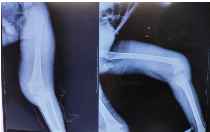
Figure 1: Pre-operative radiograph showing distal femur physeal separation.
Following pediatric consultation, empirical antibiotic therapy was initiated while awaiting surgical clearance, given the patient’s background of CP, global developmental delay, and seizure disorder. The child subsequently underwent surgical intervention under general anesthesia. Intaroperatively, the femur also showed distal femur physeal separation (Fig. 2).
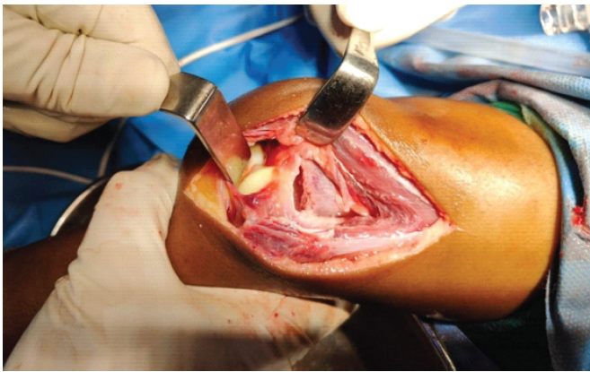
Figure 2: Intraoperative picture showing distal femur physeal separation.
The procedure included open arthrotomy, joint debridement, saucerization of the distal femur, and Kirschner wire fixation (Fig. 3). Post-operative radiographs were obtained, which showed distal femur physeal reduction with Kirschner wire fixation (Fig. 4 and 5).
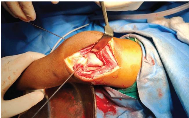
Figure 3: Intraoperative picture showing open reduction and Kirschner wire fixation.
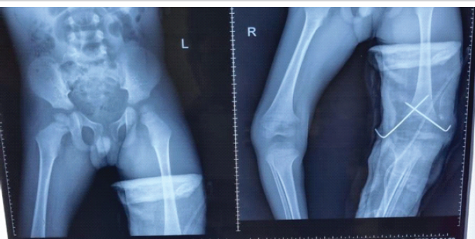
Figure 4: Post-operative anteroposterior radiograph showing fixation with Kirschner wires.
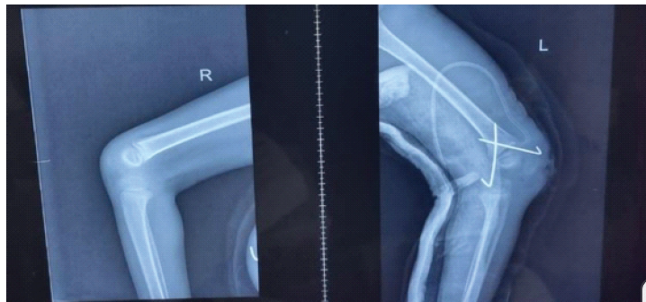
Figure 5: Post-operative lateral radiograph showing fixation with Kirschner wires.
Approximately 15 mL of blood-stained purulent fluid was drained from the knee joint. Additional purulent material was noted discharging from a cortical window created over the distal femur. Intraoperative samples were sent for GeneXpert testing, histopathological examination, acid-fast bacilli staining and culture, Gram stain, and routine pus culture and sensitivity.
Postoperatively, the patient was transferred to the pediatric department for a 6-week course of intravenous antibiotics. Notably, all microbiological and histopathological investigations returned negative for any specific organism or pathology.
The child’s post-operative recovery was uneventful. He was monitored in the pediatric department and completed a 6-week course of intravenous antibiotics without any complications. On follow-up in the orthopedics outpatient department, the patient showed no signs of fever, pain, or swelling in the affected limb. Although orthopedic symptoms had resolved, the child continued to receive ongoing care from the pediatric department for CP, West syndrome, and seizure disorder.
SA in children is a serious condition that, if not diagnosed and managed promptly, can result in various complications including growth plate damage, joint destruction, and long-term functional impairment. Among these, physeal separation remains one of the rarest complications [5]. Physeal separations are uncommon and may involve either the proximal or distal femoral physis [5]. The simultaneous occurrence of a physeal fracture and infection is particularly rare, with complete epiphyseal separation described as an unusual sequela of neonatal osteomyelitis or SA [6]. The hip joint is most frequently implicated in these cases [6].
Differential diagnoses of pathologic epiphyseal slip include birth trauma, non-accidental injury, and scurvy [6]. In children with CP, scurvy may occur due to nutritional deficiencies, and epiphyseal separation can present with characteristic clinical and radiological features [7]. Subperiosteal hematomas, gingival bleeding, and metaphyseal radiodensities – particularly the “white line of Frenkel,” which represents the scorbutic lattice – are classic findings [7,8]. Children with developmental delays, restrictive diets, or physical disabilities are at increased risk for nutritional deficiencies such as scurvy [8].
In neurologically impaired children, including those with CP, diagnosing SA is particularly challenging. Clinical signs may be masked by spasticity, communication barriers, or baseline motor deficits. In such scenarios, laboratory markers – such as elevated ESR and CRP – and imaging studies, including ultrasound and MRI, are crucial in making a timely diagnosis. Kocher’s criteria, which incorporate both clinical and biochemical parameters, are useful in distinguishing SA from other inflammatory or infectious processes [9].
There are only limited reports of SA-induced physeal separation in children(Table 1).
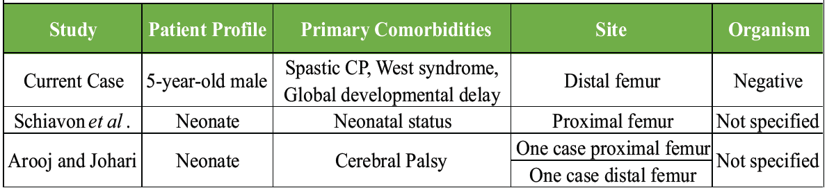
Table 1: Comparison with similar cases in the literature
Schiavon et al. described a case of septic physeal slip in a neonate managed successfully with open debridement and Kirschner wire fixation [10]. Arooj and Johari and Gupta et al. reported similar cases in children with CP and underlying scurvy [6,8]. However, to date, very few cases describe SA causing physeal slip in children with CP without nutritional deficiency, making our case particularly unique.
In our patient, the presence of CP and global developmental delay obscured the clinical presentation. Nonetheless, elevated inflammatory markers and supportive imaging findings facilitated early diagnosis. The child was successfully managed with surgical debridement, Kirschner wire fixation, and intravenous antibiotics, with an uneventful recovery.
Cases by Schiavon et al. (proximal femur physeal separation) and by Arooj and Johari (one case of proximal femur and one case of distal femur physeal slip) have been mentioned above. The unique aspect of the present case is SA leading to distal femoral physeal slip in a CP patient without documented nutritional deficiency.
Given the risk of permanent growth disturbance, we feel that patients with similar injuries should undergo regular radiographic follow-up every 3–6 months to monitor for physeal bridging, limb length discrepancy, or angular deformities.
This case underscores the importance of maintaining a high index of suspicion for rare complications in high-risk pediatric populations and highlights the value of multidisciplinary management in achieving optimal outcomes.
Despite its clinical utility, the study is primarily limited by its nature as a single case report, which restricts the ability to generalize these findings or establish definitive causation. A significant diagnostic gap exists because all microbiological investigations remained negative, preventing the identification of a specific causative organism, despite frank intraoperative purulence. In addition, the lack of MRI imaging, often considered the gold standard for detecting early osteomyelitis or subtle physeal involvement, is a limitation in the diagnostic workup. Due to the lack of long-term follow-up, potential growth disturbances, limb length discrepancies, or angular deformities could not be systematically assessed or monitored.
This case underscores the diagnostic complexity of SA in neurologically impaired children and emphasizes the necessity for heightened clinical vigilance, early advanced imaging, and prompt multidisciplinary intervention to avert rare yet devastating complications, including physeal separation and permanent growth disturbance.
In neurologically impaired, non-ambulatory children, septic arthritis can progress silently yet aggressively, predisposing to rare growth-plate injuries. Early recognition through vigilant clinical assessment, prompt imaging, and immediate multidisciplinary intervention is imperative to prevent irreversible deformity and lifelong functional disability.
References
- 1. Krogstad P. Osteomyelitis and septic arthritis. In: Feigin RD, Cherry JD, editors. Textbook of Pediatric Infectious Diseases. 6th ed. Philadelphia, PA: Saunders; 2009. p. 725-48. [Google Scholar] [PubMed]
- 2. Pääkkönen M, Peltola H. Management of a child with suspected acute septic arthritis. Arch Dis Child 2012;97:287-92. [Google Scholar] [PubMed]
- 3. Starr CL. Acute hematogenous osteomyelitis. Arch Surg 1922;4:567-87. [Google Scholar] [PubMed]
- 4. Haruno LS, Thompson CR, Osorio M, Risoli TJ, Schrader T, Rosenfeld SB. Group A streptococcal septic hip arthritis in a child with spastic triplegic cerebral palsy. J Am Acad Orthop Surg Glob Res Rev 2021;5:e20.00228. [Google Scholar] [PubMed]
- 5. Mangurten HH, Puppala B, Knuth A. Neonatal distal femoral physeal fracture requiring closed reduction and pinning. J Perinatol 2005;25:216-9. [Google Scholar] [PubMed]
- 6. Arooj AJ, Johari AN. Epiphyseal separations after neonatal osteomyelitis and septic arthritis. J Pediatr Orthop 2000;20:554-9. [Google Scholar] [PubMed]
- 7. Quiles M, Sanz TA. Epiphyseal separation in scurvy. J Pediatr Orthop 1988;8:223-5. [Google Scholar] [PubMed]
- 8. Gupta S, Kanojia R, Jaiman A, Sabat D. Scurvy: An unusual presentation of cerebral palsy. World J Orthop 2012;3:58-61. [Google Scholar] [PubMed]
- 9. Bisht RU, Burns JD, Smith CL, Kang P, Shrader MW, Belthur MV. The modified Kocher criteria for septic hip: Does it apply to the knee? J Child Orthop 2022;16:233-7. [Google Scholar] [PubMed]
- 10. Schiavon R, Borgo A, Micaglio A. Septic physeal separation of proximal femur in a newborn. J Orthop Traumatol 2009;10:105-10. [Google Scholar] [PubMed]
Related Articles in Journal of Orthopaedic Case Reports
 January 1, 2026 From Hip Effusion to Hemophilia: Unveiling the Cause of Pain in 6-Month-Old Infant. A Case Report
 November 1, 2024 Comparison of Percutaneous Versus Open Hamstring Lengthening in Patients of Spastic Diplegic Cerebral Palsy – A Randomized Controlled Trial
 November 1, 2026 Septic Arthritis of the Hip Complicating a Non-Perforated Acute Appendicitis – A Case Report and Review of the Literature
 November 1, 2026 Identical Twins with Legg-Calve-Perthes Disease: A Report of Two Cases