


Introduction
Tibial plateau fractures are a common peri-articular fractures of the proximal tibia [1]. They may result from either high- or low-energy trauma. High-energy injuries occur predominantly in adults following motor vehicle collisions or pedestrian accidents, whereas low-energy injuries are more commonly observed in patients with osteoporotic bone [2].
According to the Schatzker and Arbeitsgemeinschaft für Osteosynthese/Orthopaedic Trauma Association classification, tibial plateau fractures could be classified into complete and partial articular fractures. Complete fractures are bicondylar fractures usually caused by high-energy trauma, while partial fractures are usually caused by low-energy trauma [1,2,3].
The management of complex tibial plateau fractures is challenging and aims to restore the articular surface and limb alignment, achieve stable fixation that permits early knee mobilization, and address associated soft-tissue injuries [4]. Treatment options include open reduction and internal fixation with plates and screws or, in selected cases, external fixation [5,6,7]. Regardless of its classification, surgery remains the gold standard for its management, which consists of open reduction and internal fixation (ORIF) with plates and screws [5].
In this case, we describe a patient with a complete tibial plateau articular fracture that was treated conservatively with satisfactory clinical and functional outcomes.
Case Report
History
A 36-year-old previously healthy man, a firefighter, presented to the emergency department following a motorcycle-car collision. He presented severe pain in his left knee, especially on weight-bearing. The patient has been a waterpipe smoker for 3 years, does not suffer from any chronic or hereditary diseases, and has never undergone any surgery. The patient was 178 cm tall, weighed 76 kg, and had a body mass index of 24.0 kg/m2.
Physical examination
Examination of the knee revealed slight swelling and erythema, with no open wounds. Moderate tenderness was found during palpation. The patient could neither flex nor extend his knee. Peripheral pulses were detected, and manual examination of the foot and neuromuscular screening were normal. No soft tissue injury was found on physical exam (Lachman test was negative; McMurray and drawer tests couldn’t be done).
Investigation
Plain radiographs of the left knee revealed a linear, non-displaced metaphyseal-epiphyseal fracture of the proximal tibia.
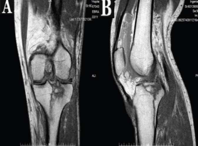
One week after the injury, magnetic resonance imaging of the left knee (Fig. 1a and b) demonstrated a large, impacted, comminuted fracture of the lateral tibial plateau, with fracture lines extending into the metaphyseal and epiphyseal regions involving the intercondylar eminence and longitudinal posterior medial tibial diaphysis. No associated soft-tissue injuries were identified.
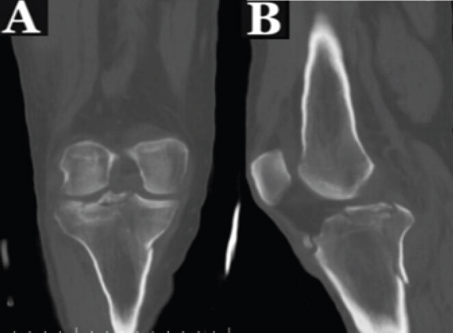
Two weeks after the injury, computed tomography (CT) demonstrated a non-displaced comminuted fracture of the proximal tibia involved the lateral and medial tibial plateau. The fracture also involved the intercondylar eminence and extended posteromedially into the proximal tibia, findings consistent with a Schatzker type V tibial plateau fracture (Fig. 2a and b).
Treatment
On the day of injury, a cricket-pad splint was applied for temporary immobilization, and surgical treatment was recommended. After being fully informed about the proposed surgical treatment and the potential risks and benefits of both surgical and non-operative management, the patient declined surgery. Therefore, a hinged knee brace was applied, initially allowing 0–30° of knee motion, with no to minimal weight-bearing for 3 weeks. The patient was also instructed to wear the brace while sleeping during this period.
The range of motion permitted by the hinged knee brace was progressively increased by 30° every 3 weeks until full knee flexion was achieved. Partial weight-bearing with two crutches was initiated at 3 weeks and progressively advanced according to clinical and radiographic findings.
Follow-up
A CT scan performed at the 1-month follow-up showed an unchanged fracture pattern, with no secondary displacement. The patient continued mobilization and weight-bearing with the assistance of two crutches and a knee brace.
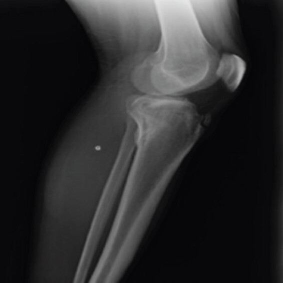
At the 4-month follow-up, full range of motion had been achieved, and radiographs demonstrated evidence of fracture consolidation, allowing removal of the brace (Fig. 3). At that time, the patient was clinically well, was able to kneel with only mild discomfort, and had returned to work.
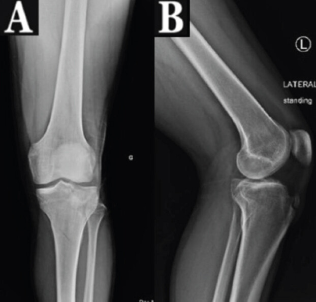
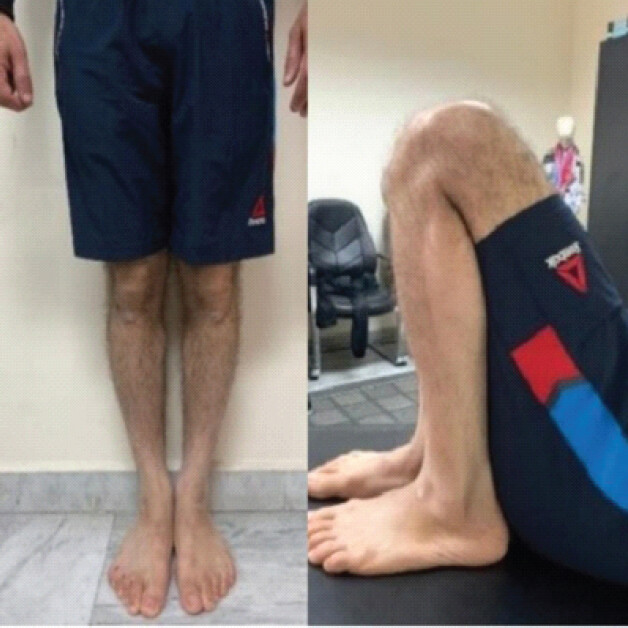
At the 2-year follow-up, radiographs of the left knee showed complete fracture healing with maintained alignment (Fig. 4); the patient demonstrated a satisfactory clinical and functional outcome, with knee flexion of 137° and an extension deficit of only 2° (Fig. 5). He had fully returned to his normal activities of daily living. The Knee Society Score was 89, and the Oxford Knee Score was 47.
Discussion
Tibial plateau fractures account for approximately 1–2% of all fractures and represent a heterogeneous group of injuries, ranging from low-energy split-depression fractures in osteoporotic bone to high-energy bicondylar fractures associated with severe comminution and soft-tissue injury [2].
The widely used Schatzker classification divides these fractures into six types according to fracture morphology, including fracture-line orientation and articular depression. Schatzker type V and VI fractures represent complex bicondylar injuries and are generally managed surgically. ORIF with plates and screws remains the standard treatment, while external fixation, including hybrid and circular constructs, may be used in selected cases, particularly when significant soft-tissue compromise is present [4,5,6].
Lee et al. [5] reported 65 patients with high-energy Schatzker Type V and VI fractures treated surgically. Partial weight-bearing was initiated 2–3 months postoperatively after radiographic evidence of callus formation. At follow-up, mean knee motion ranged from a 3.9° extension deficit to 136.3° of flexion. According to the Oxford Knee Score, outcomes were excellent in 33% of patients and good in the remaining patients.
External fixation has also demonstrated satisfactory outcomes. Babis et al. [6] reported 33 Schatzker Type V and VI fractures treated with hybrid external fixation. The frame was removed after a mean of 3.4 months, and 26 patients achieved satisfactory knee function and alignment without pain or instability. At 1 year, mean flexion was 115°, with a mean extension deficit of 5° and a mean Knee Society Score of 81.6. Pin-tract and deep infections occurred in 9.1% and 3% of patients, respectively.
Similarly, Ferreira and Marais [7] reported 54 high-energy bicondylar fractures managed with circular external fixation. Mean knee flexion was 100.5°, and the mean Knee Society Score was 81.6. Pin-site infection was the most frequent complication, occurring in 23.9% of patients.
Although satisfactory outcomes have been reported, surgical treatment carries potential risks and complications, including infection, prolonged hospital stay, knee stiffness, non-union, and the need for additional surgical procedures [8].
In contrast to these surgical approaches, our patient was managed entirely non-operatively, without incisions, percutaneous wires, screws, or external fixation. Treatment consisted of a knee motion brace and progressive weight-bearing, with discontinuation of the brace after 4 months. At the 2-year follow-up, the patient achieved 137° of knee flexion with only a 2° extension deficit, an Oxford Knee Score of 47, and a Knee Society Score of 89, indicating an excellent functional outcome.
The favorable mid-term outcome observed in this case raises the possibility that, in carefully selected patients with complex tibial plateau fractures in whom surgery is declined or contraindicated because of local or systemic factors, non-operative management may occasionally represent a viable option.
Early mechanical loading may also have contributed to fracture healing; mechanical loading is sensed by osteocytes and modulates cellular signaling pathways involved in bone formation [9,10].
Williamson et al. [11] evaluated 90 patients with tibial plateau fractures managed with restricted or minimal weight-bearing versus immediate weight-bearing as tolerated. No fixation failures occurred in either group, and although no significant difference in healing rate was demonstrated, the authors supported immediate weight-bearing in appropriately selected patients. These findings suggest that early loading may be safe under certain circumstances [12,13]. However, evidence derived from surgically stabilized fractures cannot be directly extrapolated to non-operatively managed Schatzker Type V or VI fractures.
To the best of our knowledge, reports of successful non-operative management of complex bicondylar tibial plateau fractures using a knee motion brace with early weight-bearing are extremely limited [3,4,8,11]. Non-operative management is rarely indicated due to concerns regarding fracture displacement, delayed union, joint instability, and post-traumatic osteoarthritis. Nevertheless, avoiding surgery eliminates procedure-related complications, including wound complications, deep infection, and pin-tract infection [9]. However, the favorable outcome observed in this patient should not be interpreted as evidence that conservative management is equivalent or superior to established surgical treatment.
One of the major long-term complications following tibial plateau fractures is the development of post-traumatic osteoarthritis, which is particularly relevant in younger patients because of their longer life expectancy and greater cumulative joint loading [14,15]. The development of post-traumatic osteoarthritis is influenced by several factors, including residual joint instability, malalignment, and articular incongruity [15,16]. In the present case, some degree of articular incongruity persisted despite the satisfactory mid-term clinical and functional outcome, emphasizing the importance of continued long-term clinical and radiographic surveillance.
This report has several limitations. As a single case, its findings cannot be generalized to the broader population of patients with complex bicondylar tibial plateau fractures. The absence of a control group prevents direct comparison with surgical management, and the favorable outcome may have been influenced by patient- and fracture-specific factors. Furthermore, this case cannot establish the safety or effectiveness of early weight-bearing in unstable or displaced tibial plateau fractures. Longer-term follow-up is also required to assess the durability of the clinical and radiographic outcomes.
Conclusion
Complex Schatzker Type V bicondylar tibial plateau fractures are generally managed surgically because of their complex fracture pattern and the risk of displacement, joint instability, malalignment, and post-traumatic osteoarthritis. Nevertheless, the satisfactory mid-term clinical, functional, and radiographic outcome observed in this case suggests that carefully monitored non-operative management may occasionally represent a reasonable alternative in highly selected patients who decline surgery or have local or systemic contraindications to surgical treatment. Close clinical and radiographic follow-up is essential to monitor fracture healing, alignment, joint stability, and functional recovery. However, the favorable outcome of a single case should not be interpreted as evidence of equivalence to established surgical treatment, and longer-term follow-up is required to assess the durability of the outcome, particularly with regard to the development of post-traumatic osteoarthritis.
Clinical Message
Complex Schatzker Type V bicondylar tibial plateau fractures generally require surgical management. However, when surgery is declined or contraindicated, carefully selected patients may be considered for non-operative treatment with close clinical and radiographic monitoring, recognizing that evidence supporting this approach remains limited.
Conflict of Interest:
Nil
Source of Support:
Nil
Consent
The authors confirm that informed consent was obtained from the patient for publication of this article
How to Cite this Article
El-Osta B, Hamdan A, Al Ali M, Sciarretta FV, Zmerly H. Conservative Management of a Complex Schatzker Type V Bicondylar Tibial Plateau Fracture: A Case Report With 2-Year Follow-Up. J Orthop Case Rep. 2026 November;16(11): 221-226. doi: 10.13107/jocr.2026.v16.i11.8448.
References
- Prat-Fabregat S, Camacho-Carrasco P. Treatment strategy for tibial plateau fractures: An update. EFORT Open Rev 2017;1:225-32. [Google Scholar] | [PubMed]
- Elsoe R, Larsen P, Nielsen NP, Swenne J, Rasmussen S, Ostgaard SE. Population-based epidemiology of tibial plateau fractures. Orthopedics 2015;38:e780-6. [Google Scholar] | [PubMed]
- Mthethwa J, Chikate A. A review of the management of tibial plateau fractures. Musculoskelet Surg 2018;102:119-27. [Google Scholar] | [PubMed]
- Franulic N, Koch M, Muñoz JT, Olivieri R, Gaggero N, Pesantez R. Current concepts in the management of high-energy tibial plateau fractures: A narrative review. EFORT Open Rev 2026;11:676-90. [Google Scholar] | [PubMed]
- Lee TC, Huang HT, Lin YC, Chen CH, Cheng YM, Chen JC. Bicondylar tibial plateau fracture treated by open reduction and fixation with unilateral locked plating. Kaohsiung J Med Sci 2013;29:568-77. [Google Scholar] | [PubMed]
- Babis GC, Evangelopoulos DS, Kontovazenitis P, Nikolopoulos K, Soucacos PN. High energy tibial plateau fractures treated with hybrid external fixation. J Orthop Surg Res 2011;6:35. [Google Scholar] | [PubMed]
- Ferreira N, Marais LC. Bicondylar tibial plateau fractures treated with fine-wire circular external fixation. Strategies Trauma Limb Reconstr 2014;9:25-32. [Google Scholar] | [PubMed]
- Gálvez-Sirvent E, Ibarzábal-Gil A, Rodríguez-Merchán EC. Complications of the surgical treatment of fractures of the tibial plateau: Prevalence, causes, and management. EFORT Open Rev 2022;7:554-68. [Google Scholar] | [PubMed]
- Kubiak EN, Beebe MJ, North K, Hitchcock R, Potter MQ. Early weight bearing after lower extremity fractures in adults. J Am Acad Orthop Surg 2013;21:727-38. [Google Scholar] | [PubMed]
- Fändriks A, Möller M, Tranberg R, Erichsen A. Insights into the rehabilitation journey - patients' experiences following tibial plateau fractures. Int J Qual Stud Health Well-Being 2026;21:2636758. [Google Scholar] | [PubMed]
- Williamson M, Iliopoulos E, Jain A, Ebied W, Trompeter A. Immediate weight bearing after plate fixation of fractures of the tibial plateau. Injury 2018;49:1886-90. [Google Scholar] | [PubMed]
- Joslin CC, Eastaugh-Waring SJ, Hardy JR, Cunningham JL. Weight bearing after tibial fracture as a guide to healing. Clin Biomech (Bristol) 2008;23:329-33. [Google Scholar] | [PubMed]
- Singh S. Weight bearing decisions after tibial plateau fractures: A literature review based on clinical perspective for physiotherapists in India. Physiother Res Int 2026;31:e70277. [Google Scholar] | [PubMed]
- Moreno-Degante L, Pettet-Ruiz B, Ponce-De-Léon-Dominguez J, Torres-Casado E, Chiquini-Ramírez P, Reyes-García HM. Radiographic osteoarthritis and patient-reported outcomes one year following tibial plateau fracture surgery. Cureus 2026;18:e112364. [Google Scholar] | [PubMed]
- Softness KA, Murray RS, Evans BG. Total knee arthroplasty and fractures of the tibial plateau. World J Orthop 2017;8:107-14. [Google Scholar] | [PubMed]
- Pander P, Fransen BL, Hagemans FJ, Keijser LC. Functional outcome of total knee arthroplasty following tibial plateau fractures: A systematic review. Arch Orthop Trauma Surg 2023;143:1265-74. [Google Scholar] | [PubMed]
© 2026 Journal of Orthopaedic Case Reports - Published by Indian Orthopaedic Research Group
About the Authors
-

Bassel El-Osta
-

Alaa Hamdan
-

Mariam Al Ali
-

Fabio Valerio Sciarretta
-

Hassan Zmerly
Related Articles in Journal of Orthopaedic Case Reports
 November 1, 2026 Second and Third Carpometacarpal Joint Dislocation in a Polytrauma Patient: Successful Closed Reduction and 2-Year Follow-Up
 November 1, 2026 Incidentally Detected Osseous Hemangioma of the Femoral Head: Conservative Management of a Rare Case with Review of the Literature
 October 1, 2026 A Prospective Clinical and Radiological Assessment of Osteochondral Autograft Transfer for International Cartilage Repair Society Grade IV Osteochondral Lesions of the Knee
 October 1, 2026 Functional Outcome of Lucerne Cast in Patients with Metacarpal Fracture with or without Proximal Phalanx Fracture: An Observational Prospective Study