


Introduction
Intertrochanteric fractures in elderly patients are very common in orthopedic practice [1]. Early fixation of the fracture allows early mobilization and rehabilitation, which eventually leads to early union, return to the pre-injury activity status, improved gait, and a decreased rate of fracture-related complications [2,3].
These fractures are commonly treated on a traction table. The table has a foot attachment that allows traction to be applied to the injured limb and permits manipulation of the distal fragment with respect to elevation, adduction, and abduction in order to achieve fracture reduction without an additional assistant.
An intertrochanteric fracture in a patient with a below-knee amputation (BKA) is a rare entity. Surgical management and fracture fixation in such patients are associated with various challenges, including positioning the shortened limb on a fracture table, applying traction to the lower limb, maintaining that traction until definitive fixation, and manipulating the distal stump to obtain satisfactory valgus and rotational alignment [4,5]. This case report describes a method of applying traction for reduction of an intertrochanteric femur fracture on a standard traction table using the patient’s own BK prosthesis, in a 69-year-old woman with an ipsilateral BKA.
Case Report
A 69-year-old woman presented with a history of a fall with trauma to the right hip region, complaining of pain and inability to bear weight and walk. She had undergone a BKA of the same leg 4 years earlier before the current injury for diabetic foot. Before the fall, she was able to walk and perform her day-to-day activities freely with the use of a BK prosthesis.
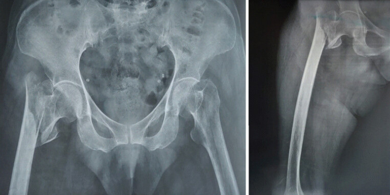
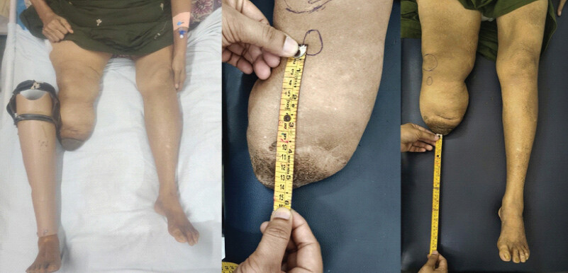
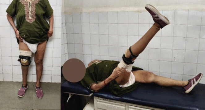
On examination, the right BK stump was externally rotated, and an active straight leg raise was not possible. The amputation stump was healthy and well healed, with no skin breach or tenderness. The residual limb measured approximately 15 cm from the tibial tubercle to the end of the stump. There was a limb shortening of approximately 30 cm due to amputation (Fig. 1). The knee had a flexion deformity of approximately 10° with further flexion of approximately 120°. The skin was well healed, supple, and free of pressure areas or ulceration. Radiographs showed a right intertrochanteric fracture with impaction and varus angulation (Fig. 2). The fracture was classified as AO/OTA 31-A2.2, with the presence of a lesser trochanter fragment. According to the Evans classification, it was categorized as Type I, Group 2, indicating a displaced intertrochanteric fracture that achieved a stable reduction with restoration of medial cortical apposition.
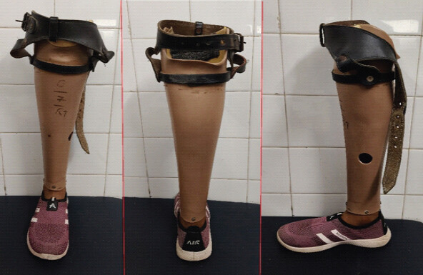
The patient had a well-fitted right transtibial (BK) prosthesis consisting of a prosthetic socket accommodating the residual limb, an external shank/pylon, and a prosthetic foot (Fig. 3). The socket of the prosthesis was to be secured around the stump of the residual limb using adjustable velcro straps. The socket provided adequate cushioning and rotational stability. The prosthetic foot had a relatively broad, stable base and was suitable for weight bearing and ambulation. The patient was accustomed to using this prosthesis for routine activities and was able to independently don and doff it.
Surgical management
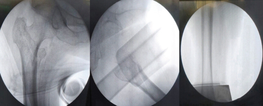
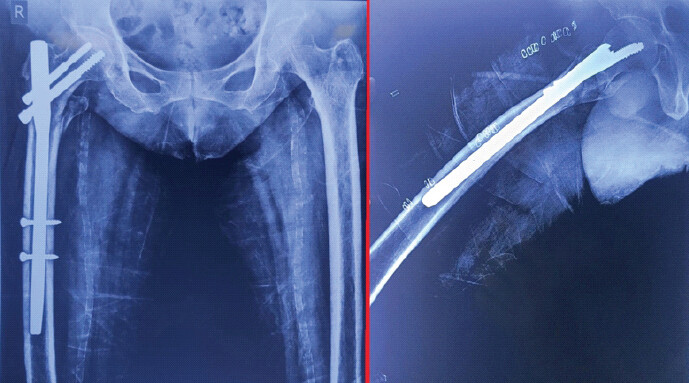
Closed reduction and internal fixation with a proximal femoral nail was planned. Achieving appropriate positioning and traction on the fracture table was technically demanding because of the shortened amputated limb. Special attention was therefore required to obtain stable positioning, traction, and adequate fracture reduction during the procedure. We planned to use the patient’s own BK prosthesis for positioning and traction application on the traction table. Intraoperative C- arm imaging showed a reduced fracture with good alignment (Fig. 4). The fracture was then fixed with a 135° proximal femoral nail measuring 11 mm by 240 mm. Post-operative radiographs showed good alignment and stable fixation with the implant (Fig. 5). There were no skin, soft-tissue, or stump-related complications attributable to the traction technique.
Traction technique
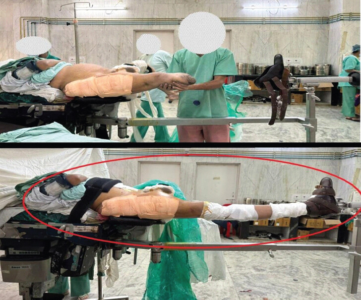
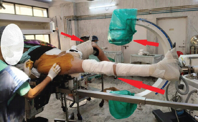
The residual limb was gently seated within the prosthetic socket and secured using the prosthesis’s Velcro straps. The portion of the residual limb proximal to the socket was additionally secured to the prosthesis using 3–4 layers of 6-inch roller bandage, followed by 2–3 circumferential layers of 5-inch-wide adhesive white tape extending over both the skin and the prosthesis. Care was taken to avoid excessive compression of the residual limb. The prosthetic foot was then secured to the standard foot attachment of the fracture table in the usual manner (Fig. 6). Before applying traction, the assembly was manually checked to ensure secure attachment and to assess for slippage. The technique relies on the prosthetic socket, straps, bandages, and adhesive tape to transmit traction. To obtain unobstructed image intensifier views during the procedure, the contralateral limb was attached to the other limb attachment of the traction table with the hip flexed to 90° and abducted (Fig. 7). Traction was applied gradually in a stepwise manner by an assistant, under fluoroscopic guidance, using the minimum traction necessary to restore limb length and correct varus malalignment. The applied traction force was not quantified. Internal rotation of the prosthetic foot was adjusted under fluoroscopy to correct rotational malalignment and achieve satisfactory fracture reduction with restoration of medial cortical continuity. The reduction and alignment were assessed fluoroscopically, and the stability of the prosthesis–table connection was checked before proceeding with surgical preparation. The stump and surrounding skin were assessed after initial traction for slippage, pressure-related changes, blanching, and swelling; however, continuous intraoperative monitoring was not performed. In our case, the prosthesis remained stable throughout the procedure, and no intraoperative skin or stump complication was observed as the surgical duration was <1 h.
Post-operative course and follow-up
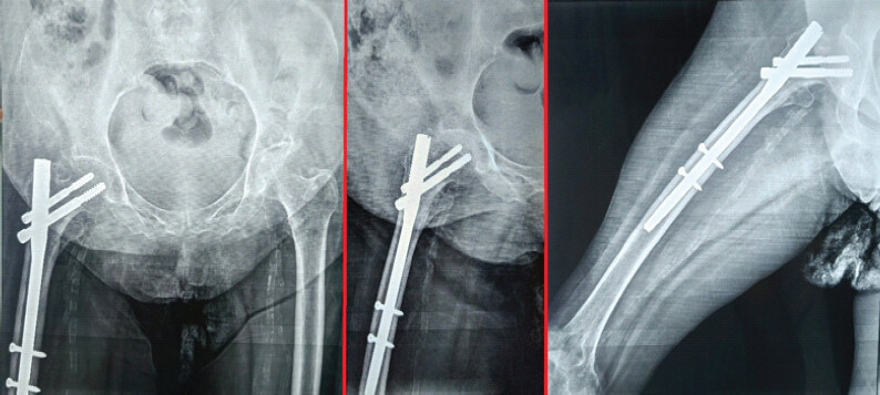
The patient was mobilized non-weight-bearing with walker support, without the prosthesis, on the 1st post-operative day. One month postoperatively, she was allowed to walk full weight-bearing using the BK prosthesis. At 6 months of follow-up, she had a good radiological outcome with no displacement and a united fracture (Fig. 8) and had returned to her pre-injury level of ambulation with the prosthesis (Fig. 9).
Discussion and Review of Literature
Intertrochanteric fractures in BK amputees pose challenges in conventional traction-table positioning. Proximal femoral nailing provides stable intramedullary fixation through a minimally invasive approach [6]. A short PFN is an acceptable option for fractures without subtrochanteric extension, whereas long nails are preferred when a longer segment of the femur requires protection [7].
Surgery is performed on a lower limb traction table, which has a foot attachment that allows traction to be applied to the injured limb and permits manipulation of the distal fragment with respect to flexion, extension, adduction, and abduction to achieve fracture reduction and alignment without additional assistants. In a patient with an amputation and a short limb, it is not possible to place the limb on the foot attachment and apply direct traction. Various methods have been described to manage this situation, including skin traction, insertion of additional pins or wires, and the use of special devices to provide indirect traction to the limb.
Anjum and McNicholas [8] and Lee et al. [9] used skin traction applied to the stump, although rotational control may be inadequate and the method can be difficult in patients with short, conical or poorly padded stumps, and in unstable fracture patterns requiring stronger traction. Direct skin traction may also lead to skin complications.
Ramseier et al., described skeletal traction using a supracondylar Steinmann pin to provide direct femoral traction, assisting reduction and its maintenance [10]. When inserted under sterile operating-room conditions and removed after the procedure, the risk of pin-tract infection is low. Nevertheless, skeletal traction remains an invasive technique and carries procedure-related risks such as pin loosening, neurovascular injury and, particularly in osteoporotic bone, distal femoral fracture or pin pull-out. These considerations should be balanced against the need for reliable traction in each individual patient.
Another described method is distal femoral external fixation secured to the traction table [11]. This provides good traction and stability, but the frame is cumbersome to adjust during surgery. Application of an external fixator also increases soft-tissue injury to the stump and carries the risk of pin-tract infection [12].
Gilmour et al., proposed a technique using a 1.8 mm wire in the distal femur tensioned to an Ilizarov half-ring [13]. With this method, manipulation of the ring together with the amputation stump is difficult because it increases the weight of the limb, and the ring construct is awkward to attach to the traction table.
Patwardhan et al., described skeletal traction using a Denham pin inserted into the femoral stump and attached to the traction table through a Böhler stirrup in a patient with an above-knee amputation [14]. The disadvantages of this method are that it is invasive.
Al-Harthy et al. proposed securing the BK stump in an inverted boot to apply traction and achieve rotational control and fracture reduction without invasive pin insertion [15]. Ochi et al. used this inverted boot technique to support a BKA stump for traction in a 97-year-old amputee, fixing the fracture with an intramedullary gamma nail [16]. McAllister et al. advocated the reverse boot technique as a simple and non-invasive method [17]. Unlike the reverse/inverted boot technique, which uses the fracture-table boot directly around the stump, our technique uses the patient’s own prosthetic socket to facilitate traction and rotational control. The reverse boot may be difficult to apply in patients with short residual limbs, restricted knee flexion, or obesity [16]. Our technique avoids specialized equipment but requires an intact, well-fitting prosthesis.
The summary of various techniques described in the literature for managing pertrochanteric hip fractures in amputation patients is presented in Table 1.
Summary of traction techniques for proximal femoral fractures in patients with lower-limb amputation
| Sr. No | Author (Ref.) | Amputation/fracture | Technique | Reported features and limitations | Comparison with present technique |
|---|---|---|---|---|---|
| 1 | Al-Harthy et al. (1997) [15] | Below-knee amputation; hip fracture | Inverted traction-table boot | Non-invasive; permits traction and rotational control. Suitability depends on stump length, knee flexion, and boot fit. | Uses the patient’s prosthesis instead of an inverted traction-table boot. |
| 2 | Ramseier et al. (2005) [10] | Below-knee amputation; pertrochanteric fracture | Supracondylar Steinmann pin traction | Provides direct skeletal traction; requires invasive pin insertion. | Avoids skeletal pin insertion. |
| 3 | Anjum and McNicholas (2006) [8] | Below-knee amputation; femoral neck fracture | Skin traction using strapping and elastic bandages attached to the traction table | Non-invasive; traction and rotational control may be limited. | Uses a prosthetic socket and foot to facilitate traction and rotational control. |
| 4 | Gamulin and Farshad (2015) [11] | Lower-limb amputation; proximal or diaphyseal femoral fracture | Rigid fixation of the residual limb to the traction-table attachment using an external- fixator construct | Provides rigid attachment and multiplanar control; requires additional fixation hardware. | Avoids a separate external-fixator construct. |
| 5 | Gilmour et al. (2017) [13] | Lower-limb amputation; proximal femoral fracture | Tensioned 1.8-mm fine wire connected to an Ilizarov half-ring and traction table | Provides traction and rotational control; requires wire insertion and additional hardware. | Avoids wire insertion and ring hardware. |
| 6 | Ochi et al. (2017) [16] | Below-knee amputation; intertrochanteric fracture | Inverted traction-table boot with the knee flexed | Allows traction and rotational control using the patella as a rotational reference; requires suitable stump length and positioning. | Uses the patient’s own prosthesis rather than an inverted boot. |
| 7 | Lee et al. (2021) [9] | Below- and above-knee amputations; intertrochanteric fractures | Modified fracture-table positioning with boot rotation and additional skin-traction bands | Satisfactory reduction reported in two cases; requires modification of positioning and skin-traction application. | Uses the existing prosthesis as the traction interface. |
| 8 | Patwardhan et al. (2024) [14] | Above-knee amputation; intertrochanteric fracture | Skeletal traction using a Denham pin and Bohler stirrup | Provides direct skeletal traction; invasive and requires careful pin placement. | Offers a non-invasive alternative in selected below-knee amputees. |
| 9 | McAllister et al. (2025) [17] | Below-knee amputation; proximal femoral fracture | Reverse-boot technique | Uses the inverted traction-table boot to accommodate the flexed knee and residual limb; requires appropriate positioning and fit. | Avoids a dedicated reverse-boot setup but requires a suitable prosthesis. |
| 10 | Present case (2026) | Below-knee amputation; intertrochanteric fracture | Patient’s own prosthesis secured to the standard traction-table foot attachment | Non-invasive and uses equipment already available to the patient; requires secure attachment, careful traction application, and assessment of the stump and skin. | Demonstrates a practical alternative in a selected patient with a well- fitting prosthesis. |
The advantages of this technique include simplicity, cost-effectiveness, use of the patient’s own prosthesis, and avoidance of additional traction devices or invasive skeletal fixation. The prosthetic foot permits conventional traction and rotational manipulation. However, careful socket fitting, secure attachment, and avoidance of excessive traction or compression are essential. In our case, the stump and prosthesis were assessed before and after initial traction for slippage and pressure-related changes; continuous intraoperative monitoring was not performed. Therefore, the possibility of transient pressure-related or soft-tissue complications cannot be excluded, and the safety of this technique requires further evaluation in larger clinical series. Limitations include additional positioning time and dependence on an intact, well-fitting prosthesis of adequate length. Further studies are required to establish the technique’s safety and reproducibility.
Conclusion
The patient’s own BK prosthesis can serve as a practical, cost -effective, non-invasive traction interface during fixation of intertrochanteric fractures in selected amputees. This technique may facilitate fracture reduction and rotational control using a standard traction table while avoiding additional invasive skeletal fixation. Careful socket fitting, secure attachment, and assessment of the stump site are essential.
Clinical Message
An intertrochanteric fracture with an ipsilateral below-knee amputation is an atypical problem faced by orthopedic surgeons. During surgery, positioning of the patient and reduction of the fracture with traction are difficult, but can be achieved with the technique described using the patient’s own prosthesis. Good alignment and stable fixation are required for a good functional outcome, and this can be obtained without resorting to skeletal pins, external fixation, or additional equipment.
Conflict of Interest:
Nil
Source of Support:
Nil
Consent:
The authors confirm that informed consent was obtained from the patient for publication of this article
How to Cite this Article
Ranjankar MC, Pawar A, Thorat P. Positioning and Traction Application Technique in Patient with Below Knee Amputation Using the Patient’s Own Prosthesis for Intertrochanteric Femur Fracture Fixation Surgery: J Orthop Case Rep. 2026 November;16(11): 159-165. doi: 10.13107/jocr.2026.v16.i11.8424.
References
- Pillai A, Eranki V, Shenoy R, Hadidi M.. Age related incidence and early outcomes of hip fractures: A prospective cohort study of 1177 patients. J Orthop Surg Res 2011;6:5. [Google Scholar] | [PubMed]
- Kaufer H, Matthews LS, Sonstegard D.. Stable fixation of intertrochanteric fractures. J Bone Joint Surg Am 1974;56:899-907. [Google Scholar] | [PubMed]
- Chun YS, Oh H, Cho YJ, Rhyu KH.. Technique and early results of percutaneous reduction of sagittally unstable intertrochanteric fractures. Clin Orthop Surg 2011;3:217-24. [Google Scholar] | [PubMed]
- Tanpure S, Chaugule C, Date J, Naikwade D.. Intertrochanteric femur fracture fixation in a patient with below-knee amputation presents a surgical dilemma: A case report. J Orthop Case Rep 2022;12:105-8. [Google Scholar] | [PubMed]
- Mitrasinovic S, Kiziridis G, Wellekens S, Roslee C, Anjum SN.. Innovative method of traction in a bilateral diaphyseal femur fracture in a polytrauma below-knee amputee. Case Rep Orthop 2019;2019:8691398. [Google Scholar] | [PubMed]
- Boldin C, Seibert FJ, Fankhauser F, Peicha G, Grechenig W, Szyszkowitz R.. The proximal femoral nail (PFN)--a minimal invasive treatment of unstable proximal femoral fractures: A prospective study of 55 patients with a follow-up of 15 months. Acta Orthop Scand 2003;74:53-8. [Google Scholar] | [PubMed]
- Hulet DA, Whale CS, Beebe MJ, Rothberg DL, Gililland JM, Zhang C. Short versus long cephalomedullary nails for fixation of stable versus unstable intertrochanteric femur fractures at a level 1 trauma center. Orthopedics 2019;42:e202-9. [Google Scholar] | [PubMed]
- Anjum SN, McNicholas MJ.. Innovative method of traction on fracture table in femoral neck fracture fixation in a below knee amputee. Injury Extra 2006;37:277-8. [Google Scholar] | [PubMed]
- Lee SM, Suh KT, Oh YK, Shin WC.. Manipulation of intertrochanteric fractures in patients with below- or above-knee amputation using a fracture table: Two case reports. Medicine (Baltimore) 2021;100:e24233. [Google Scholar] | [PubMed]
- Ramseier LE, Werner CM, Hug T, Preiss S.. Suprakondyläre extension einer pertrochantären femurfraktur bei einem patienten mit unterschenkelamputation [Supracondylar traction of a pertrochanteric femur fracture in a patient amputated below the knee. Unfallchirurg 2005;108:239-40. [Google Scholar] | [PubMed]
- Gamulin A, Farshad M.. Amputated lower limb fixation to the fracture table. Orthopedics 2015;38:679-82. [Google Scholar] | [PubMed]
- Parameswaran AD, Roberts CS, Seligson D, Voor M.. Pin tract infection with contemporary external fixation: How much of a problem?. J Orthop Trauma 2003;17:503-7. [Google Scholar] | [PubMed]
- Gilmour L, Kassam AM, Hughes AM.. Tensioned fine wire for proximal femur traction table reduction in amputees. Ann R Coll Surg Engl 2017;99:96. [Google Scholar] | [PubMed]
- Patwardhan SS, Kempanna VH, Al Najjar M.. Intertrochanteric fracture fixation using a traction table in a patient with ipsilateral above-knee amputation - a case report and review of reduction techniques. J Orthop Case Rep 2024;14:178-82. [Google Scholar] | [PubMed]
- Al-Harthy A, Abed R, Campbell AC.. Manipulation of hip fracture in the below-knee amputee. Injury 1997;28:570. [Google Scholar] | [PubMed]
- Ochi H, Baba T, Hamanaka T, Ozaki Y, Watari T, Homma Y. Safe and effective reduction technique for intertrochanteric fracture with ipsilateral below-knee amputated limb. Case Rep Orthop 2017;2017:2672905. [Google Scholar] | [PubMed]
- McAllister R, Franklin M, Hyder N.. Reverse boot technique for applying traction in below knee amputees. Ann R Coll Surg Engl 2025;107:309. [Google Scholar] | [PubMed]
© 2026 Journal of Orthopaedic Case Reports - Published by Indian Orthopaedic Research Group
About the Authors
-

Mangesh C Ranjankar
-

Akshay Pawar
-

Pratik Thorat
Related Articles in Journal of Orthopaedic Case Reports
 November 1, 2026 Retrieval of a Broken Cannulated Drill Bit from the Femoral Neck during Proximal Femoral Nailing: Case Report of a Novel Lever Technique Using a Depth Gauge
 August 1, 2026 Radiological Outcomes of Reamed Short Proximal Femoral Nailing in Intertrochanteric Fractures in Patients Aged 50 Years and Above
 August 1, 2026 Evaluation of Orthopedic Outcomes of Proximal Femoral Nailing for Intertrochanteric Fractures and Predictive Value of Pre-operative Glycemic Status, Neutrophil-to-Lymphocyte Ratio and Platelet-to-Lymphocyte Ratio: A Prospective Study
 June 1, 2026 Pseudoaneurysm of the Medial Femoral Circumflex Artery following Surgical Fixation of Trochanteric Femur Fracture: A Case Report