


Introduction
Intraoperative instrument breakage is a recognised complication in orthopaedic and trauma surgery. Among all instruments, drill bits represent the most frequently broken, a finding attributed in part to the practice of reusing them across multiple procedures. Pichler et al. reported an overall instrument breakage rate of 0.35% across 11,856 procedures, with none of the patients experiencing significant long-term sequelae from retained fragments [1]. A larger study by Haberal and Beyaz involving 12,601 procedures found a breakage rate of 0.28% overall, rising to 0.64% specifically in fracture fixation – 7.44 times higher than in elective surgery [2]. Price et al. further demonstrated that drill bits accounted for 11 of 14 total breakages over a 2-year surveillance period, and documented that patients were often not informed and documentation was frequently deficient [3]. Dharmshaktu et al. demonstrated that drill bits were the most frequently broken orthopaedic instruments in a retrospective study of elective procedures, with prolonged use and material fatigue identified as potential contributing factors [4].
Although the literature does not demonstrate significant long-term adverse effects from retained drill bit fragments, leaving them in situ carries tangible risks: Potential migration into adjacent structures, interference with planned implant placement, increased susceptibility to peri-implant infection, and medicolegal exposure [1,2,5]. Migration of broken drill bits has been documented in the context of acetabular fracture fixation and hip arthroplasty [5]. The femoral neck is a particularly challenging anatomical location for retrieval due to its limited access, restricted working space, and proximity to neurovascular structures. Suboptimal screw placement in proximal femoral nailing (PFN) – typically defined as a tip-apex distance (TAD) exceeding 25 mm or non-central screw position – is a well-established predictor of implant cut-out and fixation failure [6,7].
Several techniques for retrieving broken cannulated drill bits from the femoral neck have been previously described. Bassi et al. popularized a multi-K-wire technique in which a 1 mm K-wire with a right-angle bent tip is passed through the lumen of the broken drill bit to hook onto its far end, with two additional K-wires “stuffed” into the cannulation to facilitate traction [8]. Al-Kharouf et al. described using the hooked tip of a depth gauge, engaging the edge of the broken drill bit and employing clockwise and counterclockwise rotation to retrieve it through the proximal reaming tract [9]. Chouhan and Sharma reported a “push-back” technique using a narrow Langenbeck retractor, applicable where the broken fragment is longer than the bone diameter [10].
We describe a novel modification of the depth gauge technique – a lever method – that achieves more secure mechanical engagement of the broken fragment and facilitates safe, efficient retrieval without additional instruments, incisions, or significant extension of operative time.
Case Report
A male patient in his mid-sixties presented to our unit following a low-energy fall at home, sustaining a left intertrochanteric femur fracture. He had no significant comorbidities. After pre-operative optimization, he was taken to the theatre for closed reduction and internal fixation using a standard PFN.
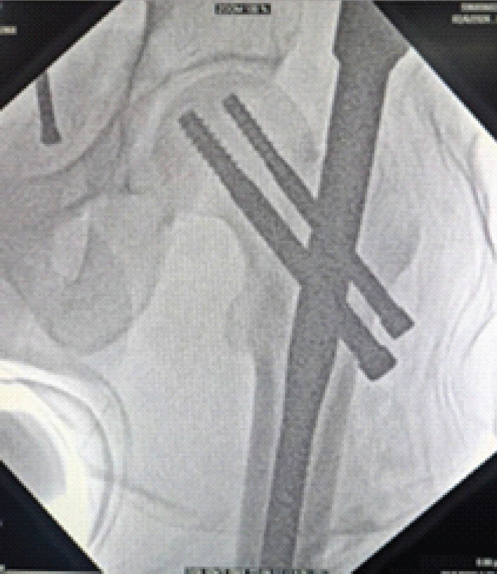
The patient was positioned in hemilithotomy position on a fracture table. Closed reduction was achieved under fluoroscopic guidance. The intramedullary nail was inserted through a standard trochanteric entry point, and two guide wires were placed into the femoral neck and head using the targeting jig and biplanar fluoroscopic confirmation. During reaming over the inferior (compression) guide wire with an 8 mm cannulated drill bit, the drill bit fractured at mid-shaft, leaving the distal portion lodged within the reamed canal in the femoral neck (Fig. 1).
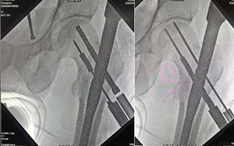
Retrieval of the broken fragment was necessary before proceeding, as its position directly obstructed placement of the compression screw at the desired TAD. Leaving it in situ would have necessitated a significantly suboptimal screw trajectory, increasing the risk of fixation failure.
Surgical technique
The proximal segment of the fractured drill bit, still attached to the drill chuck, was removed, followed by careful withdrawal of the guide wire through the cannulation of the broken fragment. A standard depth gauge from the PFN instrumentation set, which features a small hook at its distal tip, was then selected.
Under continuous fluoroscopic guidance, the depth gauge was carefully advanced through the reamed canal and through the cannulation of the broken drill bit until its hooked tip emerged from the far (medial) end (Fig. 2). The depth gauge was then rotated axially so that its hooked tip was oriented to face medially.
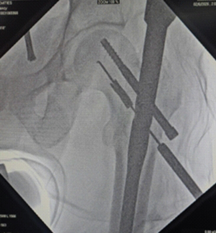
The key technical step – the lever maneuver – was then performed: While maintaining the hooked tip in its medial orientation, the proximal (external) arm of the depth gauge was levered firmly in the lateral direction, away from the limb (Fig. 3). This created a lever-arm effect that drove the hooked distal tip against the medial wall of the broken drill bit, establishing firm mechanical engagement with the inner surface of the fragment.
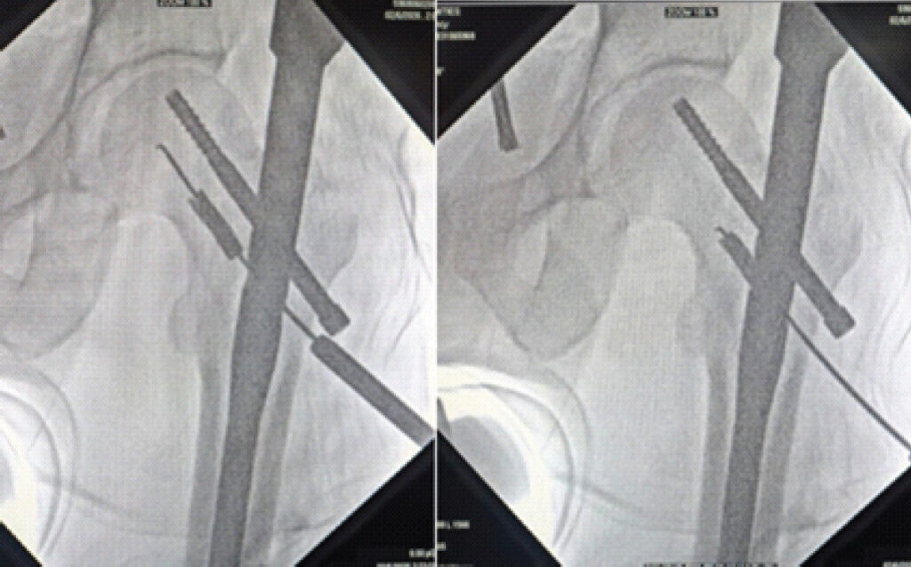
Fluoroscopic confirmation of hook engagement was obtained before applying steady, axial traction along the instrument axis. The broken 8 mm cannulated drill bit was retrieved in its entirety through the nail locking hole without any additional soft tissue dissection or cortical disruption (Fig. 4).
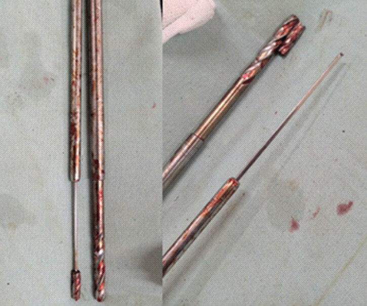
The guide wire was reinserted over the original trajectory, the canal was reamed with a new 8 mm cannulated drill bit, and the compression screw was placed in the optimal central-inferior position with a TAD of <25 mm (Fig. 5). The remainder of the procedure was uneventful.
Discussion
Drill bit breakage during surgery is more common than the published literature suggests, as it is frequently underreported. Price et al. found documentation deficiencies in a significant proportion of cases and noted that patients were often not informed of the incident [3]. Real-world incidences are therefore likely to exceed published figures. Drill bits are the most frequently broken instrument in orthopaedic surgery, with repeated reuse across multiple procedures a key contributing factor, as material fatigue from cyclic loading significantly reduces the number of cycles to failure [3,4,8].
While no significant long-term adverse effects have been demonstrated from retained drill bits in well-embedded positions, the situation is markedly different when the fragment obstructs planned implant placement. In PFN, suboptimal compression screw positioning – particularly placement outside the central or inferior quadrant of the femoral head or with a TAD exceeding 25 mm – is strongly associated with screw cut-out, a potentially catastrophic complication especially in elderly osteoporotic bone [6,7]. In such circumstances, retrieval is essential. Additional concerns include the potential for migration [5], peri-implant infection, and the medicolegal risks of a retained foreign body in the absence of a compelling clinical justification [8].
The technical challenge of retrieval from the femoral neck has led to multiple described techniques. Bassi et al.’s multi-K-wire technique is well-established for larger-bore cannulated drill bits but requires considerable fluoroscopic time and technical precision in loading multiple K-wires into the narrow cannulation [8]. Al-Kharouf et al.’s technique of hooking onto the edge of the broken bit using a depth gauge tip and applying twisting traction is simpler but relies on surface engagement, which may be insufficient for deeply embedded or tightly wedged fragments [9]. Chouhan and Sharma’s push-back technique is limited by the requirement that the broken piece must exceed the diameter of the bone, making it inapplicable in many scenarios involving the femoral neck [10].
Our technique offers several advantages over these approaches. First, by passing the depth gauge tip through the entire length of the broken drill bit’s cannulation and emerging at the far end, we achieve intraluminal engagement rather than relying on surface-edge hooking, thereby providing a secure hold. Secondly, the lever maneuver – orienting the hook medially and levering the external arm laterally – exploits a mechanical advantage that actively drives the hook against the inner wall of the fragment before traction is applied. This two-step process (engage then extract) reduces the risk of the hook disengaging mid-procedure. Thirdly, the entire technique is performed using instruments already present in the standard PFN nailing set, with no requirement for additional specialized equipment, no extension of the surgical incision, and no violation of cortical bone beyond what has already been reamed.
A prerequisite for this technique, shared with the Al-Kharouf depth gauge method, is that the drill bit must be cannulated – the central lumen through which the depth gauge tip is passed is essential. This technique is therefore not applicable to solid drill bits. The use of continuous biplanar fluoroscopy during the retrieval steps is also strongly recommended to confirm correct hook orientation, engagement, and to monitor fragment position during traction.
From a preventative standpoint, departments should consider implementing a policy for the retirement of drill bits after a specified number of uses or upon demonstrating macroscopic evidence of wear. Quality checks prior to each use and single-use drill bits for critical high-stress applications may reduce the incidence of intraoperative breakage. Any instance of instrument breakage should be documented fully in the operative notes and the patient informed as part of post-operative disclosure [1,3].
Conclusion
We describe a novel lever technique using a standard PFN-set depth gauge for the retrieval of a broken 8 mm cannulated drill bit from the femoral neck. The technique achieves secure intraluminal engagement of the fragment through a mechanical lever mechanism, enabling safe and time-efficient extraction without additional incisions, specialized instrumentation, or cortical violation. Removal of the broken fragment in this context was essential to allow optimal compression screw placement and to reduce the risk of implant failure. We recommend this technique in comparable intraoperative scenarios and advocate surgeon familiarity with the full repertoire of described retrieval methods.
Clinical Message
Broken drill bits are a rare intraoperative complication in orthopaedic surgery, and their retrieval can be technically challenging, particularly when the fragment is deeply embedded within bone. This case highlights a simple and reproducible retrieval technique that allowed safe removal of the fragment while preserving bone stock and maintaining stable fracture fixation.
Conflict of Interest:
Nil
Source of Support:
Nil
Consent
The authors confirm that informed consent was obtained from the patient for publication of this article
How to Cite this Article
Patil RN, Bhat VGK, Mittal N, Khandelwal A, Sudhanva GN. Retrieval of a Broken Cannulated Drill Bit from the Femoral Neck during Proximal Femoral Nailing: Case Report of a Novel Lever Technique Using a Depth Gauge. J Orthop Case Rep. 2026 November;16(11): 116-120. doi: 10.13107/jocr.2026.v16.i11.8408.
References
- Pichler W, Mazzurana P, Clement H, Grechenig S, Mauschitz R, Grechenig W. Frequency of instrument breakage during orthopaedic procedures and its effects on patients. J Bone Joint Surg Am 2008;90:2652-4. [Google Scholar] | [PubMed]
- Haberal B, Beyaz S. Effects of orthopaedic instruments breakage during surgery: A minimum five-year follow-up. Ann Clin Anal Med 2021;12:443-6. [Google Scholar] | [PubMed]
- Price MV, Molloy S, Solan MC, Sutton A, Ricketts DM. The rate of instrument breakage during orthopaedic procedures. Int Orthop 2002;26:185-7. [Google Scholar] | [PubMed]
- Dharmshaktu GS, Adhikari N, Mourya P, Bhandari SS, Singh P. Intraoperative instrument breakage during orthopaedic elective procedures: A retrospective single-centre experience. J Orthop Spine 2020;8:80-5. [Google Scholar] | [PubMed]
- Jamot S, Razif A, Azhar MM, Abbas AA. Migration of a broken drill bit after acetabular fracture fixation. A case report. Malays Orthop J 2011;5:56-9. [Google Scholar] | [PubMed]
- Baumgaertner MR, Curtin SL, Lindskog DM, Keggi JM. The value of the tip-apex distance in predicting failure of fixation of peritrochanteric fractures of the hip. J Bone Joint Surg Am 1995;77:1058-64. [Google Scholar] | [PubMed]
- Pervez H, Parker MJ, Vowler S. Prediction of fixation failure after sliding hip screw fixation. Injury 2004;35:994-8. [Google Scholar] | [PubMed]
- Bassi JL, Pankaj M, Navdeep S. A technique for removal of broken cannulated drill bit: Bassi's method. J Orthop Trauma 2008;22:56-8. [Google Scholar] | [PubMed]
- Al-Kharouf KF, Abbas K, Anjum S, Khan FI. Removal of broken cannulated drill bit. Cureus 2021;13:e19706. [Google Scholar] | [PubMed]
- Chouhan DK, Sharma S. A simple technique for the removal of a broken intramedullary drill bit. J Orthop Trauma 2010;24:576-8. [Google Scholar] | [PubMed]
© 2026 Journal of Orthopaedic Case Reports - Published by Indian Orthopaedic Research Group
About the Authors
-

Rakshith N Patil
-

Vikram G K Bhat
-

Naman Mittal
-

Akshit Khandelwal
-

G N Sudhanva
Related Articles in Journal of Orthopaedic Case Reports
 November 1, 2026 Positioning and Traction Application Technique in Patient with Below Knee Amputation Using the Patients Own Prosthesis for Intertrochanteric Femur Fracture Fixation Surgery: A Case Report with Review of Literature
 August 1, 2026 Radiological Outcomes of Reamed Short Proximal Femoral Nailing in Intertrochanteric Fractures in Patients Aged 50 Years and Above
 August 1, 2026 Evaluation of Orthopedic Outcomes of Proximal Femoral Nailing for Intertrochanteric Fractures and Predictive Value of Pre-operative Glycemic Status, Neutrophil-to-Lymphocyte Ratio and Platelet-to-Lymphocyte Ratio: A Prospective Study
 June 1, 2026 Pseudoaneurysm of the Medial Femoral Circumflex Artery following Surgical Fixation of Trochanteric Femur Fracture: A Case Report