

Posterior sternoclavicular physeal fracture dislocations in adolescents may be safely managed nonoperatively in select subacute cases with stable outcomes.
Mr. Benjamin T Whittaker, Medical Student, BS, 801 N Rutledge St, Springfield, IL 62702. Email: bwhittaker65@siumed.edu
Abstract
Introduction: Posterior sternoclavicular (SC) injuries are clinically significant due to their proximity to vital mediastinal structures. Rapid diagnosis of these injuries is critical, yet challenging, as plain radiographs often fail to detect posterior displacement. Current guidelines call for immediate closed versus open reduction of SC injuries. However, there remains a paucity of literature reporting on outcomes following management in subacute presentations. In skeletally immature patients, most injuries involving the medial clavicle represent physeal fractures rather than true dislocations. This allows for greater remodeling potential, allowing for conservative management in select cases.
Case Report: A 14-year-old male presented to the clinic 1 month after sustaining an injury to his left shoulder during a football tackle. On examination, the patient demonstrated a posterior step-off of the SC joint. Imaging was used to confirm the diagnosis of a medial clavicular physeal fracture with posterior displacement. The patient demonstrated clinical stability and a lack of progressive symptoms, and the decision was made to proceed with non-operative management with observation. The patient returned to activity without limitation.
Conclusion: This case highlights that subacute posterior SC physeal dislocations in adolescents may be managed nonoperatively with favorable outcomes.
Keywords: Sternoclavicular joint, physeal injury, posterior sternoclavicular dislocation, pediatric trauma, non-operative management, subacute diagnosis.

The sternoclavicular (SC) joint is the primary articulation between the axial skeleton and the upper limb, providing stability while protecting nearby mediastinal structures. Posterior dislocation of the SC joint is rare, accounting for approximately 1% of all joint dislocations and <5% of shoulder girdle injuries [1,2]. These injuries typically result from high-energy trauma directed to the shoulder or medial clavicle [3].
Patients often present with pain, asymmetry, and restricted motion, and life-threatening complications may occur due to compression of the trachea, esophagus, or nearby neurovascular structures [4,5,6,7]. Diagnosis is challenging because conventional radiographs often miss posterior SC dislocations [8,9,10,11]. Computed tomography (CT) is considered the gold standard for diagnosis and surgical planning [12,13].
In skeletally immature patients, these injuries often represent physeal fractures rather than true dislocations, as the medial clavicular physis does not fully fuse until the mid-20s [14,15]. Standard of care involves prompt reduction (open or closed) [13,16,17,18], but there is a paucity of literature reporting on outcomes following non-operative management in subacute presentations.
We present the case of a 14-year-old male with a posterior physeal fracture-dislocation treated nonoperatively with a successful return to activity. Written informed consent for publication was obtained from the patient and family.
A 14-year-old male presented to the orthopedic clinic 1 month after sustaining an injury to his left shoulder during a football tackle. He experienced immediate shoulder pain, was evaluated in the emergency department, and had initial radiographs of the clavicle and shoulder that were read as negative (Fig. 1).
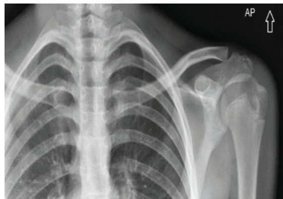
Figure 1: Initial anteroposterior radiograph obtained on the day of injury, interpreted as negative for osseous abnormality by an outside facility.
The patient self-treated with sling immobilization and gradually returned to basketball. Four weeks later, he underwent evaluation with orthopedics when he still was experiencing some intermittent shoulder pain and his parents noted some clavicle asymmetry.
On examination, the left SC joint demonstrated a visible posterior step-off with tenderness at the joint. He was neurovascularly intact with full shoulder range of motion. He was not complaining of any cardiopulmonary symptoms. A serendipity view radiograph revealed asymmetry of the SC joints and notable posterior dislocation of the left SC joint (Fig. 2). CT confirmed a medial clavicular physeal fracture with posterior displacement and mild mass effect on the left subclavian and internal jugular veins (Fig. 3).
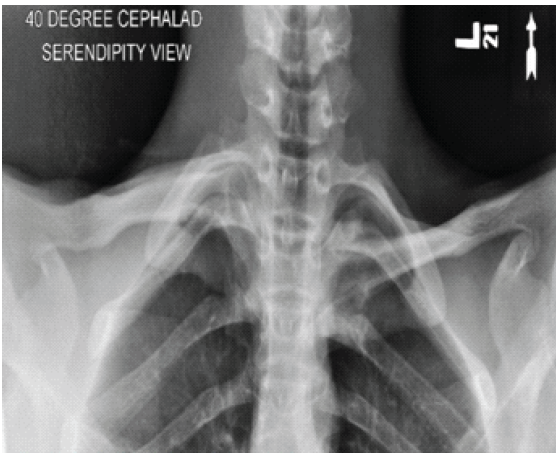
Figure 2: Serendipity view obtained at 1 month post-injury demonstrating posterior sternoclavicular dislocation.
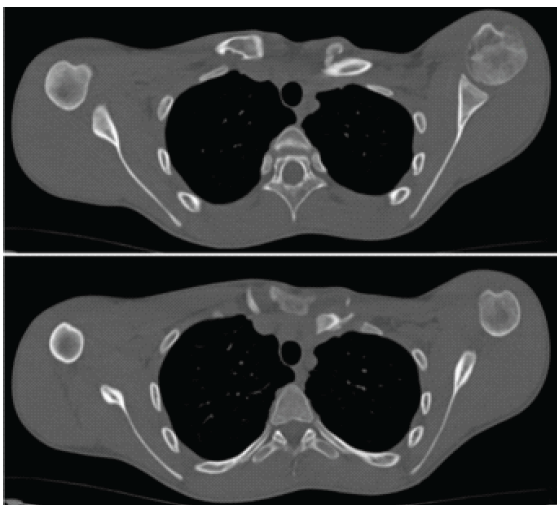
Figure 3: Two axial-plane computed tomography images performed 1 month after injury demonstrating a medial clavicular epiphyseal fracture with posterior displacement.
Multidisciplinary discussion of risks and benefits of operative versus non-operative management with cardiothoracic surgery and two different shoulder/upper extremity specialists was reviewed with the patient and family. Given clinical stability and lack of progressive symptoms, the shared decision was made with the patient and his family to proceed with observation and collision sport restriction.
The patient gradually returned to contact sports without limitation. Follow-up at 20 months post-injury revealed asymptomatic full range of motion and strength with minimal clinical asymmetry on physical examination. Repeat CT demonstrated minimal persistent posterior displacement with some remodeling (Figs. 4 and 5). There was no evidence of neurovascular compromise or functional limitation at final follow-up.
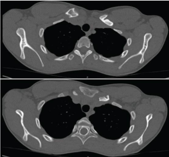
Figure 4: Two axial-plane computed tomography images performed 20 months after injury demonstrating a healed clavicle fracture with mild persistent posterior displacement.
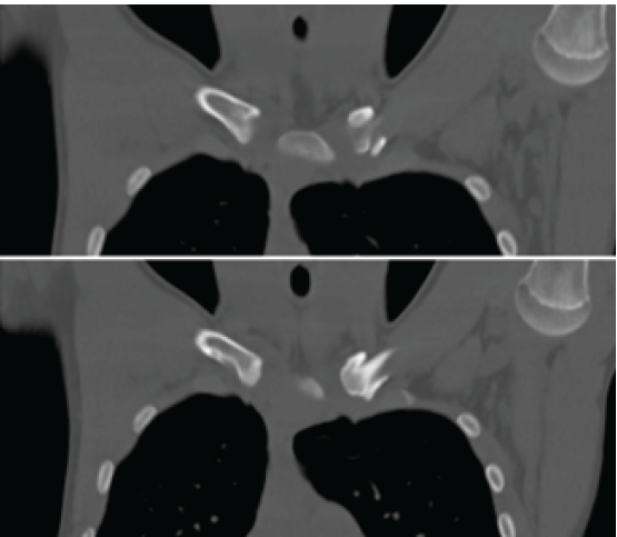
Figure 5: Coronal plane computed tomography performed 20 months after injury showing a healed physeal fracture with no significant changes in the position of the clavicle.
Final follow-up occurred at 27 months to assess further changes in alignment. The patient maintained favorable functional outcomes without limitations. A single serendipity view was obtained showing significant remodeling, with improved alignment of the SC joint (Fig. 6).
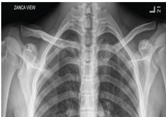
Figure 6: Serendipity view obtained at 27 months after the injury showing significant remodeling with improved alignment of the sternoclavicular joint.
Posterior SC joint injuries are rare, but clinically significant due to their proximity to vital mediastinal structures. They account for <1% of all joint dislocations and may result in life-threatening complications such as tracheal, esophageal, or vascular compression [1]. Rapid diagnosis and management are therefore critical. However, the diagnosis may be delayed because standard radiographs often fail to detect posterior displacement. Advanced imaging with CT remains the gold standard for diagnosis and assessing mediastinal involvement [12,13]. However, if advanced imaging is not readily available, an alternative option is to obtain a serendipity view radiograph. This view utilizes 40° of cephalic tilt and provides a true caudocephalic view of both the SC joints and the medial clavicles, thus improving visualization in the case of displacement.
In skeletally immature patients, most injuries involving the medial clavicle represent physeal fractures rather than true dislocations, since the medial physis may remain unfused until the early to mid-20s. The ossification of the medial clavicular epiphysis begins in adolescence (ages 14–16 years), partial fusion occurs between ages 16 and 26, and complete fusion typically is not achieved until approximately 21–26 years of age, with some individuals showing fusion as late as age 27 [19]. Because the medial clavicular physis closes later than most other growth plates, it retains greater remodeling potential, making it uniquely amenable to non-operative management in select cases. This physeal growth contributes approximately 80% of clavicular longitudinal growth [20].
Traditional guidelines emphasize urgent closed or open reduction of posterior SC joint injuries regardless of physeal status due to the risk of catastrophic complications [16,17,18]. However, literature describing outcomes of non-operative observation in subacute presentations remains sparse. A few case reports and small series suggest that when patients are clinically stable without airway, vascular, or neurologic compromise, conservative management may be reasonable [21]. The remodeling potential inherent in the adolescent medial clavicle may support functionally acceptable outcomes even when imaging remains abnormal.
This case expands the limited evidence base by reporting a 14-year-old male who sustained a posterior medial clavicular physeal fracture-dislocation diagnosed approximately 1 month post-injury. He was managed nonoperatively, with successful functional recovery and no cardiopulmonary or neurovascular compromise after nearly 2 years of follow-up, despite persistent posterior displacement on imaging.
In adolescents, posterior SC physeal fracture-dislocations pose a significant diagnostic challenge and can be a life-threatening injury if missed or mismanaged. However, injuries identified in the subacute phase may be safely managed nonoperatively if the patient is clinically stable and asymptomatic. The late physeal closure of the medial clavicle and high remodeling potential can support favorable long-term outcomes. Still, more research is needed to define selection criteria, evaluate remodeling over time, and assess reinjury risk in this rare injury pattern.
This case supports that posterior SC fracture-dislocations identified in the subacute phase may be managed nonoperatively in select cases with positive outcomes. The current gold standard of care is prompt open or closed reduction of the joint, with limited prior evidence to guide management in subacute cases. This case demonstrates that the patient was able to be spared from life-threatening risks associated with operative callus resection, reduction, and fixation of the joint while maintaining full functionality.
References
- 1. Chaudhry S. Pediatric posterior sternoclavicular joint injuries. J Am Acad Orthop Surg 2015;23:468-75. [Google Scholar] [PubMed]
- 2. Nettles JL, Linscheid RL. Sternoclavicular dislocations. J Trauma 1968;8:158-64. [Google Scholar] [PubMed]
- 3. Hellwinkel JE, McCarty EC, Khodaee M. Sports-related sternoclavicular joint injuries. Phys Sportsmed 2019;47:253-61. [Google Scholar] [PubMed]
- 4. Mirza AH, Alam K, Ali A. Posterior sternoclavicular dislocation in a rugby player as a cause of silent vascular compromise: A case report. Br J Sports Med 2005;39:e28. [Google Scholar] [PubMed]
- 5. Nakayama E, Tanaka T, Noguchi T, Yasuda J, Terada Y. Tracheal stenosis caused by retrosternal dislocation of the right clavicle. Ann Thorac Surg 2007;83:685-7. [Google Scholar] [PubMed]
- 6. Jain S, Monbaliu D, Thompson JF. Thoracic outlet syndrome caused by chronic retrosternal dislocation of the clavicle. Successful treatment by transaxillary resection of the first rib. J Bone Joint Surg Br 2002;84:116-8. [Google Scholar] [PubMed]
- 7. Gardner MA, Bidstrup BP. Intrathoracic great vessel injury resulting from blunt chest trauma associated with posterior dislocation of the sternoclavicular joint. Aust N Z J Surg 1983;53:427-30. [Google Scholar] [PubMed]
- 8. Jacob M, Snashall J, Dorfman A, Shesser R. X-ray-negative posterior sternoclavicular dislocation after minor trauma. Am J Emerg Med 2013;31:260.e3-5. [Google Scholar] [PubMed]
- 9. Albors LM, Lee RJ. Chronic posterior sternoclavicular dislocation in a contact athlete: A case report. JBJS Case Connect 2020;10:e0032. [Google Scholar] [PubMed]
- 10. Walia P, Fredette R, Mortimer E. Missed medial clavicular physeal fracture-posterior dislocation complicated by brachiocephalic pseudoaneurysm: A case report. JBJS Case Connect 2024;14:e24.00180. [Google Scholar] [PubMed]
- 11. Carmichael KD, Longo A, Lick S, Swischuk L. Posterior sternoclavicular epiphyseal fracture-dislocation with delayed diagnosis. Skeletal Radiol 2006;35:608-12. [Google Scholar] [PubMed]
- 12. Wirth MA, Rockwood CA Jr. Acute and chronic traumatic injuries of the sternoclavicular joint. J Am Acad Orthop Surg 1996;4:268-78. [Google Scholar] [PubMed]
- 13. Groh GI, Wirth MA. Management of traumatic sternoclavicular joint injuries. J Am Acad Orthop Surg 2011;19:1-7. [Google Scholar] [PubMed]
- 14. Lee JT, Nasreddine AY, Black EM, Bae DS, Kocher MS. Posterior sternoclavicular joint injuries in skeletally immature patients. J Pediatr Orthop 2014;34:369-75. [Google Scholar] [PubMed]
- 15. Kim WG, Laor T, Jarrett DY. Physeal injuries of the clavicle: Pediatric counterparts to adult acromioclavicular and sternoclavicular joint separations. Pediatr Radiol 2023;53:1513-25. [Google Scholar] [PubMed]
- 16. Ting BL, Bae DS, Waters PM. Chronic posterior sternoclavicular joint fracture dislocations in children and young adults: Results of surgical management. J Pediatr Orthop 2014;34:542-7. [Google Scholar] [PubMed]
- 17. Ingoe HM, Mohammed K, Malone AA, Beadle G, Sharpe T, Cockfield A, et al. Traumatic posterior sternoclavicular joint dislocation – current aspects of management. Injury 2023;54:110983. [Google Scholar] [PubMed]
- 18. Ciatti C, Masoni V, Maniscalco P, Asti C, Puma Pagliarello C, Caggiari G, et al. Management options for traumatic posterior sternoclavicular joint dislocation: A narrative review with a single institution’s experience. J Clin Med 2024;13:5428. [Google Scholar] [PubMed]
- 19. Kreitner KF, Schweden FJ, Riepert T, Nafe B, Thelen M. Bone age determination based on the study of the medial extremity of the clavicle. Eur Radiol 1998;8:1116-22. [Google Scholar] [PubMed]
- 20. Hughes JL, Newton PO, Bastrom T, Fabricant PD, Pennock AT. The clavicle continues to grow during adolescence and early adulthood. HSS J 2020;16 Suppl 2:372-7. [Google Scholar] [PubMed]
- 21. Yeh GL, Williams GR Jr. Conservative management of sternoclavicular injuries. Orthop Clin North Am 2000;31:189-203. [Google Scholar] [PubMed]
Related Articles in Journal of Orthopaedic Case Reports
 May 1, 2026 Spontaneous Remodeling of Posterior Sternoclavicular Joint Dislocations in Skeletally Immature Patients: A Case Report
 September 1, 2026 Angular Improvement with Bracing Over 7 Years in Anterolateral Tibial Bowing without Neurofibromatosis Type 1: A Case Report
 August 1, 2026 Generalized Tetanus Following Traditional Manipulation of an Open Supracondylar Humerus Fracture in a Child: A Rare Case Report
 August 1, 2026 Spiked Washer–Augmented Screw Fixation for Bilateral Tibial Tuberosity Avulsion Fractures in an Adolescent: A Case Report