


Introduction
Congenital tibial bowing is a spectrum of deformities present at birth, classified by the direction of angulation into anterolateral, posteromedial, and anteromedial patterns, each with a distinct natural history [1,2,3]. The anterolateral pattern is the least benign of the three. Anterolateral tibial bowing (ALTB) is not a positional deformity but a localized dysplasia in which the periosteum is the primary site of disease and the osseous abnormality is secondary. Fibrous hamartoma replaces healthy periosteum, increasing osteoclasis and reducing osteogenesis. The affected segment therefore bows progressively and, in most reported series, eventually fractures. This dysplasia is encountered most often in the setting of neurofibromatosis type 1 (NF1).
NF1 affects approximately one in 2,662 live births [4], and the same periosteal disease underlies its tibial manifestations. Loss of neurofibromin activates rat sarcoma/mitogen-activated protein kinase signaling, impairs osteoblast maturation, and accelerates osteoclast-mediated resorption, which accounts for both the progressive deformity and the failure of union [5, 6]. The end point of that failure is congenital pseudarthrosis of the tibia (CPT), which develops in 2–6% of children with NF1 [1]. CPT follows a fracture through the dysplastic segment that does not unite, so that a false joint forms at the apex. Preventing this outcome is the object of treatment in ALTB [7].
Bowing without a genetic association is frequently described as rare. In one consecutive operative series of patients with ALTB, approximately 30% carried no genetic diagnosis [8]. In a separate cohort of children with ALTB, nine of 40 (22.5%) were NF1 negative [9].
The majority of these patients require surgical management, and few cases have been reported to resolve without operation. In a series of 43 children with congenital anterolateral bowing, five (12%) corrected without progressing to fracture [10]. Among the unilateral cases, limb length inequality was the sole residual deformity at a mean follow-up of 58 months. Angulation and shortening therefore do not follow the same course. We report a child with prenatally detected ALTB and no genetic association in whom prolonged bracing accompanied improvement of the angular deformity, while limb length inequality progressed independently.
Case Report
An 8-year-old Hispanic boy has been followed in our pediatric orthopedic clinic since 1 year of age. He was referred with congenital bowing of the tibia detected on a level II obstetric sonogram at 27 weeks of gestation, which showed isolated right tibial bowing with otherwise normal fetal anatomy.
The mother tested positive for Zika virus in the second trimester. Serial fetal assessment every 3–4 weeks showed growth appropriate for gestational age and normal middle cerebral artery Doppler studies. No microcephaly, intracranial calcification, ventriculomegaly, or arthrogryposis was identified prenatally or on postnatal examination and neurodevelopmental follow-up, and the tibial deformity remained the sole structural abnormality. No family history of NF1 was reported.
At 3 years of age, examination showed symmetric hip abduction, normal femoral anteversion, moderate internal tibial torsion, right anterolateral tibial deformity, and a clinically estimated limb length discrepancy of 2 cm. There were no cafe-au-lait macules, axillary or inguinal freckling, cutaneous neurofibromas, or Lisch nodules. Chromosomal microarray identified only a 360 kb gain of uncertain significance at 7p15.3 involving DNAH11 and CDCA7L, a locus with no established phenotype in the heterozygous state and no relation to skeletal dysplasia; no copy number abnormality of NF1 (17q11.2) was found.
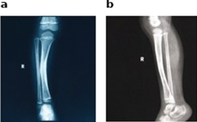
Radiographs demonstrated anterolateral bowing at the mid-diaphysis of the tibia without fracture (Fig. 1). The medullary canal remained patent at the apex, without hourglass constriction, cystic change, or cortical breach, corresponding to Paley type 1 [7]. Magnetic resonance imaging showed normal ossification for age, without discrete lesions, cortical disruption, pathological periosteum, or pseudarthrosis. No low-density line, the linear intracortical radiolucency associated with impending fracture, was identified on any radiograph obtained during the period of follow-up.
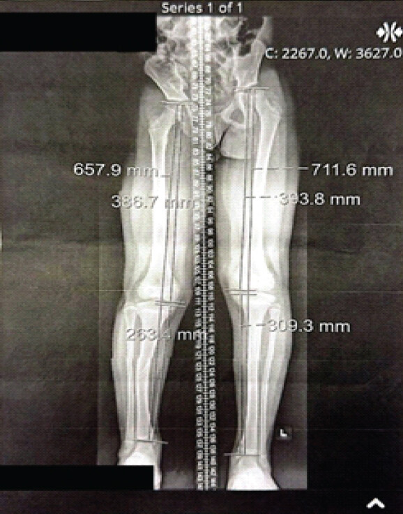
He was managed non-operatively throughout with a right total contact orthosis worn continuously and a shoe lift. Serial radiographs showed progressive improvement in anterior angulation without bone defect, and a standing lower extremity scanogram at latest follow-up confirmed the improved alignment (Fig. 2). The clinical appearance of the limb improved markedly in the assessment of the treating surgeons and the family. He remains ambulatory and pain-free, with no fracture, pseudarthrosis, or ankle valgus. Limb length discrepancy measured 4.4 cm at latest follow-up and corrective surgery is now warranted.
Discussion
We report an 8-year-old boy with congenital anterolateral bowing of the tibia detected on a second-trimester obstetric sonogram, in whom no genetic association could be identified, and who was managed non-operatively from infancy with a total contact orthosis and a graduated shoe lift. Over more than 7 years of follow-up, the tibia did not fracture, no low-density line appeared on any radiograph, and the angular deformity improved both radiographically and esthetically. The limb length inequality followed an independent course, progressing from 2 cm at presentation to 4.4 cm despite uninterrupted bracing, and now requires an operative plan of its own. The interest of this case lies in that dissociation, since a tibia that remained intact and straightened over 7 years of growth offered no protection against shortening of the affected segment.
The best comparable entity previously reported is a group of ten patients with bowing accompanied by ipsilateral duplication of the hallux without NF1 association [11,12]. The bow resolved spontaneously in all of them, and none progressed to pseudarthrosis, yet every child developed a limb length discrepancy and several required lengthening or contralateral epiphysiodesis to address it [11]. Angulation and shortening therefore diverged in that group as they did in our patient. However, our patient does not represent the aforementioned condition, since that condition is recognizable at birth by the duplicated toe. Other non-syndromic presentations reinforce the heterogeneity of the entity: The delta tibia, a triangular anterolateral angulation with a thickened concave cortex, follows a comparatively benign course [13], and isolated cases of anterolateral bowing without NF1 continue to be reported with variable outcomes [14].
A comparable natural history to that of our patient is described in the series of Tuncay et al. [10]. Of 43 children evaluated for anterolateral bowing, five corrected spontaneously without fracture, none of whom had NF1 or other congenital anomalies. Limb length discrepancy was their only residual deformity and reached 4.8 cm at a mean follow-up of 58 months.
Anterolateral bowing in children without NF1 has rarely been described. A negative genetic workup is often taken to be reassuring in itself, but the available evidence does not support this. Comparisons between patients with and without NF1 have shown no difference in the onset of bowing or fracture, tissue pathology, or union after surgery [15,16]. Paley has summarized the outcomes as equivalent in both groups [7]. The distinction is also less clean than it appears, since somatic NF1 inactivation has been identified in the periosteum of 21% of patients who did not meet clinical criteria for neurofibromatosis [17]. Tian et al. studied 40 children with bowing and found no association between NF1 status and the time to fracture, with five of the nine NF1-negative children going on to fracture [9]. A negative workup therefore carries no prognostic weight of its own, and the dysplastic tibia can fracture and the deformity progress whether or not NF1 is present.
Management of the intact bowed tibia aims to prevent the fracture that converts a manageable deformity into pseudarthrosis. Bracing remains first line, while prophylactic bypass grafting [18] and distal tibial guided growth [19, 20] are reserved for tibiae that deteriorate under observation; once fracture occurs, the reconstructive burden rises steeply [8,21,22]. Our patient required none of these, since his tibia straightened under bracing and remained intact. His remaining problem is length, and the operation therefore belongs on the sound limb. Corrective surgery has been planned, and the choice between contralateral epiphysiodesis and ipsilateral lengthening depends on the discrepancy projected to skeletal maturity [23].
This report carries the limitations of a single case. No causal claim can be made for the orthosis, since spontaneous correction under observation alone is well documented [10] and prospective evidence for the effectiveness of bracing is lacking [5], so what we observed may represent natural history rather than treatment effect. Angular correction was judged by the treating surgeons and the family rather than measured by blinded observers. The patient has not reached skeletal maturity, so neither the durability of the correction, the final inequality, nor the adequacy of the planned procedure can yet be stated.
Conclusion
In this child with ALTB and no identified genetic association, non-operative management was accompanied by improvement of the angular deformity and freedom from fracture through 7 years of skeletal growth, while limb length inequality progressed independently. Bracing acted on the bow but not on the shortening, and correction of the discrepancy now requires a separate operation. Whether bracing or early operative correction is preferable in the pre-fracture tibia remains unsettled, and a single case does not resolve it.
Clinical Message
Anterolateral tibial bowing produces two deformities with different mechanisms and different responses to treatment. Bracing may protect the dysplastic segment and allow the bow to improve, but it has no effect on limb shortening, which arises from growth inhibition in the affected segment. An intact, straightening tibia should not be taken as evidence that secondary changes are being averted. Limb length should be projected to skeletal maturity from the first consultation and planned for as a separate procedure.
Conflict of Interest:
Nil
Source of Support:
Nil
Consent:
The authors confirm that informed consent was obtained from the patient for publication of this article
References
- Siebert MJ, Makarewich CA. Anterolateral tibial bowing and congenital pseudoarthrosis of the tibia: Current concept review and future directions. Curr Rev Musculoskelet Med 2022;15:438-46. [Google Scholar] | [PubMed]
- Wright J, Hill RA, Eastwood DM, Hashemi-Nejad A, Calder P, Tennant S. Posteromedial bowing of the tibia: A benign condition or a case for limb reconstruction?. J Child Orthop 2018;12:187-96. [Google Scholar] | [PubMed]
- Hamdy RC, Makhdom AM, Saran N, Birch J. Congenital fibular deficiency. J Am Acad Orthop Surg 2014;22:246-55. [Google Scholar] | [PubMed]
- Lee TJ, Chopra M, Kim RH, Parkin PC, Barnett-Tapia C. Incidence and prevalence of neurofibromatosis type 1 and 2: A systematic review and meta-analysis. Orphanet J Rare Dis 2023;18:292. [Google Scholar] | [PubMed]
- Stevenson DA, Little D, Armstrong L, Crawford AH, Eastwood D, Friedman JM. Approaches to treating NF1 tibial pseudarthrosis: Consensus from the Children's tumor foundation NF1 bone abnormalities consortium. J Pediatr Orthop 2013;33:269-75. [Google Scholar] | [PubMed]
- Bergoug M, Doudeau M, Godin F, Mosrin C, Vallée B, Bénédetti H. Neurofibromin Structure, Functions and Regulation. Cells 2020;9:2365. [Google Scholar] | [PubMed]
- Paley D. Congenital pseudarthrosis of the tibia: Biological and biomechanical considerations to achieve union and prevent refracture. J Child Orthop 2019;13:120-33. [Google Scholar] | [PubMed]
- Shannon CE, Huser AJ, Paley D. Cross-union surgery for congenital pseudarthrosis of the tibia. Children (Basel) 2021;8:547. [Google Scholar] | [PubMed]
- Tian J, Yang G, Xie Y, Xu L, Yin L, Ouyang Y. Extent of the low-density line is associated with time to fracture in children with congenital anterolateral bowing of the tibia: A retrospective survival analysis. Front Pediatr 2026;14:1836191. [Google Scholar] | [PubMed]
- Tuncay IC, Johnston CE 2nd, Birch JG. Spontaneous resolution of congenital anterolateral bowing of the tibia. J Pediatr Orthop 1994;14:599-602. [Google Scholar] | [PubMed]
- Han J, Qu L, Li Y, Luo J, Cao J, Zhao W. A benign form of congenital anterolateral bowing of the tibia associated with ipsilateral polydactyly of the hallux: Case report and literature review. Am J Med Genet A 2012;158A:1742-9. [Google Scholar] | [PubMed]
- Bressers MM, Castelein RM. Anterolateral tibial bowing and duplication of the hallux: A rare but distinct entity with good prognosis. J Pediatr Orthop B 2001;10:153-7. [Google Scholar] | [PubMed]
- Currarino G, Herring JA, Johnston CEJr, Birch JG. An unusual form of congenital anterolateral tibial angulation-the delta tibia. Pediatr Radiol 2003;33:346-53. [Google Scholar] | [PubMed]
- Mastantuoni G, Aulisa AG, Giordano M, Savignoni P, Toniolo RM, Falciglia F. Anterolateral congenital tibial bowing: Case report. Front Pediatr 2023;11:966358. [Google Scholar] | [PubMed]
- Zhu G, Zheng Y, Liu Y, Yan A, Hu Z, Yang Y. Identification and characterization of NF1 and non-NF1 congenital pseudarthrosis of the tibia based on germline NF1 variants: Genetic and clinical analysis of 75 patients. Orphanet J Rare Dis 2019;14:221. [Google Scholar] | [PubMed]
- Borzunov DY, Chevardin AY, Mitrofanov AI. Management of congenital pseudarthrosis of the tibia with the Ilizarov method in a paediatric population: Influence of aetiological factors. Int Orthop 2016;40:331-9. [Google Scholar] | [PubMed]
- Zheng Y, Zhu G, Liu Y, Zhao W, Yang Y, Luo Z. Case series of congenital pseudarthrosis of the tibia unfulfilling neurofibromatosis type 1 diagnosis: 21% with somatic NF1 haploinsufficiency in the periosteum. Hum Genet 2022;141:1371-83. [Google Scholar] | [PubMed]
- Ofluoglu O, Davidson RS, Dormans JP. Prophylactic bypass grafting and long-term bracing in the management of anterolateral bowing of the tibia and neurofibromatosis-1. J Bone Joint Surg Am 2008;90:2126-34. [Google Scholar] | [PubMed]
- Kennedy J, O'Toole P, Baker JF, Moore D. Guided growth: A novel treatment for anterolateral bowing of the tibia. J Pediatr Orthop 2017;37:e326-8. [Google Scholar] | [PubMed]
- Laine JC, Novotny SA, Weber EW, Georgiadis AG, Dahl MT. Distal tibial guided growth for anterolateral bowing of the tibia: Fracture may be prevented. J Bone Joint Surg Am 2020;102:2077-86. [Google Scholar] | [PubMed]
- Kale A, Patil VS, Singh PJr, Raithatha H, Shah M, Aggarwal R. Congenital pseudoarthrosis of tibia with anterolateral bowing treated with ilizarov ring fixator: A case report.. Cureus 2023;15:e47615. [Google Scholar] | [PubMed]
- Jahmani R, Alorjani M. Anterolateral bowing of congenital pseudoarthrosis of tibia treated by percutaneous osteotomy and gradual correction using taylor spatial frame, then late insertion of a fussier-duval nail: A case report. Am J Case Rep 2018;19:426-30. [Google Scholar] | [PubMed]
- Frommer A, Niemann M, Gosheger G, Eveslage M, Toporowski G, Laufer A. Temporary proximal tibial epiphysiodesis for correction of leg length discrepancy in children-should proximal fibular epiphysiodesis be performed concomitantly?. J Clin Med 2021;10:1245. [Google Scholar] | [PubMed]
© 2026 Journal of Orthopaedic Case Reports - Published by Indian Orthopaedic Research Group
About the Authors
-

José Pablo Bibiloni-Lugo
-

Hiram E. Luigi-Martínez
-

Joseph Salem-Hernández
-

Antonio Fernández-Perez
-

Miguel A. Gonzalez-Ugarte
-

Samuel Fernandez-Lopez
Related Articles in Journal of Orthopaedic Case Reports
 October 1, 2026 Short-Term Clinicoradiological Outcomes of Primary Total Hip Arthroplasty With and Without Intraoperative Fluoroscopic Guidance: A Comparative Prospective Non-randomised Controlled (Quasi-experimental) Study
 October 1, 2026 Development of a Non-ossifying Fibroma in the Same Femur 6 Years after Limb-Salvage Surgery for Osteosarcoma: A Case Report
 October 1, 2026 Isolated Posterior Malleolus Fracture with Early Symptom Resolution: A Case Report
 October 1, 2026 Recurrent Ventral Scapular Osteochondroma Presenting with Pseudo-winging in an Adolescent: A Case Report and Follow-up of a Previously Published Case