

Even in non-perforated appendicitis, hematogenous dissemination of Salmonella Typhi can result in septic arthritis seeding a distant joint. Therefore, persistent post-operative sepsis or the onset of new joint symptoms should prompt urgent clinical imaging and prompt surgical intervention.
Dr. Olusegun Olugboyega Edang Ogunbanjo, Department of Orthopaedics, Steve Biko Academic Hospital, Voortrekker Road, Gezina, Pretoria, South Africa. E-mail: olusegunoemd@gmail.com OR u11010852@tuks.co.za
Abstract
Introduction: Septic arthritis is an orthopedic emergency, such that without prompt intervention, cartilage destruction, permanent disability, or death may occur. Although Salmonella Typhi is a common cause of enteric fever, its spread to the native hip joint in a healthy child, following non‑perforated appendicitis, is exceptionally rare.
Case Report: We describe a unique case and review the literature to draw lessons for clinical practice. A previously healthy 13‑year‑old boy presented with 10 days of diarrhea, 7 days of abdominal pain and weakness, and difficulty mobilizing. Examination revealed fever, diffuse abdominal tenderness without peritonitis, and decreased range of motion of the left hip. Laboratory tests showed elevated septic markers. Abdominal ultrasound confirmed non‑perforated appendicitis, which was treated via an emergency appendectomy. Persistent fever and left hip pain prompted an orthopedic consultation. A hip ultrasound demonstrated a complex effusion prompting an arthrotomy, which yielded purulent fluid culturing Salmonella Typhi. Despite culture-targeted antibiotics (ceftriaxone), persistently high inflammatory markers and computed tomography evidence of a complex collection around the hip warranted an additional debridement. Supportive care and physiotherapy yielded a good functional outcome at 2-week follow‑up.
Conclusion: This case highlights that even uncomplicated appendicitis can result in hematogenous spread of Salmonella Typhi to a distant joint in an immunocompetent child. Persisting signs of sepsis or new joint symptoms after abdominal surgery should prompt immediate imaging and an arthrotomy. Early diagnosis using clinical criteria, surgical re-intervention when needed, and coordinated multidisciplinary management are vital in preserving joint integrity and improving outcomes in such atypical presentations.
Keywords: Salmonella Typhi, Septic Arthritis, Appendicitis, Appendicectomy, Child, Postoperative Complications

Septic arthritis (SA) occurs when joint inflammation manifests secondary to an infectious etiology, commonly bacterial and rarely by other uncommon pathogens [1]. Delayed diagnosis and improper treatment can cause severe and rapid joint damage and lifelong disability, so timely management is essential, and SA is an orthopedic emergency [1,2]. SA is widely monoarticular, involving a single large joint such as the hip or knee. If left untreated, SA can be fatal, with an in-hospital mortality rate ranging between 7% and 20% [3,4,5]. Early diagnosis and treatment are crucial for preserving joint function [1,2,6].
The estimated yearly incidence of SA worldwide is 4–20/100,000 patients [1,6,7]. Staphylococcus aureus is the commonest bacterial cause of SA [4,7]. However, in sub-Saharan Africa, there is a high prevalence of infection due to Salmonella species, particularly in children. This is likely related to the fact that Salmonella is the most frequently isolated organism in cases of pediatric bacteremia in these areas [8,9]. A child’s age, immune status, vaccination, and socioeconomic status may contribute to the septic process [2,9,10,11].
Acute appendicitis and SA are seldom associated in the literature. The latter may follow hematogenous spread of infection from a perforated appendix or misdiagnosis due to overlapping symptoms [11,12,13]. No case(s) of SA have been documented in English literature following non-perforated appendicitis. This case supports transient bacteremia causing joint seeding without perforation [6]. Gastrointestinal sources should be considered in SA of unclear origin [6,11]. We present our case of SA of the left hip resulting from unperforated appendicitis and a review of the literature on the topic.
This case report has been compiled in line with the SCARE guidelines [14].
A 13-year-old African male was admitted on April 21, 2024 with complaints of generalized body weakness, diarrhea lasting for 10 days, abdominal pain for 7 days, inability to walk, and a background of significant weight loss. He had no history of chronic illnesses or prior surgeries and was confirmed to be human immunodeficiency virus-negative.
Clinical examination
The patient was ill-looking, febrile (temperature: 38°), tachycardic (pulse rate: 110), and normotensive (blood pressure: 118/72). On abdominal examination, he had diffuse tenderness with guarding and rigidity but no signs of peritonitis (negative Rovsing’s and obturator tests). Examination of the left hip revealed tenderness with restricted range of motion (ROM) (both active and passive).
Initial bloodwork revealed a normal white cell count (WCC = 3.36 × 10⁹/L) but a raised C-reactive protein (CRP = 135 mg/L). Abdominal ultrasound revealed features consistent with acute appendicitis. An emergency appendectomy performed on the same day revealed grade 1 appendicitis without perforation or abscess formation, and the abdomen was closed in layers.
Histopathology assessment confirmed an acute suppurative appendicitis with numerous microabscesses forming in the lamina propria as well as the muscularis propria and an acute inflammatory infiltrate on the serosal surface.
Despite the appendectomy and empiric intravenous antibiotics (cloxacillin 1 g intravenously, 6 hourly), the patient remained febrile and weak, with the persistent inability to walk, a normal WCC of 5.21 × 10⁹/L, and a raised CRP of 236 mg/L; therefore, on day 9 the orthopedic unit was consulted due to ongoing hip pain.
Subsequent investigations
Left hip ultrasound revealed a complex effusion of the left hip, suggestive of SA.
Management
The patient underwent an open arthrotomy and drainage of the left hip the day after consultation (Day 10). The anterior approach revealed 30 mL of purulent fluid. Postoperatively, empiric antibiotics were escalated to ceftriaxone, as guided by specimen culture yielding growth of Salmonella Typhi (sensitive to cefotaxime and ceftriaxone). Histopathology showed thickened synovial tissue with mixed inflammatory infiltrates and fibrino-suppurative exudate. Tuberculosis culture and the Auramine O stain were both negative for Mycobacterium tuberculosis.
Post-arthrotomy course
Repeat imaging on day 15: Soft-tissue collection (3 × 5 cm) deep to the left groin scar, without bony involvement. Imaging-computed tomography (Fig. 1) on day 21: Complex collection extending into the left iliopsoas and peri-femoral muscles, with permeative changes in the bilateral ischial and iliac bones.
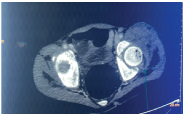
Figure 1: Axial computed tomography image of the left hip demonstrating joint effusion (green arrow).
On day 27, the clinical picture worsened. The patient experienced severe pain around the left hip, and he became tachycardic (heart rate = 123 bpm) with a raised WCC of 13.15 × 10⁹/L and a CRP of 104 mg/L. A second open arthrotomy and debridement were done, with a finding of 100 mL of murky fluid, which cultured Ertapenem-sensitive Escherichia coli.
Postoperatively, on day 32, blood results improved with a WCC of 7.21 × 10⁹/L and a CRP of 80 mg/L. On day 34, the patient was taken to the theater for a relook, and no pus was found intraoperatively. (Fig. 2).
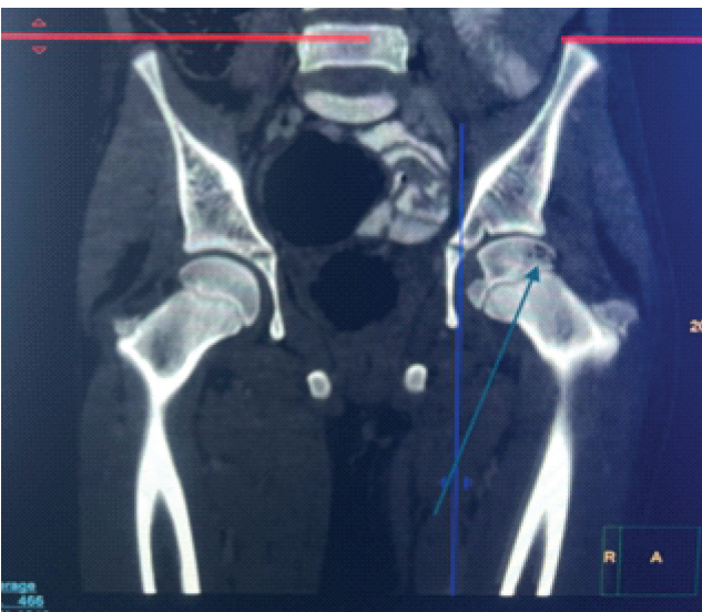
Figure 2: Coronal computed tomography image of the left hip demonstrating joint effusion and femoral head permeative changes (green arrow).
Supportive care and nutritional support were instituted. The patient had a total of 32 days of ceftriaxone intravenously, and he was discharged home on day 36. Physiotherapist-guided mobilization was started before discharge, and at 2-week follow-up, the patient showed significant clinical improvement, with painless ambulation and a good ROM of the hip.
The pathogenesis of SA constitutes an invasion of the synovial membrane of the hip joint by bacteria and a subsequent inflammatory process, which produces the characteristic purulent synovial fluid observed at arthrocentesis. SA can result from hematogenous spread secondary to bacteremia or directly from penetrating joint trauma and/or corticosteroid joint injections [9]. The pathogenesis of bacteremia and sepsis because of a non-perforated appendix is still unknown; however, it has been suggested that this occurs due to microorganism release systematically from the lumen of the intestine [6,10,11,15]. We present a previously healthy 13-year-old male who developed SA of the hip in the context of appendicitis.
The diagnosis of SA is established by meeting clinical and biochemical features described by the Modified Kocher Criteria, imaging, and synovial fluid analysis. The Kocher Criteria consist of factors that differentiate it from transient synovitis, such as inability to bear weight, fever >38.5°C, serum WCC > 12,000/mm3; erythrocyte sedimentation rate >40 mm/h and a CRP >20 mg/L [2,16].
Radiological imaging in the form of ultrasound and magnetic resonance imaging are good non-invasive diagnostic tools without radiation exposure in the evaluation of SA [2]. Joint effusion on ultrasound is seen in 91% of patients with SA; however, its limitation lies in differentiating purulent, sterile, or hemorrhagic effusion. Synovial fluid analysis revealing a positive culture and a positive Gram stain of the joint fluid or a WBC count in the joint fluid of >50,000/mm3 is highly suggestive of SA [2,9,15,16]. Our patient was febrile, could not bear weight, and had a raised CRP at presentation. Based on this alone, the patient had an 82.6% probability of SA [7,9]. Ultrasound imaging revealed a complex left hip effusion suggestive of SA. Synovial fluid assessment post-initial arthrotomy of the left hip cultured Salmonella Typhi.
Salmonella is a Gram-negative bacterium. There are two subclasses, namely typhoidal and non-typhoidal [15,17]. Typhoidal species are known to cause enteric fever. Infection of the bones and joints by Salmonella is rare, and limited cases have been reported [17]. Salmonella has been reported to cause mono-arthritis of the hip joint, with few reported cases affecting the knee joint [11,17]. In all cases, hematogenous spread is the route of inoculation for Salmonella SA. Furthermore, 10–15% of cases are blood culture positive [11,17].
Our patient had a 10-day history of gastrointestinal symptoms, which suggests an enteric infection preceding his musculoskeletal symptoms. Salmonella Typhi was then isolated from an arthrotomy specimen, and this supports the likelihood of hematogenous spread from the gastrointestinal tract. The Harris Hip Score (HHS) is a beneficial tool to evaluate hip function. It is objective and standardized and therefore helpful in assessing hip function and monitoring progression over time. Grading of HHS ranges from 1 to 100, grouped as follows: Very bad (1–31), bad (31–55), good (56–75), and very good (76–100) [18,19]. Our patient had an HHS of 76, indicating very good hip function at 2 weeks of follow-up.
Literature Review
Only a few cases of this similitude are reported in English literature (Table 1) [11,12,13].
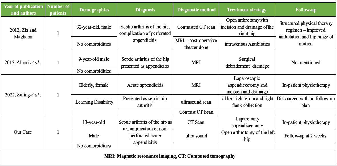
Table 1: Cases of septic arthritis of the hip and appendicitis: Review of literature
The case report presents a rare clinical scenario of SA of the hip complicating non-perforated appendicitis due to Salmonella Typhi in a previously healthy 13-year-old male. The case demonstrates that bacteria can spread hematogenously from the gastrointestinal tract without appendiceal perforation, resulting in distant joint seeding in immunocompetent individuals. Early diagnosis, prompt intervention, and holistic multidisciplinary management are vital in preventing permanent disability and achieving good functional outcomes in atypical presentations of septic arthritis.
This case highlights that in a child with persistent fever and new joint symptoms following a non-perforated appendicectomy, clinicians should maintain a high index of suspicion for hematogenous septic arthritis. In endemic areas, Salmonella Typhi should be considered, and urgent imaging with prompt surgical drainage is warranted.
References
- 1. D’Angelo F, Monestier L, Zagra L. Active septic arthritis of the hip in adults: What’s new in the treatment? A systematic review. EFORT Open Rev 2021;6:164-72. [Google Scholar] [PubMed]
- 2. Donders CM, Spaans AJ, Van Wering H, Van Bergen CJ. Developments in diagnosis and treatment of paediatric septic arthritis. World J Orthop 2022;13:122-30. [Google Scholar] [PubMed]
- 3. Wu KA, Kugelman DN, Seidelman JL, Seyler TM. Native joint septic arthritis. Antibiotics (Basel) 2024;13:596. [Google Scholar] [PubMed]
- 4. Horowitz DL, Katzap E, Horowitz S, Barilla-LaBarca ML. Approach to septic arthritis. Am Fam Physician 2011;84:653-60. [Google Scholar] [PubMed]
- 5. García-Arias M, Balsa A, Mola EM. Septic arthritis. Best Pract Res Clin Rheumatol 2011;25:407-21. [Google Scholar] [PubMed]
- 6. Faraji-Goodarzi M. Sepsis after non-perforated acute appendicitis. Clin Case Rep 2019;7:520-3. [Google Scholar] [PubMed]
- 7. Iseri Nepesov M, Kilic O, Sali E, Yesil E, Akar A, Kaman A, et al. Pathogens in pediatric septic arthritis: A multi-center study in Turkiye (PEDSART study). Children (Basel) 2024;11:134. [Google Scholar] [PubMed]
- 8. a. Lavy CB. Septic arthritis in Western and sub-saharan African children – a review. Int Orthop 2007;31:137-44. [Google Scholar] [PubMed]
- 9. Momodu II, Savaliya V. Septic Arthritis Internet. StatPearls. Treasure Island (FL): StatPearls Publishing; 2023. Available from: https://www.ncbi.nlm.nih.gov/books/nbk538176 [Last accessed: 20/08/2025]. [Google Scholar] [PubMed]
- 10. Shalman A, Acker A, Shalman A, Frank D, Borer A, Koyfman L, et al. Septic arthritis of the hip joint due to Bacteroides fragilis in a paraplegic patient. Access Microbiol. 2019;1:e000071. [Google Scholar] [PubMed]
- 11. Zia M, Maghami N. Septic arthritis of the hip, an unreported complication of perforated appendicitis: A case report. Int J Surg Case Rep 2020;72:281-4. [Google Scholar] [PubMed]
- 12. Zuling E T, Gana T, Al-Mukhtar A. A case report of appendicitis masquerading as septic arthritis. J Clin Med Surg 2022;2:1049. [Google Scholar] [PubMed]
- 13. Alhajri S, Chorny V. Septic arthritis of hip joint mimicking appendicitis. Chest 2017;152:A303. [Google Scholar] [PubMed]
- 14. Sohrabi C, Mathew G, Maria N, Kerwan A, Franchi T, Agha RA, et al. The SCARE 2023 guideline: Updating consensus surgical case report (SCARE) guidelines. Int J Surg 2023;109:1136-40. [Google Scholar] [PubMed]
- 15. Lo I, Chang H. Salmonella septic arthritis in a patient with a hip implant: A case report. Int J Gerontol 2018;12:150-2. [Google Scholar] [PubMed]
- 16. Valisena S, De Marco G, Vazquez O, Cochard B, Steiger C, Dayer R, et al. The Kocher-Caird criteria for pediatric septic arthritis of the hip: Time for a change in the Kingella era? Microorganisms 2024;12:550. [Google Scholar] [PubMed]
- 17. Gharib MH, Alebbi S, Rashid F, Elhaj MF, Zahirb FZ, Al Emadi S. Salmonella-related septic arthritis in an immunocompetent adult: A case report. Cureus 2022;14:e21030. [Google Scholar] [PubMed]
- 18. Lee SY, Park SJ, Gim JA, Kang YJ, Choi SH, Seo SH, et al. Correlation Between Harris Hip Score and Gait Analysis through Artificial Intelligence Pose Estimation in Patients After Total Hip Arthroplasty. Research Square [Preprint]; 2022. [Google Scholar] [PubMed]
- 19. Ramadanov N, Voss M, Hable R, Hakam HT, Prill R, Salzmann M, et al. Postoperative Harris hip score versus Harris hip score difference in hip replacement: What to report? Orthop Surg 2025;17:3-21. [Google Scholar] [PubMed]
Related Articles in Journal of Orthopaedic Case Reports
 November 1, 2026 Septic Arthritis Complicating Growth Plate Integrity in Pediatric Cerebral Palsy: A Rare Presentation
 September 1, 2026 Right Knee Ureaplasma Septic Arthritis in an Immunocompromised Host, Requiring Right Hip Disarticulation for Definitive Source Control
 September 1, 2026 Serratia Marcescens Septic Arthritis of the Ankle in a Compromised Host: Ring Fixator Arthrodesis as Salvage, a Case Report
 July 1, 2026 Tuberculosis of the Elbow in a Chronic Kidney Disease Patient with Arteriovenous Fistula: A Case Report