

Sterile-glove-assisted negative pressure wound therapy provides a simple and adaptable method for maintaining a seal across hand and forearm anatomy.
Joaquin Miguel del Mundo, Kirk Kerkorian School of Medicine at UNLV, Las Vegas, Nevada, United States. E-mail: delmuj1@unlv.nevada.edu
Abstract
Introduction: Negative pressure wound therapy (NPWT) is widely used in the management of complex wounds following trauma, infection, and debridement. However, application to the hand can be technically challenging because of the complex anatomy of the digits, web spaces, and transitions between the dorsal and palmar surfaces. This can make maintenance of an airtight seal difficult. We describe a sterile glove-assisted NPWT technique used in the management of an extensive hand and forearm wound secondary to necrotizing fasciitis.
Case Report: A 59-year-old woman presented with a painful, erythematous wound involving the left hand and forearm. In <4 h, her mental status greatly declined and she was ultimately diagnosed with necrotizing fasciitis. Wound cultures grew Strep pyogenes and Candida albicans, and the patient was treated with fluconazole and serial operative debridement. Following debridement, NPWT was applied using foam over the wound and a sterile glove over the hand. The glove was incorporated into an occlusive dressing using adhesive drapes and gauze extending proximally along the extremity, creating an airtight seal. The dressing was connected to continuous negative pressure. This technique was used during staged wound management before definitive split-thickness skin grafting.
Conclusion: Sterile glove-assisted NPWT provided a practical and simple method of maintaining negative pressure across complex hand and forearm anatomy during staged treatment.
Keywords: Negative pressure wound therapy, wound vac, hand wound, upper extremity wound, necrotizing fasciitis, sterile glove, soft-tissue reconstruction.

Negative pressure wound therapy (NPWT) is a widely used treatment modality for soft-tissue injuries. These injuries primarily include trauma, burns, infections, and post-operative complications [1,2,3]. NPWT promotes wound healing through multiple mechanisms, including decreasing the wound surface area, accelerating granulation tissue formation by inducing microdeformations that stimulate cellular proliferation, and minimizing bacterial burden and edema with constant or intermittent evacuation of exudate from the wound [4,5,6,7,8,9]. These benefits make NPWT particularly useful in many settings; however, application to the hand can be technically challenging due to the irregular topography of the digits, web spaces, palm creases, as well as the multiple motion segments of the many carpal and interphalangeal joints [5,6]. These non-uniform surfaces not only make application difficult but also create multiple potential sites for seal loss, leading to a decrease in the efficacy of the wound therapy [10].
We present a case of extensive hand and forearm necrotizing fasciitis treated with serial debridement and staged reconstruction in which a sterile glove-assisted NPWT technique was used to facilitate application and maintenance before final coverage.
A 59-year-old woman with a past medical history of hypothyroidism, hypertension, major depressive disorder, dementia, and opioid use disorder presented with a left hand and arm wound. She reported significant pain, blistering, and swelling in her hand starting 2 days before presenting to the emergency department (ED). She denied any recent trauma. While in the ED, she was hemodynamically stable. Physical examination demonstrated a left dorsal hand wound with erythema and skin breakdown. Significant findings on laboratory analysis included white blood cell count elevated to 17.23 k/mm3, sodium 134 mmol/L, blood urea nitrogen of 32 mg/dL, creatinine of 1.88 mg/dL, erythrocyte sedimentation rate of 52 mm/h, and C-reactive protein >200 mg/L. Her LRINEC score was 10. She underwent X-rays and magnetic resonance imaging of her left hand and forearm which showed soft tissue swelling without evidence of osteomyelitis. She was emergently taken back to the operating room for incisional debridement. In total, she had 4 debridements managed with sterile-glove-assisted NPWT. Intraoperative cultures grew Strep pyogenes and Candida albicans. She received Unasyn 3 g in the ED, then every 6 h for 6 days when admitted, linezolid 600 mg every 12 h for 7 days, and fluconazole 400 mg daily for 10 days, which was transitioned to augmentin 875 mg twice a day for 5 days upon discharge from the hospital. The patient’s final surgery was a split-thickness skin graft of her left hand and left forearm, which occurred on day 16 of her hospitalization. She was discharged on day 18 and instructed to follow up with Wound Care. Her first outpatient follow-up occurred 3 days after discharge. During this initial outpatient visit, she reported less pain and underwent her first skin graft dressing takedown. At this visit, she had a punctate area of tendon exposed. Bacitracin and Xeroform were applied to the area, and she was instructed to continue with daily Bacitracin Xeroform changes. Her second outpatient follow-up was 2 weeks later and she was found to have 90% incorporation of her graft. She had a total of 8 wound care visits for dressing changes. Her last wound care visit was 28 days after discharge and she endorsed tolerating the graft and major improvement in her mobility with no complications noted.
Surgical technique
After debridement, appropriate hemostasis, and thorough irrigation, the extremity is dried with lap sponges. All areas of the skin that are able to be closed should be approximated with non-absorbable suture such as nylon (Fig. 1).
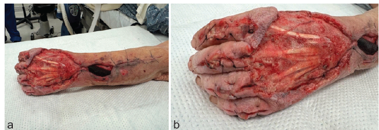
Figure 1: (a) Dorsal view of left upper extremity with significant soft tissue defect requiring negative pressure wound therapy, (b) More distal view of the same extremity demonstrating extent of digital involvement.
A 4 cm large pore foam sponge should be trimmed to the appropriate thickness and shape; we cut the sponge to 2 cm thick and used it to cover all exposed, fascia, and subcutaneous tissue. If there are exposed tendons, it is recommended you use a small-pore foam sponge. You may then secure the sponge in place with staples or suture. All other approximated surgical incisions should be covered with a non-adherent petrolatum gauze and dry sterile gauze pads secured in place with staples or sutures (Fig. 2).
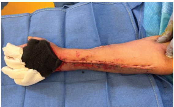
Figure 2: Dorsal view of left upper extremity after gauze and foam placement.
Once adequate coverage of all surgical incisions and wounds is confirmed, a large sterile glove should be placed over the hand (Fig. 3).
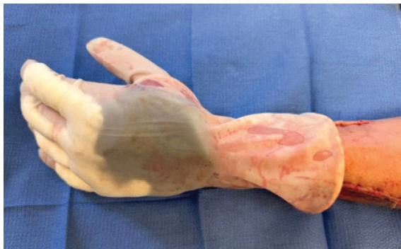
Figure 3: Dorsal View of left hand after sterile glove was applied.
An adhesive drape is then cut into strips roughly 6 cm wide. Each strip is placed longitudinally starting distal to the cuff of the glove and extending proximally to the desired level. This should be repeated with each strip having a 1 cm overlap until there is a seal circumferentially around the cuff of the glove (Fig. 4).
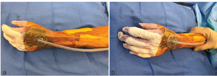
Figure 4: (a) Dorsal view of left upper extremity after a seal was made with an adhesive drape and initiation of Negative pressure wound therapy, (b) More distal view of the same extremity demonstrating fingertip coverage.
Optionally, at this point, if the surgeon prefers the patient’s fingertips to be free, they may cut the fingers off the glove and use an adhesive drape to create a seal at the skin-glove interface. At this point, the remainder of the NPWT application follows standard application instructions. A hole is cut in the adhesive drape and glove if needed, and the adhesive vacuum tubing pad is applied. The desired level of negative pressure is selected; we recommend −125 mmHg continuously. This technique is safe for multiple-day applications and may also be employed for bedside wound vac changes until the patient is ready for definitive coverage (Fig. 5). In addition, a splint may be placed over the dressing if needed.
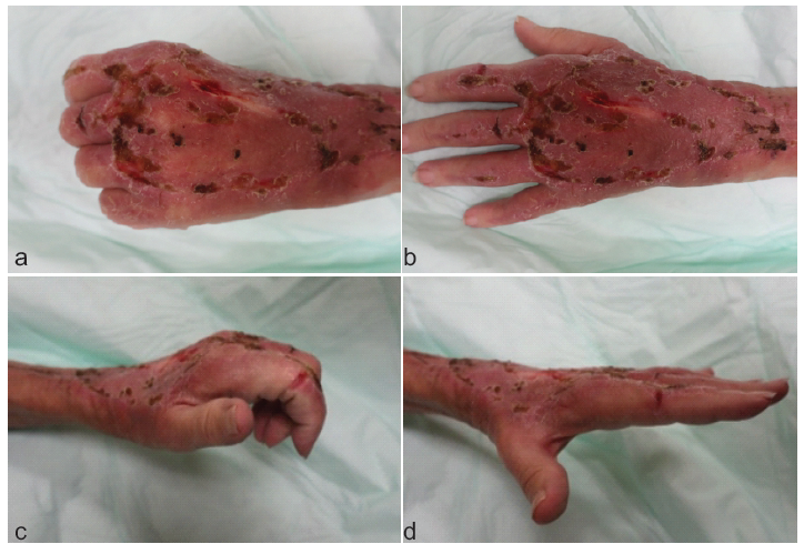
Figure 5: Images demonstrating motion and soft tissue healing of left upper extremity at six weeks status post index surgery.
There are many options for managing complex wounds on the hands; however, NPWT is a mainstay for managing complex wounds. The benefits of NPWT have been demonstrated in wounds with significant tissue loss, exposed critical structures, increased risk of infection, and need for multi-step reconstruction [1,2,3].
Although NPWT remains a popular option for temporizing a complex wound, there remains to be a standard approach to applying a vacuum dressing to the hand. We believe that the key reason for this is due to the comparatively complex nature of applying NPWT to the hand as compared to other aspects of the body. In addition, hand wounds themselves occur in a variety of shapes and locations that often require a more individualized approach for successful application. Proper dressing configuration and amount of negative pressure applied to the wound are essential to adequate NPWT and can be particularly challenging in the hand due to its vascularity and anatomy [11,12]. Several authors have attempted to solve this issue by describing novel NPWT application techniques using adhesive constructs, gauze-based dressings, and “mitten” configurations to maintain an adequate seal, stabilize grafting, and preserve functional positioning [13,14,15]. Other techniques such as the ones described by Sreelesh and Bhandari in 2016 and Klein et al. in 2022 have shown to be effective; however, these techniques have limitations that restrict their generalizability. Some considerations are limited access to materials such as collagen powders, reluctance to place black foam directly on intact skin as it may cause friction, painful irritation, and ultimately skin breakdown, and no described ability to allow the access to digits to monitor perfusion [16,17].
The technique we used differs from previous applications of NPWT in that the sterile glove primarily functions as an outer sealing envelope rather than wound-contact material. This allows the foam to be selectively placed over the wound and sensitive neurovascular structures within it. The glove also eliminates the need to individually seal each interdigital space while also being airtight. Similar to the other described techniques is the potential risk of limiting vascular supply to the hand and digits from the compressive effects of a circumferential vacuum dressing; however, another unique advantage of this technique is that the glove fingertips can be easily removed and individually sealed, preserving access for neurovascular monitoring.
There are limitations to this technique. To be employed successfully, a surgeon must operate at a facility that has access to wound vacuum devices and ideally has ancillary staff that can manage these devices on the floor. The facility must also carry sterile gloves in a large enough size that can fit loosely on a patient’s hand and accommodate swelling. Some institutions do not regularly stock glove sizes larger than 8.5 or 9 which may limit the employment of this technique to patients with smaller hands. In addition, if a patient’s wounds or surgical incisions extend distally past the interphalangeal joints, it limits the ability to remove the glove fingertips and maintain an airtight seal.
This case demonstrates that sterile glove-assisted NPWT can provide a practical and adaptable method for managing complex hand wounds where conventional dressings and standard NPWT application are limited by complexity. Using the sterile glove as an outer sealing envelope, this technique creates an airtight domain around the hand while preserving access for neurovascular assessment.
In this patient, the technique allowed for multiple quick straightforward NPWT applications, decreasing OR time and blood loss. Sterile glove-assisted NPWT represents a useful option for management of a wide variety of hand and forearm wounds.
While NPWT is a well-documented tool in wound management, complex anatomy can limit its application. This case report demonstrates the successful use of sterile-glove-assisted NPWT.
References
- 1. Huang C, Leavitt T, Bayer LR, Orgill DP. Effect of negative pressure wound therapy on wound healing. Curr Probl Surg 2014;51:301-31. [Google Scholar] [PubMed]
- 2. Liu C, Xie H, Wei P, Gong T, Wu G, Xu Z, et al. Clinical study of early rehabilitation training combined with negative pressure wound therapy for the treatment of deep partial-thickness hand burns. Front Surg 2023;10:1040407. [Google Scholar] [PubMed]
- 3. Patel S, Nischal SA, Kale KM, Dubb A, Prasad SK, Refai D. Incisional negative pressure wound therapy for prevention of surgical site infection: A systematic review and meta-analysis. JAMA Surg 2026;161:1024-32. [Google Scholar] [PubMed]
- 4. Robert N. Negative pressure wound therapy in orthopaedic surgery. Orthop Traumatol Surg Res 2017;103:S99-103. [Google Scholar] [PubMed]
- 5. Quacinella MA, Yong TM, Obremskey WT, Stinner DJ. Negative pressure wound therapy: Where are we in 2022? OTA Int 2023;6:e247. [Google Scholar] [PubMed]
- 6. Lesiak AC, Shafritz AB. Negative-pressure wound therapy. J Hand Surg Am 2013;38:1828-32. [Google Scholar] [PubMed]
- 7. Webb LX, Pape HC. Current thought regarding the mechanism of action of negative pressure wound therapy with reticulated open cell foam. J Orthop Trauma 2008;22:S135-7. [Google Scholar] [PubMed]
- 8. Borgquist O, Ingemansson R, Malmsjo M. Wound edge microvascular blood flow during negative-pressure wound therapy: Examining the effects of pressures from -10 to -175 mmHg. Plast Reconstr Surg 2010;125:502-9. [Google Scholar] [PubMed]
- 9. Birke-Sorensen H, Malmsjo M, Rome P, Hudson D, Krug E, Berg L, et al. Evidence-based recommendations for negative pressure wound therapy: Treatment variables (pressure levels, wound filler and contact layer)–steps towards an international consensus. J Plast Reconstr Aesthet Surg 2011;64:S1-16. [Google Scholar] [PubMed]
- 10. Collinge C, Reddix R. The incidence of wound complications related to negative pressure wound therapy power outage and interruption of treatment in orthopaedic trauma patients. J Orthop Trauma 2011;25:96-100. [Google Scholar] [PubMed]
- 11. Kairinos N, Solomons M, Hudson DA. Negative-pressure wound therapy I: The paradox of negative-pressure wound therapy. Plast Reconstr Surg 2009;123:589-98. [Google Scholar] [PubMed]
- 12. Kairinos N, Voogd AM, Botha PH, Kotze T, Kahn D, Hudson DA, et al. Negative-pressure wound therapy II: Negative-pressure wound therapy and increased perfusion. Just an illusion? Plast Reconstr Surg 2009;123:601-12. [Google Scholar] [PubMed]
- 13. Kang GCW, Yam A. Vacuum-assisted closure of a large palmar defect after debriding a midpalmar tuberculous abscess. Int Wound J 2008;5:45-8. [Google Scholar] [PubMed]
- 14. Taylor CJ, Chester DL, Jeffery SL. Functional splinting of upper limb injuries with gauze-based topical negative pressure wound therapy. J Hand Surg Am 2011;36:1848-51. [Google Scholar] [PubMed]
- 15. Kasukurthi R, Borschel GH. Simplified negative pressure wound therapy in pediatric hand wounds. Hand (N Y) 2010;5:95-8. [Google Scholar] [PubMed]
- 16. Sreelesh LS, Bhandari PL. An easy technique for negative-pressure wound therapy for extremities using collagen powder and sterile gloves. Indian J Surg 2017;79:81-3. [Google Scholar] [PubMed]
- 17. Klein DS, Yingling JM, Patel P, Capo JT. Vacuum-assisted therapy for combined volar-dorsal soft-tissue defects of the hand: A case report. Adv Skin Wound Care 2022;35:57-61. [Google Scholar] [PubMed]
Related Articles in Journal of Orthopaedic Case Reports
 November 1, 2026 Limb Salvage in Limb-Threatening Necrotizing Fasciitis of the Diabetic Foot: A Case Report
 October 1, 2026 Degloving in Disguise: Managing a Delayed Spinal Morel-Lavallée Lesion after Thoracic Fracture-Dislocation
 October 1, 2026 Early Surgical Escalation and Structured Multidisciplinary Pathways Improve Limb-Salvage Outcomes in Necrotizing Fasciitis: A Two-Cycle Quality Improvement Project
 May 1, 2026 Segmental Achilles Tendon Loss With Skin Defect in an Open Calcaneal Fracture Managed Without Tendon Reconstruction: A Case Report