


Introduction
Necrotizing fasciitis (NF) is a rare but fulminant infection of the deep soft tissues that progresses rapidly along fascial planes, causing widespread necrosis, systemic sepsis, and multiorgan failure. Published mortality rates range from 20% to 40%, with higher figures consistently associated with diagnostic delay, inadequate initial debridement, and failure to involve senior decision-makers at the point of care [1]. Despite its severity, the early clinical presentation of NF is frequently indistinguishable from that of non-necrotizing cellulitis, and no single diagnostic test carries sufficient sensitivity to exclude the diagnosis confidently [2].
Orthopedic units bear particular responsibility for managing NF, since the majority of cases involve the lower extremity, where musculoskeletal involvement and the proximity of fascial compartments amplify the risk of limb loss [3]. The Laboratory Risk Indicator for Necrotizing Fasciitis (LRINEC) score provides a structured hematological screen. It remains the most widely validated bedside risk-stratification tool for distinguishing NF from other soft-tissue infections. However, its sensitivity for early disease is imperfect, and it should not be used in isolation [4]. Adjunctive bedside tools – most notably the finger-sweep test – and cross-sectional imaging with computed tomography (CT) contribute meaningful diagnostic information, particularly in atypical presentations [5].
Early surgical debridement is the single most consistently reported determinant of survival and limb salvage in NF, yet translating this evidence into consistent practice remains difficult in the emergency setting, where diagnostic uncertainty and variable escalation pathways often delay operative intervention [6,7]. Formalized quality improvement (QI) frameworks have increasingly been applied to surgical emergency pathways to reduce variation, accelerate decision-making, and improve documentation. Still, their application to NF care specifically remains limited [8,9].
The rationale for this study arises directly from this gap: while the association between surgical delay and poor outcome in NF is well established, little evidence exists on whether a structured, protocol-driven diagnostic and escalation pathway can measurably shorten that delay within a routine orthopedic service, rather than in a specialist or research setting [10,11]. A locally-led, protocol-based QIP therefore offered a pragmatic way to test this within existing service constraints, using a unit’s own baseline practice as the comparator rather than an external control.
The objectives of this study were therefore twofold: first, to determine whether implementation of a structured diagnostic checklist, mandatory LRINEC and finger-sweep documentation, and early multidisciplinary team (MDT) activation reduced the time from presentation to surgical decision in patients with suspected or confirmed NF; and second, to evaluate the impact of this pathway on limb-salvage and mortality outcomes, comparing a baseline audit cycle against a second cycle following intervention.
Materials and Methods
Study design and setting
This was a two-cycle, retrospective, single-center QI project (QIP), structured as a before-and-after comparison of a consecutive case series rather than a randomized or blinded trial. It was conducted at a tertiary orthopedic center in Newport, United Kingdom, between June 2022 and October 2024. The unit manages the full spectrum of lower-limb NF and acts as a regional referral center for complex soft-tissue infections. Cycle 1 (June 2022–March 2023) established baseline practice, and Cycle 2 (April 2023–October 2024) assessed the impact of structured interventions. All patients were identified through electronic health records and specialist referral logs. The study was conducted under the auspices of institutional service evaluation and did not require formal ethics committee review; all data were anonymized before analysis.
Sample size
No formal a priori power calculation was performed, as this project was designed as a QI audit of consecutive, naturally occurring NF presentations rather than a hypothesis-driven comparative study with a pre-specified effect size. Sample size in each cycle was therefore determined pragmatically by the total number of patients meeting inclusion criteria within each cycle’s fixed data-collection window (9 and 8 months respectively), giving 14 patients in Cycle 1 and 13 in Cycle 2. This is acknowledged as a limitation for the secondary outcomes in the Discussion, where the modest sample size limits statistical power to detect smaller effects.
Patient identification and data extraction
All patients aged ≥18 years admitted under orthopedic care with a confirmed or clinically suspected diagnosis of NF during the study period were included. Confirmation required either intraoperative findings consistent with NF (gray necrotic fascia, lack of bleeding, tissue plane dissection without resistance) or microbiological/histopathological evidence. Cases managed conservatively without operative exploration were included if the clinical diagnosis was substantiated by imaging and clinical progression.
Data extraction encompassed: patient demographics and comorbidities; anatomical site of infection; causative organisms and antibiotic sensitivities; LRINEC score at presentation; performance and outcome of finger-sweep test; CT imaging results; time from initial presentation to first senior clinical review; time from presentation to operative decision; type and number of surgical procedures; length of hospital stay; and final clinical outcome (recovery, major amputation, or in-hospital death).
Cycle 1: Baseline assessment
Cycle 1 (June 2022–March 2023) was conducted prior to any structured intervention and reflected existing departmental practice for suspected NF. No diagnostic checklist, mandatory documentation standard, or fixed escalation timeframe was in place; LRINEC scoring, finger-sweep testing, CT imaging, and MDT referral were performed at individual clinician discretion rather than as protocolized steps. Data on time to senior review, time to surgical decision, diagnostic test use, and clinical outcomes were collected retrospectively for this cycle to establish a baseline against which the Cycle 2 intervention bundle could be measured. Findings from Cycle 1 – in particular the low rates of LRINEC and finger-sweep documentation and the long interval to surgical decision – were reviewed by the multidisciplinary group and directly informed the design of the structured intervention bundle described below.
QI interventions (Cycle 2)
Following completion of Cycle 1, a series of targeted interventions were developed collaboratively with orthopedic, emergency medicine, infectious diseases, microbiology, and plastic surgery teams:
-
Implementation of a structured NF diagnostic checklist at the point of initial senior review
-
Mandatory documentation of LRINEC score within two hours of presentation
-
Standardized recording of finger-sweep test performance and result
-
Mandatory CT documentation with confirmation of whether gas, fascial edema, or soft-tissue emphysema was identified
-
Direct escalation to consultant orthopedic surgeon within 4 h of presentation if NF was suspected
-
Compulsory early MDT activation including infectious diseases, microbiology, and plastic surgery
-
Interactive education sessions for emergency department nursing and junior medical staff on NF recognition and escalation triggers.
Statistical analysis
Descriptive statistics were used to summarize patient demographics and baseline characteristics. Categorical variables were compared between cycles using Chi-squared or Fisher’s exact tests, as appropriate. Continuous variables were assessed using an independent samples t-test or Mann–Whitney U test depending on data distribution. Statistical significance was defined as P < 0.05. All analyses were performed using Statistical Package for the Social Sciences Statistics version 27.0 (IBM Corp., Armonk, NY, USA). Results are presented with 95% confidence intervals where applicable.
Results
Patient demographics and baseline characteristics
A total of 27 patients were included across both cycles (Cycle 1: n = 14; Cycle 2: n = 13). The mean age was 52.1 ± 11.3 years, with a slight male predominance (63%). Diabetes mellitus was the most prevalent comorbidity, present in 70% of cases. Lower-limb infection predominated in both cycles (78.6% and 84.6%, respectively), and polymicrobial infection was identified in the majority of cultured specimens. Baseline characteristics were comparable between cycles, with no statistically significant intergroup differences (Table 1).
Baseline patient demographics and microbiology by cycle
| Characteristic | Cycle 1 (n=14) | Cycle 2 (n=13) | P-value |
|---|---|---|---|
| Age, years (mean±SD) | 51.4 ± 12.1 | 52.9 ± 10.6 | 0.72 |
| Male sex, n (%) | 9 (64.3) | 8 (61.5) | 0.87 |
| Diabetes mellitus, n (%) | 10 (71.4) | 9 (69.2) | 0.89 |
| Immunosuppression, n (%) | 4 (28.6) | 3 (23.1) | 0.71 |
| Peripheral vascular disease, n (%) | 3 (21.4) | 2 (15.4) | 0.66 |
| Obesity (BMI >30), n (%) | 5 (35.7) | 4 (30.8) | 0.76 |
| Lower limb, n (%) | 11 (78.6) | 11 (84.6) | 0.66 |
| Upper limb, n (%) | 2 (14.3) | 1 (7.7) | 0.55 |
| Perineum/trunk, n (%) | 1 (7.1) | 1 (7.7) | 0.95 |
| Polymicrobial infection, n (%) | 8 (57.1) | 7 (53.8) | 0.84 |
| Group A Streptococcus, n (%) | 4 (28.6) | 4 (30.8) | 0.89 |
| Culture negative, n (%) | 2 (14.3) | 2 (15.4) | 0.93 |
Groups were compared using Chi-squared or Fisher’s exact test for categorical variables and an independent samples t-test for continuous variables. No significant differences between cycles. SD: Standard deviation, BMI: Body mass index
Diagnostic timeliness and documentation
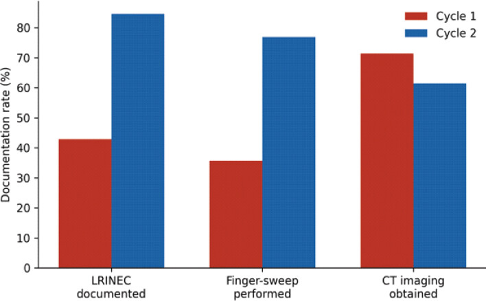
Following QIP implementation, significant improvements were observed across all documentation metrics (Table 2). LRINEC score utilization nearly doubled, from 42.9% in Cycle 1 to 84.6% in Cycle 2 (P = 0.01). Finger-sweep test performance rose from 35.7% to 76.9% (P = 0.02). Time to first senior clinical review fell from a mean of 4.2 ± 1.8 h to 2.1 ± 0.9 h (P = 0.003). Most strikingly, the mean time from presentation to operative decision decreased from 11.5 ± 3.2 h in Cycle 1 to 6.8 ± 2.1 h in Cycle 2 (P = 0.002), representing a 41% reduction. MDT involvement increased from 50.0% to 92.3% (P = 0.01). CT imaging use appeared marginally lower in Cycle 2; however, this reflected documentation inconsistency rather than a true reduction in practice.
Diagnostic metrics and timeliness by audit cycle
| Diagnostic metric | Cycle 1 (n=14) | Cycle 2 (n=13) | P-value |
|---|---|---|---|
| LRINEC score documented, n (%) | 6 (42.9) | 11 (84.6) | 0.01* |
| LRINEC ≥6 (high risk), n (%) | 4 (66.7) | 9 (81.8) | 0.41 |
| LRINEC mean score (±SD) | 5.8±2.3 | 7.2±1.9 | 0.09 |
| Finger-sweep test performed, n (%) | 5 (35.7) | 10 (76.9) | 0.02* |
| Positive finger-sweep, n (%) | 4 (80.0) | 9 (90.0) | 0.54 |
| CT imaging obtained, n (%) | 10 (71.4) | 8 (61.5) | 0.57 |
| Gas on CT, n (%) | 6 (60.0) | 5 (62.5) | 0.91 |
| Time to senior review, hours (mean±SD) | 4.2±1.8 | 2.1±0.9 | 0.003* |
| Time to surgical decision, hours (mean±SD) | 11.5±3.2 | 6.8±2.1 | 0.002* |
| MDT involvement, n (%) | 7 (50.0) | 12 (92.3) | 0.01* |
*
Statistically significant (P<0.05). LRINEC: Laboratory risk indicator for necrotizing fasciitis, CT: Computed tomography, SD: Standard deviation, MDT: Multidisciplinary team
Clinical outcomes
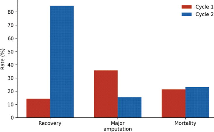
Cycle 2 demonstrated marked improvements in limb-salvage outcomes (Table 3). Recovery rates – defined as discharge home with functional limb preservation – rose from 14.3% in Cycle 1 to 84.6% in Cycle 2 (P < 0.001). Major limb amputation rates fell from 35.7% to 15.4% (P = 0.04). Limb-salvage debridement rates trended upward (42.9% vs. 61.5%; P = 0.15), though this did not reach statistical significance, likely reflecting the modest sample size. In-hospital mortality remained comparable across both cycles (21.4% vs. 23.1%; P = 0.91), consistent with the observed persistence of fulminant disease in a subset of patients despite improved surgical timeliness. Secondary outcomes including intensive care admission rate, median number of debridement procedures, and length of hospital stay showed numerical improvement in Cycle 2, though none reached statistical significance (Figs. 1, 2, 3).
Clinical outcomes by audit cycle
| Outcome | Cycle 1 (n =14) | Cycle 2 (n =13) | P-value |
|---|---|---|---|
| In-hospital mortality, n (%) | 3 (21.4) | 3 (23.1) | 0.91 |
| Recovery (discharged home), n (%) | 2 (14.3) | 11 (84.6) | <0.001* |
| Major limb amputation,n (%) | 5 (35.7) | 2 (15.4) | 0.04* |
| Limb-salvage debridement, n (%) | 6 (42.9) | 8(61.5) | 0.15 |
| Intensive care admission, n (%) | 8 (57.1) | 7 (53.8) | 0.84 |
| Total debridement procedures (median) | 2.5 (1–5) | 2.0 (1–4) | 0.48 |
| Hospital length of stay, days (mean±SD) | 28.4±11.6 | 22.1±9.3 | 0.08 |
| Wound closure/skin graft, n (%) | 5 (35.7) | 8(61.5) | 0.15 |
| Re-operation rate, n (%) | 4 (28.6) | 3 (23.1) | 0.72 |
| Discharge to rehabilitation, n (%) | 4 (28.6) | 8(61.5) | 0.07 |
*
Statistically significant (P<0.05). Values represent n (%) unless otherwise stated
![Figure 1: Mean time to surgical decision (h) in Cycle 1 (pre-quality improvement project [QIP]) versus Cycle 2 (post-QIP). Error bars represent±standard deviation. The 41% reduction was statistically significant (P=0.002).](https://jocr.co.in/wp/wp-content/uploads/599FF1-converted.jpg)
Discussion
This two-cycle QIP demonstrates that structured, protocol-driven interventions can substantially reduce the time from presentation to operative decision and significantly improve limb-salvage rates in orthopedic NF management. The 41% reduction in time to surgical decision is clinically meaningful, given that necrotizing infection can progress within hours [1], that organized systems of care are considered central to its management [2], and that surgical delay is consistently associated with higher mortality [3–5]. The Infectious Diseases Society of America guidelines call for urgent surgical inspection and debridement when a necrotizing infection is suspected [6]; however, most published series reporting favorable outcomes document surgical exploration considerably earlier [7–9]. Our data support the premise that reducing this interval – even within a service where care was already relatively prompt – yields tangible benefits in terms of limb salvage.
The dramatic improvement in recovery rates (14.3–84.6%) warrants careful interpretation. Cycle 1 data likely captured a period of suboptimal practice, characterized by diagnostic hesitancy, delayed escalation, and inconsistent documentation. Diagnostic hesitancy is well described in necrotizing soft-tissue infection, as the early features are often difficult to separate from cellulitis [10]. The QIP interventions addressed these deficiencies directly and systematically. Similar patterns have been reported in other QI studies of NF pathways, where multidisciplinary activation and mandatory documentation of diagnostic tools produced step-change improvements in process metrics and secondary clinical outcomes [11,12]. Published guidance likewise favors a multidisciplinary, multiparametric assessment over reliance on any single sign or test [13,14]. The reduction in major amputation from 35.7% to 15.4% aligns with data suggesting that earlier fasciotomy and debridement reduce the extent of irreversible tissue loss and preserve viable limb segments [15,16]. Baseline severity of illness also influences survival and limb outcomes, and the two cycles should be compared with that in mind [17].
Improvements in LRINEC documentation (42.9–84.6%) and finger-sweep test performance (35.7–76.9%) reflect the utility of formalized checklists in emergency surgical settings. The LRINEC score, originally derived and validated by Wong et al. as a six-parameter hematological scoring system to distinguish NF from severe cellulitis and other soft-tissue infections [18], remains the most widely adopted bedside risk-stratification tool in this context. However, its predictive accuracy remains contested, with reported sensitivities ranging from 68 to 80% across different populations [19,20], and it has also been examined in a tropical tertiary referral population [21]. Newer early warning models have been proposed to address these limitations [22]. Its structured use nonetheless appears to enhance clinical vigilance and reduce the risk of missed early cases. The finger-sweep test, while highly specific when positive, requires senior operator experience for reliable interpretation [23]. The increased documentation rates achieved here suggest that education and protocol mandation can overcome the barriers to its uptake in routine practice. Cross-sectional imaging may assist when the diagnosis is uncertain. MRI has been reported to help separate necrotizing fasciitis from cellulitis [24], CT has been used to define deep infection, although the reported experience is largely in head and neck disease [25], and combining clinical and radiological assessment has been reported to improve diagnostic accuracy [26]. Imaging should not delay surgical exploration when clinical suspicion is high.
Unchanged mortality across cycles (21.4% vs. 23.1%) was not unexpected, and mirrors published literature showing that NF mortality is driven predominantly by host factors – including severity of sepsis at presentation, comorbid immunosuppression, and the virulence of infecting organisms – rather than by modifiable process variables alone [27,28]. Our cohort carried a high burden of diabetes mellitus (70%), a recognized independent predictor of NF mortality [29]. It is plausible that further reductions in mortality will require parallel optimization of perioperative resuscitation, early sepsis bundle adherence, and post-operative critical care pathways – elements that lie beyond the scope of the current QIP but represent clear targets for future audit cycles.
This study has limitations inherent to its retrospective QIP design and modest sample size, which limit statistical power for secondary outcomes and preclude definitive causal inference; as noted above, no a priori sample size calculation was performed, and the cohort size instead reflects consecutive real-world caseload within each fixed audit window. The two cycles were not contemporaneous, introducing the possibility of secular trends in NF management affecting outcomes. Compliance with individual QIP interventions was not formally tracked, and the relative contribution of each component cannot be disaggregated from the bundle effect. Future work should prospectively evaluate individual intervention components, incorporate patient-reported outcome measures, and include longer follow-up to assess functional recovery beyond the acute admission.
Conclusion
NF demands urgent, coordinated surgical care. This two-cycle QIP demonstrates that the introduction of structured diagnostic pathways, mandatory clinical documentation, and early multidisciplinary escalation can produce clinically significant reductions in operative delay and major amputation rates within an orthopedic service. These improvements are achievable with low-cost, high-impact interventions targeting process compliance rather than infrastructure. Mortality reduction will likely require additional focus on perioperative sepsis management and critical care integration. Continued audit cycles and prospective evaluation of individual pathway components are recommended to sustain and extend these gains.
Clinical Message
Early recognition, prompt senior surgical escalation, and structured multidisciplinary management of NF can substantially reduce delays to operative decision-making and may improve limb-salvage outcomes. A simple protocol incorporating mandatory diagnostic assessment, early consultant involvement, and MDT activation can be implemented within routine orthopedic practice.
Conflict of Interest:
Nil
Source of Support:
Nil
Consent:
The authors confirm that informed consent was obtained from the patient for publication of this article
How to Cite this Article
Venkatesan A, Baid M, Aldoori O, Abdalla M. Early Surgical Escalation and Structured Multidisciplinary Pathways Improve Limb-Salvage Outcomes in Necrotizing Fasciitis: A Two-Cycle Quality Improvement Project. Journal of Orthopaedic Case Reports 2026 October;16(10): 599-605.
References
- Yuan KC, Shen SH, Chen CC. Rapid progression in necrotising fasciitis: Implications for early surgical intervention. Int Orthop 2018;42:1863-70. [Google Scholar] | [PubMed]
- Hakkarainen TW, Kopari NM, Pham TN, Evans HL. Necrotizing soft tissue infections: Review and current concepts in treatment, systems of care, and outcomes. Curr Probl Surg 2014;51:344-62. [Google Scholar] | [PubMed]
- McHenry CR, Piotrowski JJ, Petrinic D, Malangoni MA. Determinants of mortality for necrotizing soft-tissue infections. Ann Surg 1995;221:558-63. discussion 563-5.. [Google Scholar] | [PubMed]
- Goh T, Goh LG, Ang CH, Wong CH. Early diagnosis of necrotizing fasciitis. Br J Surg 2014;101:e119-25. [Google Scholar] | [PubMed]
- Lee CY, Kuo LT, Peng KT. Clinical predictors for mortality in necrotising fasciitis. J Trauma 2019;87:742-8. [Google Scholar] | [PubMed]
- Stevens DL, Bisno AL, Chambers HF, Dellinger EP, Goldstein EJ, Gorbach SL. Practice guidelines for the diagnosis and management of skin and soft tissue infections: 2014 Update by the infectious diseases society of America. Clin Infect Dis 2014;59:e10-52. [Google Scholar] | [PubMed]
- Stevens DL, Bryant AE. Necrotizing soft-tissue infections. N Engl J Med 2017;377:2253-65. [Google Scholar] | [PubMed]
- Wong CH, Chang HC, Pasupathy S, Khin LW, Tan JL, Low CO. Necrotizing fasciitis: Clinical presentation, microbiology, and determinants of mortality. J Bone Joint Surg Am 2003;85:1454-60. [Google Scholar] | [PubMed]
- Shiroff AM, Leitman IM, Ott DA. Early operative management improves survival in necrotising soft-tissue infection. J Trauma Acute Care Surg 2020;88:77-84. [Google Scholar] | [PubMed]
- Lancerotto L, Tocco I, Salmaso R, Vindigni V, Bassetto F. Necrotizing fasciitis: Classification, diagnosis, and management. J Trauma Acute Care Surg 2012;72:560-6. [Google Scholar] | [PubMed]
- Kahle AC, Novak-Frazer L, Lau R, Gomez K, O'Brien C, Kelsey J. Bedside tests in the assessment of necrotising fasciitis. Emerg Med J 2018;35:193-6. [Google Scholar] | [PubMed]
- Bossuyt PM, Reitsma JB, Bruns DE, Gatsonis CA, Glasziou PP, Irwig L. STARD 2015: An updated list of essential items for reporting diagnostic accuracy studies. Ann Intern Med 2015;163:291-6. [Google Scholar] | [PubMed]
- Endorf FW, Cancio LC, Klein MB. Necrotizing soft-tissue infections: Clinical guidelines. J Burn Care Res 2009;30:769-75. [Google Scholar] | [PubMed]
- Morgan MS. Diagnosis and management of necrotising fasciitis: A multiparametric approach. J Hosp Infect 2010;75:249-57. [Google Scholar] | [PubMed]
- Hadeed GJ, Smith J, O'Keeffe T, Barber J, Shiroff A, Leitman IM. Early surgical intervention and its impact on patients presenting with necrotising soft tissue infections. Plast Reconstr Surg 2016;137:429-38. [Google Scholar] | [PubMed]
- Faraklas I, Saffle JR, Cochran A. Lower extremity necrotising fasciitis in a burn centre population: A comparison of the extremities. J Burn Care Res 2010;31:257-63. [Google Scholar] | [PubMed]
- Yilmazlar T, Ozturk E, Alsoy A, Ozguc H. Necrotizing soft tissue infections: APACHE II score, dissemination, and survival. World J Surg 2007;31:1858-62. [Google Scholar] | [PubMed]
- Wong CH, Khin LW, Heng KS, Tan KC, Low CO. The LRINEC (laboratory risk indicator for necrotizing fasciitis) score: A tool for distinguishing necrotizing fasciitis from other soft tissue infections. Crit Care Med 2004;32:1535-41. [Google Scholar] | [PubMed]
- Thomas AH, Meyer MA, Roberts CM, Guo H, Okonkwo O, Aungier S. An evaluation of the LRINEC score in predicting outcomes for patients presenting with a soft tissue infection. ANZ J Surg 2019;89:80-5. [Google Scholar] | [PubMed]
- Neeki MM, Dong F, Au C, Toy J, Khoshab N, Lee C. Evaluating the laboratory risk indicator to differentiate cellulitis from necrotising fasciitis in the emergency department. West J Emerg Med 2017;18:684-9. [Google Scholar] | [PubMed]
- Holland MJ. Application of the laboratory risk indicator in necrotising fasciitis (LRINEC) score to patients in a tropical tertiary referral centre. Anaesth Intensive Care 2009;37:588-92. [Google Scholar] | [PubMed]
- Jong CH, Wong SL, Lim KH, Ahmad Kamal AI. Development and validation of an early warning scoring model for necrotising fasciitis. Injury 2021;52:763-9. [Google Scholar] | [PubMed]
- Hassan K, Bhatt D, Treffalls J, Bhatt S. Finger test in clinical evaluation of necrotising fasciitis. Int J Surg 2018;57:1-4. [Google Scholar] | [PubMed]
- Schmid MR, Kossmann T, Duewell S. Differentiation of necrotizing fasciitis and cellulitis using MR imaging. AJR Am J Roentgenol 1998;170:615-20. [Google Scholar] | [PubMed]
- Becker M, Zbären P, Hermans R, Becker CD, Marchal F, Kurt AM. Necrotizing fasciitis of the head and neck: Role of CT in diagnosis and management. Radiology 1997;202:471-6. [Google Scholar] | [PubMed]
- Haines T, Hamaji A, Guo L, Tran T, Riedl O, Prochazka H. Combining clinical and radiological assessment to improve the diagnostic accuracy of NF. Eur J Trauma Emerg Surg 2018;44:543-8. [Google Scholar] | [PubMed]
- Sarani B, Strong M, Pascual J, Schwab CW. Necrotizing fasciitis: Current concepts and review of the literature. J Am Coll Surg 2009;208:279-88. [Google Scholar] | [PubMed]
- Chan T, Yaghoubian A, Rosing D, Kaji A, Lee S, Donahue T. Low sensitivity of physical examination findings in necrotising soft-tissue infection is improved with laboratory values. Am Surg 2010;76:1349-52. [Google Scholar] | [PubMed]
- Cheung JP, Fung B, Ip WY. Necrotising fasciitis of the upper and lower limb: Outcome and prognostic factors. J Orthop Surg (Hong Kong) 2013;21:139-43. [Google Scholar] | [PubMed]
© 2026 Journal of Orthopaedic Case Reports - Published by Indian Orthopaedic Research Group
About the Authors
-

Aakaash Venkatesan
-

Mahal Baid
-

Omar-Aldoori
Related Articles in Journal of Orthopaedic Case Reports
 October 1, 2026 Development of a Non-ossifying Fibroma in the Same Femur 6 Years after Limb-Salvage Surgery for Osteosarcoma: A Case Report
 October 1, 2026 Soft-Tissue Closure through Acute Deformation and Gradual Correction Using Taylor Spatial Frame in a Gustilo-Anderson Type IIIB Open Tibial Pilon Fracture: A Case Report
 September 1, 2026 Reimagining Orthopedic Practical Examinations: The Expanding Role of Objective Structured Clinical Examination
 September 1, 2026 Limb Salvage for Entire Tibial Ewing Sarcoma in a 4-year-Old Child Using Extracorporeal Irradiation and Free Flap Reconstruction: A Case Report and Surgical Technique