


Introduction
Congenital pseudarthrosis of the tibia (CPT) is a rare dysplastic condition of the lower extremity, presenting with an incidence of approximately 1 in 150,000 live births, and is frequently associated with neurofibromatosis type 1 [1]. The natural history of the disease is characterized by anterolateral bowing, spontaneous or trauma-induced fracture, and localized biological inability to achieve osseous union [2]. This recalcitrance is driven by a highly cellular, osteoclastogenic fibrous hamartoma that acts as a mechanical and biological barrier to osteogenesis [3].
Treatment is almost exclusively surgical and predominantly performed during early childhood utilizing combinations of intramedullary rodding, vascularized fibular grafting, and circular external fixation [4]. However, the management of recurrent, multiply operated CPT presenting in adulthood poses severe reconstructive hurdles [5]. Adult patients may present with substantial limb-length discrepancy (LLD), disuse osteopenia, and multiplanar deformity, including ankle valgus associated with concomitant fibular pseudarthrosis [2].
Deep infection involving a reconstructed CPT segment is rarely described and introduces an additional reconstructive challenge, as radical debridement may further increase pre-existing bone loss and LLD while limiting the use of internal fixation. We report the salvage of a multiply operated adult CPT complicated by graft-site fracture and deep infection. The reconstructive strategy spatially separated the treatment of the infected dysplastic pseudarthrosis from the generation of new bone through a healthy proximal metaphyseal segment.
Case Report
An 18-year-old male presented with instability of the right lower extremity, angular deformity, and functional limb shortening secondary to CPT. He had no family history of neurofibromatosis, and on physical examination, the patient had no café-au-lait macules, axillary or inguinal freckling, or cutaneous or plexiform neurofibromas. Further, genetic testing for neurofibromatosis type 1 was negative. He had undergone four previous surgical procedures starting in early childhood; however, detailed operative records, exact implant choices, prior union/failure episodes, and baseline rehabilitation records were unavailable, precluding determination of the exact overall duration of the pseudarthrosis.
Clinical and radiographic evaluation demonstrated a mobile, Crawford Type IV atrophic pseudarthrosis involving the mid-to-distal tibial diaphysis, with obliteration of the medullary canal and a marked flexion deformity at the non-union site (Fig. 1). The affected limb demonstrated a 5 cm LLD and an associated ankle valgus deformity with lateral distal tibial angle (LDTA) of 80°, and the patient was skeletally mature. Disuse osteopenia was noted on plain radiographs; DEXA Z-score was −0.8 (bone mineral density within the expected range for age).
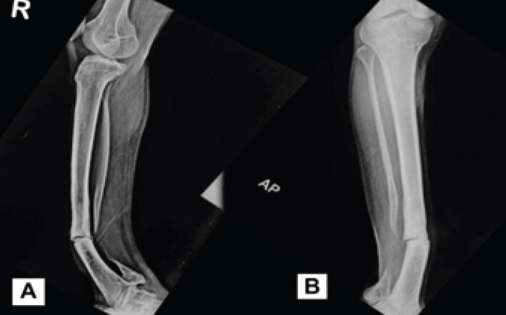
Initial reconstructive stages
At initial presentation, an Ilizarov circular external fixator was applied to provide axial compression across the pseudarthrosis. Despite maintained compression, serial radiographs at 4 months demonstrated no progressive osseous consolidation. The frame was maintained for a total of 6 months before the reconstructive strategy was revised (Fig. 2).
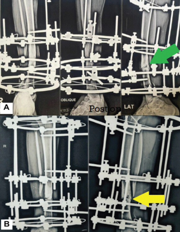
Following the removal of the initial frame, the reconstructive strategy was revised to incorporate biological augmentation. The pseudarthrosis was exposed, and pathological tissue was excised. A structural autologous onlay tricortical iliac crest bone graft was applied across the pseudarthrosis. During the same procedure, a supramalleolar corrective osteotomy was performed at the distal tibial metaphysis, near the center of rotation of angulation, to correct the associated ankle valgus deformity. The LDTA improved from 80° pre-operatively to 89° following correction (Fig. 3).
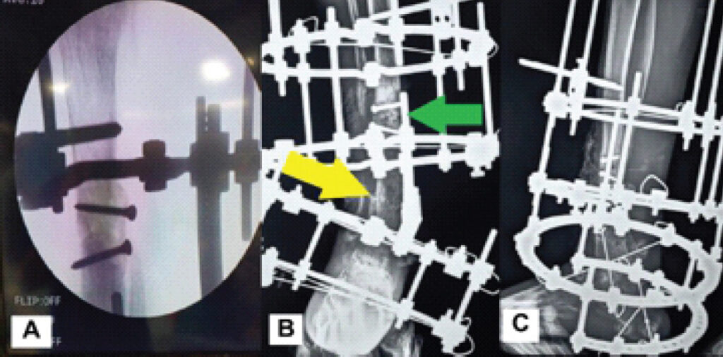
Progressive radiographic incorporation of the graft was observed, with radiographic union evident at approximately 3 months. Full weight bearing was initiated, and at 18 weeks of weight bearing, the patient sustained a fracture through the reconstructed graft site (Fig. 4).
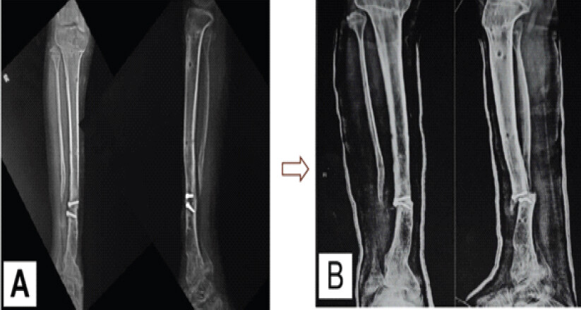
Within 2 weeks of the fracture, the patient developed fever, local warmth, wound breakdown, purulent discharge, and a draining sinus at the reconstructed segment. Deep tissue and bone specimens obtained for microbiological evaluation grew methicillin-sensitive Staphylococcus aureus, confirming deep infection involving the reconstructed pseudarthrosis.
Definitive salvage reconstruction
Following confirmation of deep infection, definitive limb-salvage reconstruction was undertaken. Radical debridement was performed with excision of infected graft material, pathological tissue, and non-viable bone until healthy bleeding bone margins were obtained. Approximately 2 cm of bone was resected. The resulting defect was managed by acute 2 cm shortening and direct compression of the debrided pseudarthrosis.
A multilevel Ilizarov circular external fixator was applied, and acute intraoperative compression was achieved across the distal debrided pseudarthrosis. During the same procedure, a proximal tibial metaphyseal corticotomy was performed through healthy bone to permit subsequent distraction osteogenesis (Fig. 5). The fibula was transported alongside the distal tibia, maintaining the critical anatomical relationship at the distal tibiofibular syndesmosis.
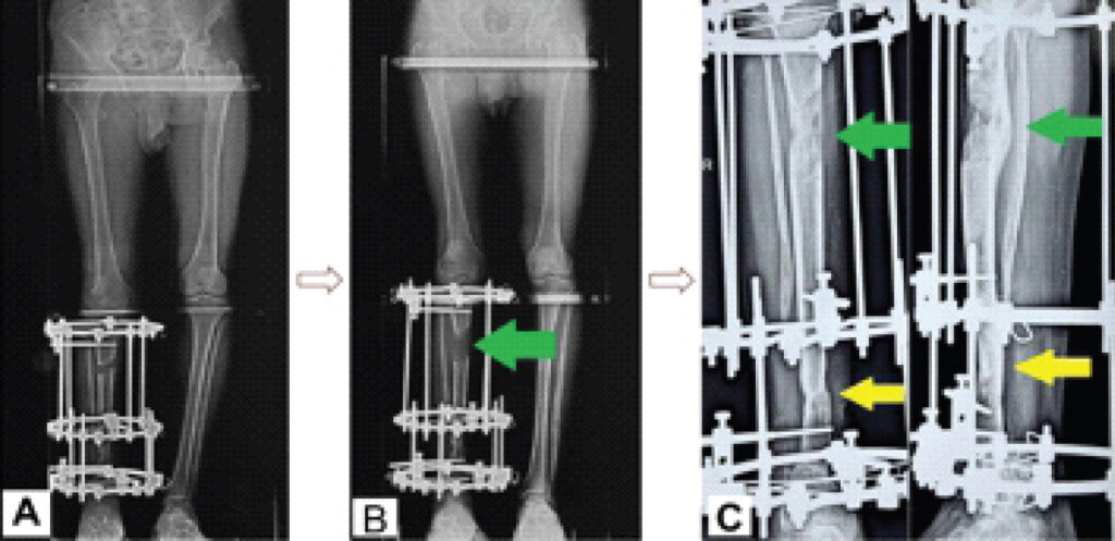
Following a latency period of 10 days, distraction was initiated at a rate of 1 mm/day in four increments of 0.25 mm. A total of 7 cm of lengthening was achieved, addressing both the pre-existing 5 cm LLD and the additional 2 cm shortening created by radical debridement and acute compression. Thus, osseous regeneration was deliberately transferred to a healthy proximal metaphyseal segment, spatially separate from the infected and dysplastic distal pseudarthrosis.
Culture-directed intravenous cefazolin was administered for 7 days , followed by oral amoxicillin–clavulanate for a further 6 weeks. Clinical surveillance demonstrated resolution of the purulent discharge, wound healing, and absence of recurrent clinical signs of infection.
Frame course and complications
The definitive circular external fixator was maintained for approximately 10 months, corresponding to an external fixation index (EFI) of 43 days/cm. Frame-related complications were recorded systematically throughout treatment. The patient experienced three episodes of Grade I/II pin-tract infections (Checketts-Otterburn classification), which were successfully managed with local pin-site care and short courses of oral antibiotics without requiring wire/pin removal. Knee joint stiffness developed during the late distraction phase, which resolved with targeted physical therapy. No neurovascular compromise, pin loosening, or compartment syndrome occurred. Frame tolerance was satisfactory, and the patient maintained partial weight-bearing ambulation with crutches throughout the consolidation phase.
Outcome and follow-up
Serial radiographs demonstrated progressive consolidation of the proximal tibial regenerate and osseous union across the compressed distal pseudarthrosis. White blood cell counts, erythrocyte sedimentation rate and C-reactive protein showed a declining trend. The definitive circular external fixator was maintained for 10 months. Before frame removal, the construct was dynamized for a week, and the patient demonstrated painless full weight-bearing. After removal of the frame, a Sarmiento brace was given for 3 weeks. Radiographic evidence of bridging cortices and satisfactory regenerate consolidation was confirmed before removal of the frame.
Based on total external fixation duration of approximately 10 months and 7 cm of lengthening, the EFI was approximately 43 days/cm. Final radiographs demonstrated mature union at the distal pseudarthrosis and consolidation of the proximal regenerate.
At 24-month follow-up, the patient maintained radiographic union without clinical recurrence of infection. Limb-length equality was restored, with no residual LLD and a residual angular deformity of 2° (Fig. 6). The patient reported no pain or limps and had returned to daily activities and work. Residual ankle stiffness persisted with 10° dorsiflexion and 25° plantarflexion. According to the Association for the Study and Application of the Method of Ilizarov criteria, the bone result was graded excellent, and the functional result was graded Good [6].
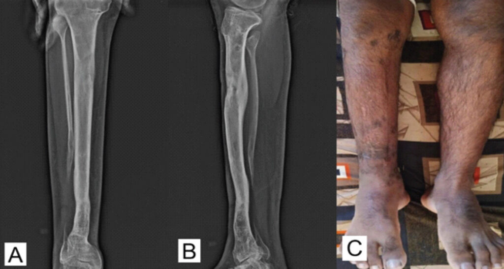
Functionally, the patient reported no limb pain or limp and successfully returned to daily activities and employment. However, residual ankle joint stiffness persisted, with 10° dorsiflexion and 25° plantarflexion. While this restriction altered late-stance phase ankle mechanics, the patient compensated effectively with footwear modifications and reported no severe functional disability during routine daily living.
Discussion
CPT has a global incidence of approximately 1 in 150,000 live births [1]. It affects the middle and distal thirds of the tibial diaphysis, with involvement of the proximal third being rare and limited to only a handful of documented cases globally [7]. The natural history of the disease varies slightly depending on the morphological subtype, but it is universally unpredictable. In dysplastic forms as observed in this patient’s Crawford Type IV atrophic presentation anterolateral bowing is evident at birth, accompanied by a narrow tibia with an hourglass appearance and an obstructed medullary cavity, which fractures when the child begins ambulating [8]. In contrast, in cystic forms, bowing typically appears before the 1st year of life [9].
The eradication of CPT requires overcoming a hostile local biological environment characterized by diminished osteogenesis, impaired vascularity, and increased osteoclastic resorption [2,3]. When complicated by adult presentation and active osteomyelitis, the reconstructive algorithm narrows substantially, pushing the limits of limb salvage.
The principal reconstructive lesson of this case is the distinct utility of circular external fixation to simultaneously address the mechanical, biological, and infectious elements of a failing CPT limb. While contemporary literature strongly advocates for combined hybrid fixation (intramedullary coupled with extramedullary fixation) to prevent late refracture in standard pediatric CPT [4,5], the presence of active deep infection made immediate intramedullary fixation undesirable.
Circular external fixation provides stable fixation while permitting controlled axial loading and avoiding placement of internal implants within the previously infected medullary environment [10]. In this case, the isolated failure of the initial Ilizarov compression highlighted that mechanical stability alone cannot overcome the cellular deficit of CPT without aggressive biological resection of the hamartoma [2]. The subsequent graft fracture highlighted the severe mechanical vulnerability of the dysplastic bone subject to physiological loading before full structural consolidation.
By relocating osteogenesis to a healthy proximal metaphyseal segment, distraction osteogenesis generated regenerate bone outside the infected and dysplastic zone while permitting acute shortening and compression at the debrided pseudarthrosis [11]. Unlike an infected non-union, a dysplastic CPT segment typically resists consolidation under compression due to the biological and mechanical barrier of the cellular fibrous hamartoma.
However, once the infected tissues and the pathological hamartoma were radically resected to healthy bleeding osseous margins, the previously dysplastic distal segment behaved comparably to a standard infected non-union under acute compression. By relocating the demand for de novo bone generation to the proximal metaphysis through distraction osteogenesis, this bifocal strategy successfully bypassed the hostile local environment of the CPT. [12] Consequently, this case demonstrates that the established compression-distraction technique is a highly effective limb-salvage strategy for complex, infected adult CPT when active infection renders internal fixation unsuitable.
Limitations
This case report has several limitations. First, as a single-case report, the findings represent proof-of-concept in a complex salvage setting and cannot be generalized to the broader adult CPT population. Second, no comparator group was available to evaluate alternative strategies. Third, although 24-month follow-up demonstrated sustained union and clinical infection control, longer surveillance is required given the lifelong risk of refracture, recurrent deformity, and late infection. Finally, complete records of the four previous surgeries were unavailable.
Conclusion
Adult CPT complicated by graft-site fracture and deep infection presents a complex limb-salvage challenge. In this case, radical debridement and culture-directed antimicrobial therapy were combined with acute compression of the infected pseudarthrosis and proximal distraction osteogenesis through healthy metaphyseal bone. Spatially separating infection control from bone generation achieved union, restored limb length, and preserved the limb at 24-month follow-up. This compression–distraction strategy may represent a reconstructive option in selected complex CPT cases in which active infection limits internal fixation.
Clinical Message
The successful management of adult CPT needs a planned, multi-targeted approach. Surgeons must aggressively resect the entire pathological hamartoma, rigorously correct all mechanical axes, including fibular pseudarthrosis and ankle valgus, and employ stabilization techniques. The Ilizarov method, through its tension-stress effect and distraction osteogenesis, is useful to salvage limbs threatened by a combination of non-union, massive limb shortening, and deep bone infection.
Conflict of Interest:
Nil
Source of Support:
Nil
Consent
The authors confirm that informed consent was obtained from the patient for publication of this article
How to Cite this Article
Dussa A, Gandhi K, Jagdale S, Kelkar S. Compression of the Diseased Segment and Regeneration through Healthy Bone for Salvage of Infected Recurrent Congenital Pseudarthrosis of the Tibia in an Adult: A Case Report. J Orthop Case Rep 2026 November;16(11): 54-60. doi: 10.13107/jocr.2026.v16.i11.8386.
References
- Pannier S. Congenital pseudarthrosis of the tibia. Orthop Traumatol Surg Res 2011;97:750-61. [Google Scholar] | [PubMed]
- Laufer A, Frommer A, Gosheger G, Roedl R, Schiedel F, Broeking JN. Reconstructive approaches in surgical management of congenital pseudarthrosis of the tibia. J Clin Med 2020;9:4132. [Google Scholar] | [PubMed]
- Banchhor H, Chimurkar V. Congenital pseudoarthrosis of the tibia: A narrative review. Cureus 2022;14:e32501. [Google Scholar] | [PubMed]
- Borzunov DY, Chevardin AY, Mitrofanov AI. Management of congenital pseudarthrosis of the tibia with the Ilizarov method in a paediatric population: Influence of aetiological factors. Int Orthop 2016;40:331-9. [Google Scholar] | [PubMed]
- Agashe MV, Song SH, Refai MA, Park KW, Song HR. Congenital pseudarthrosis of the tibia treated with a combination of Ilizarov's technique and intramedullary rodding. Acta Orthop 2012;83:515-22. [Google Scholar] | [PubMed]
- Paley D, Catagni MA, Argnani F, Villa A, Benedetti GB, Cattaneo R. Ilizarov treatment of tibial nonunions with bone loss. Clin Orthop Relat Res 1989;241:146-65. [Google Scholar] | [PubMed]
- Li Y, Li J, Gan T, Geng Q, Ma X, Yin S. Ilizarov distraction for congenital pseudoarthrosis of the tibia in adults. Orthop Surg 2024;16:2191-201. [Google Scholar] | [PubMed]
- Vaidya SV, Aroojis A, Mehta R, Agashe MV, Dhawale A, Bansal AV. Short term results of a new comprehensive protocol for the management of congenital pseudarthrosis of the tibia. Indian J Orthop 2019;53:736-44. [Google Scholar] | [PubMed]
- Kamath SK, Madegowda A, Baghel A, Singh KA, Shah HH. Tibial lengthening in congenital pseudoarthrosis of the tibia: A scoping review. BMC Musculoskelet Disord 2026;27:307. [Google Scholar] | [PubMed]
- Horn J, Steen H, Terjesen T. Epidemiology and treatment outcome of congenital pseudarthrosis of the tibia. J Child Orthop 2013;7:157-6. [Google Scholar] | [PubMed]
- Ohnishi I, Sato W, Matsuyama J, Yajima H, Haga N, Kamegaya M. Treatment of congenital pseudarthrosis of the tibia: A multicenter study in Japan. J Pediatr Orthop 2005;25:219-24. [Google Scholar] | [PubMed]
- Kale A, Patil VS, Singh PJr, Raithatha H, Shah M, Aggarwal R. Congenital pseudoarthrosis of tibia with anterolateral bowing treated with ilizarov ring fixator: A case report. Cureus 2023;15:47615. [Google Scholar] | [PubMed]
© 2026 Journal of Orthopaedic Case Reports - Published by Indian Orthopaedic Research Group
About the Authors
-

Abhishek Dussa
-

Ketankumar Gandhi
-

Swapnil Jagdale
-

Sagar Kelkar
Related Articles in Journal of Orthopaedic Case Reports
 November 1, 2026 Immediate Limb Lengthening Following Vascular Repair of Iatrogenic Arterial Lesions – A Report of Two Cases
 October 1, 2026 Soft-Tissue Closure through Acute Deformation and Gradual Correction Using Taylor Spatial Frame in a Gustilo-Anderson Type IIIB Open Tibial Pilon Fracture: A Case Report
 August 1, 2026 Management of Infected Non-union of Long Bones Using Antibiotic Cement-Coated Intramedullary Nails: A Prospective Case Series
 July 1, 2025 Circular Fixation, Endless Possibilities: Rebuilding a Shattered Tibia using the Ilizarov Technique – A Case Report