


Introduction
Stress fractures occur in younger individuals with normal bone composition that undergo atypical mechanical loading during physical activity [1]. These injuries most commonly are observed in the weight-bearing bones of the lower leg and foot [2]. The underlying pathophysiology results from repetitive loading forces from overuse that result in osteoclastic activity outpacing osteoblastic activity in the natural bone remodeling response to the stressor [1,3]. With continual imbalance in bone remodeling, microfractures occur and can progress to a complete fracture [3].
The femoral neck is an uncommon site of stress fracture injury, composing only 5% of all stress fractures in young adults, 3% of all stress fractures in athletes, and 3.5-8% of all stress fractures in military recruits [4,5,6]. Femoral neck stress fractures (FNSF) are associated with significant morbidity, especially with a delay in diagnosis and treatment. Risk factors identified include female sex, nutritional deficiency, poor bone mineral density, decreased physical fitness or muscle tone concomitant with sudden increased physical activity, smoking, or chronic corticosteroid use [3,7]. Two genetic predispositions have been identified: Calcitonin receptor C allele and Vitamin D receptor C-A allele [8]. These genetic variations increase susceptibility to stress fractures through abnormal bone architecture. Both receptors influence bone mineral density and subsequently fracture risk through mechanisms affecting osteoclast activity, bone remodeling signal pathways, and calcium absorption efficiency. In individuals lacking either or both alleles, there was 3 times greater risk of FNSF [9].
The most common clinical presentation of FNSF is an acute atraumatic onset of severe groin pain and inability to bear weight, especially following strenuous physical activity. With a 14-week average delay in diagnosis and a poor prognosis in adolescents, prompt recognition and treatment of a FNSF is clinically important [10,11]. However, diagnostic challenges occur because radiographic findings may be unremarkable at the time of injury, and abnormal findings may take weeks to months to be evident on imaging [1]. As many as 90% of initial radiographs and 50% of repeat radiographs at 4–6 weeks demonstrated no evidence of the stress fracture [9]. Advanced imaging with computed tomography (CT) scans, magnetic resonance imaging (MRI), and technetium bone scan can be helpful in identifying the fracture. Physicians must integrate clinical suspicion based on history and physical exam into the entirety of the clinical framework for optimal opportunity of an early diagnosis.
Bilateral femoral neck fractures are exceedingly rare, with only a few cases identified in young athletes. We are reporting on a military recruit who sustained simultaneous bilateral femoral neck fractures during basic training.
Case Report
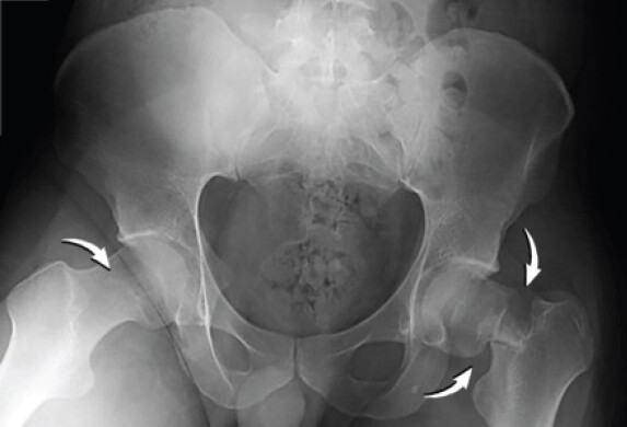
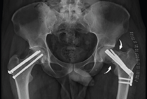
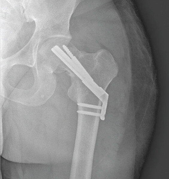
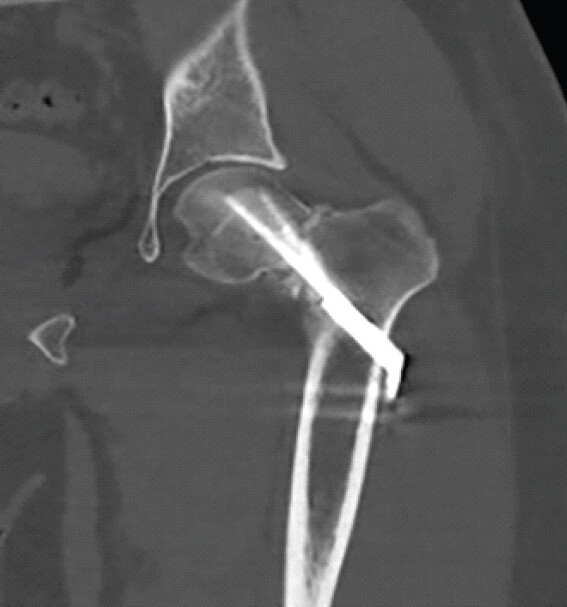
A 22-year-old newly recruited male military trainee sustained bilateral FNSF. While in training 3 weeks prior, he complained of severe bilateral anterior groin pain. He presented to the medical unit and was originally diagnosed with bilateral muscle strains, treated conservatively, and instructed to remain in training activities as tolerated. Two days later, he jumped from an obstacle course and landed with his feet flat on the ground, resulting in a non-displaced right femoral neck fracture (Garden’s type I and Pauwel’s type I) and a displaced left (Garden’s type IV and Pauwel’s type III) femoral neck fracture (Fig. 1). In review of his pertinent medical history, he did not have any predisposing conditions, documented history of osteoporosis, Vitamin D or calcium deficiency, or other significant past medical history. He was not taking any medications. However, with routine labs to assess bone health, the patient had a Vitamin D deficiency (12.1 ng/mL) and mildly low testosterone (9.3 nmol/L). The right fracture was treated surgically using closed reduction with percutaneous screw fixation, and the left fracture was treated using open reduction and fixation with a Femoral Neck System (DePuy Synthes) and supplemental lag screw (Fig. 2). Two weeks postoperatively, radiographs demonstrated varus malalignment of the left femur measuring 110° (Fig. 3 and 4).
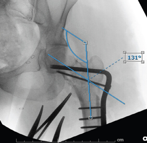
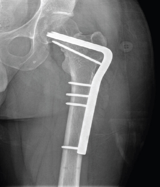
The patient was referred to orthopedic trauma for fracture realignment and revision of the fixation device. A 20° intertrochanteric valgus osteotomy was performed. The osteotomy and fracture were stabilized using a blade plate inserted into the proximal osteotomy segment. In addition, a cortical screw was placed across the femoral neck fracture to compress and stabilize the fracture. A tensioning device was utilized in the distal femur to plate the femoral shaft and compress the osteotomy site. Multi-planer fluoroscopy confirmed adequate reduction and placement of implants (Fig. 5). Following the revision surgery, the patient returned to weight-bearing as tolerated for 3 months. His fractures went on to heal without complications (Fig. 6).
Discussion
Military recruits, elderly individuals with predisposing conditions, and athletes, particularly runners, are at higher risk for FNSF [9]. When these injuries do occur in military recruits and runners, they are typically associated with strenuous activities, such as jumping and long-distance running [12].
Although military recruits undergo a rigorous series of medical, physical, and fitness evaluations to join the military, screening of bone health and metabolic deficiencies is not routinely performed prior to basic training. However, this is notable considering the prevalence of Vitamin D deficiency among young adults. In the United States, of individuals ages 20–39, an estimated 7.6% were at risk of Vitamin D deficiency (<30 nmol/L), and 23.8% were at risk for Vitamin D inadequacy (30–49 nmol/L), which were the highest among any age group [13]. Therefore, the contribution to increased risk of fracture from these deficiencies underscores the need for improved screening protocols through assessment and lab monitoring prior to precipitating activities.
In active young adults, the consequences of missed diagnosis or prolonged intervention of femoral neck fractures often result in unfavorable clinical outcomes including non-union, femoral shortening, avascular necrosis, and hardware failure [14]. In accurately identifying the fracture with both high specificity and sensitivity, MRI is considered the gold standard for diagnosis, detecting proximal femur fractures approximately 97–100% of the time [4,14]. In addition, emphasis on careful assessment of the radiographic fracture characteristics, including posterior tilt, is essential to determine appropriate fixation [14]. Correction of posterior tilt to 5° or less before fixation has proven to reduce the rate of secondary revision surgery by 3.4 times [14].
While non-operative management can be considered in select cases, surgical intervention is recommended in FNSF of young individuals, even in non-displaced fractures [4]. Consensus on surgical treatment of these fractures typically involves placement of cannulated screws or sliding hip screws, with no significant difference in surgical outcomes and revision rates between methods [14,15,16].
With prolonged delay in intervention, the risk of fracture displacement increases significantly and is associated with an overall increased likelihood of avascular necrosis [11,14]. In displaced fractures with varus alignment, intertrochanteric valgus osteotomy is an effective technique to restore alignment, promote union, and preserve femoral head vascular supply [4,17]. Among fractures that remained in varus alignment after fixation, 71.4% developed avascular necrosis during follow-up [12]. Therefore, early diagnosis, prompt anatomical reduction, and adequate fixation are important to reduce the incidence of complications.
Post-operative management of younger patients can be difficult, as they tend to be less adherent to non-weight-bearing status after fixation due to their active lifestyles [12]. Therefore, patient education and close follow-up are essential.
Conclusion
Simultaneous bilateral FNSF are rare, especially in healthy young adults. Early diagnosis requires a high index of suspicion, especially in the setting of inconclusive initial imaging. Advanced imaging plays a critical role in early diagnosis, and prompt surgical intervention with appropriate fixation is necessary to decrease the incidence of complications such as non-union, femoral shortening, avascular necrosis, and hardware failure.
Although there is no consensus on optimal surgical intervention, closed reduction with percutaneous screw fixation or open reduction with dynamic hip screw are used with no significant difference in surgical outcomes or revision rates. With post-surgical complications occurring in 10–40% of cases, optimizing initial surgical intervention is important, preventing the necessity for surgical revision. While compliance with post-operative weight-bearing status is difficult in young adults, it is important for optimal recovery and rehabilitation.
In addition, this case emphasizes an important gap in current military medical evaluation protocol. Recruits undergo extensive physical and medical examinations before training, but routine assessment of bone health and metabolic deficiencies is not consistently performed. Given the sudden and intense physical rigor of basic training, these overlooked deficiencies may predispose otherwise healthy individuals to stress-related injuries.
Clinical Message
Implementing targeted screening protocols, including baseline bloodwork evaluation and risk stratification, may facilitate early identification and optimization of high-risk individuals prior to strenuous activity to prevent avoidable injuries and improve outcomes in military populations. Proactive measures such as nutritional supplementation, gradual conditioning programs, and education on alarm symptoms could reduce the incidence of stress fractures and improve overall musculoskeletal preparation.
Conflict of Interest:
Nil
Source of Support:
Nil
Consent:
The authors confirm that informed consent was obtained from the patient for publication of this article
How to Cite this Article
Jones A, Floyd J. Bilateral Femoral Neck Stress Fractures in a Healthy Young Military Trainee: A Case Report and Review of Literature. J Orthop Case Rep. 2026 November;16(11): 137-141. doi: 10.13107/jocr.2026.v16.i11.8416.
References
- Jones BH, Thacker SB, Gilchrist J, Kimsey CDJr, Sosin DM.. Prevention of lower extremity stress fractures in athletes and soldiers: A systematic review. Epidemiol Rev 2002;24:228-47. [Google Scholar] | [PubMed]
- Schroeder JD, Trigg SD, Capo Dosal GE.. Bone stress injuries: Diagnosis and management. Am Fam Physician 2024;110:592-600. [Google Scholar] | [PubMed]
- Tenforde AS, Matzkin EG.. Letter to the Editor: Femoral neck stress fractures: An updated review: Addressing biological and biomechanical factors results in optimal management. J Am Acad Orthop Surg 2022;32:e568-9. [Google Scholar] | [PubMed]
- Naik MA, Sujir P, Tripathy SK, Vijayan S, Hameed S, Rao SK.. Bilateral stress fractures of femoral neck in non-athletes: A report of four cases. Chin J Traumatol 2013;16:113-7. [Google Scholar] | [PubMed]
- Onibere OA, Sugathan HK.. A case report of missed femoral neck stress fracture. SICOT J 2015;1:2. [Google Scholar] | [PubMed]
- Robertson GA, Wood AM.. Femoral neck stress fractures in sport: A current concepts review. Sports Med Int Open 2017;1:E58-68. [Google Scholar] | [PubMed]
- Biz C, Berizzi A, Crimì A, Marcato C, Trovarelli G, Ruggieri P.. Management and treatment of femoral neck stress fractures in recreational runners: A report of four cases and review of the literature. Acta Biomed 2017;88:96-106. [Google Scholar] | [PubMed]
- Bernstein EM, Kelsey TJ, Cochran GK, Deafenbaugh BK, Kuhn KM.. Femoral neck stress fractures: An updated review. J Am Acad Orthop Surg 2022;30:302-11. [Google Scholar] | [PubMed]
- Jalan D, Rathore KS, Elhence A, Kumar Yadav S, Maley DK.. Stress fracture of bilateral neck of femur in a healthy non-athletic young adult - a case report with review of literature. Cureus 2020;12:e11193. [Google Scholar] | [PubMed]
- Johansson C, Ekenman I, Törnkvist H, Eriksson E.. Stress fractures of the femoral neck in athletes. The consequence of a delay in diagnosis. Am J Sports Med 1990;18:524-8. [Google Scholar] | [PubMed]
- Lee CH, Huang GS, Chao KH, Jean JL, Wu SS.. Surgical treatment of displaced stress fractures of the femoral neck in military recruits: A report of 42 cases. Arch Orthop Trauma Surg 2003;123:527-33. [Google Scholar] | [PubMed]
- Naranje S, Sezo N, Trikha V, Kancherla R, Rijal L, Jha R.. Simultaneous bilateral femoral neck stress fractures in a young military cadet: A rare case report. Eur J Orthop Surg Traumatol 2012;22 Suppl 1:103-6. [Google Scholar] | [PubMed]
- Herrick KA, Storandt RJ, Afful J, Pfeiffer CM, Schleicher RL, Gahche JJ. Vitamin D status in the United States, 2011-2014. Am J Clin Nutr 2019;110:150-7. [Google Scholar] | [PubMed]
- Okike K, Hasegawa IG.. Current trends in the evaluation and management of nondisplaced femoral neck fractures in the elderly. J Am Acad Orthop Surg 2021;29:e154-64. [Google Scholar] | [PubMed]
- Fixation using Alternative Implants for the Treatment of Hip fractures (FAITH) Investigators. Fracture fixation in the operative management of hip fractures (FAITH): An international, multicentre, randomised controlled trial. Lancet 2017;389:1519-27. [Google Scholar] | [PubMed]
- Watson A, Zhang Y, Beattie S, Page RS.. Prospective randomized controlled trial comparing dynamic hip screw and screw fixation for undisplaced subcapital hip fractures. ANZ J Surg 2013;83:679-83. [Google Scholar] | [PubMed]
- Diwanji SR, Kong IK, Cho SG, Seon JK, Yoon TR.. Displaced stress fracture of the femoral neck treated by valgus subtrochanteric osteotomy: 2 case studies. Am J Sports Med 2007;35:1567-70. [Google Scholar] | [PubMed]
© 2026 Journal of Orthopaedic Case Reports - Published by Indian Orthopaedic Research Group
About the Authors
-

Aneesa Jones
-

John Floyd
Related Articles in Journal of Orthopaedic Case Reports
 December 1, 2025 The use of an Antiprotrusio Cage in Acetabular Reconstruction for Periprosthetic Transverse Fracture through a Direct Anterior Approach: A Case Report
 October 1, 2026 Functional Outcome of Femoral Neck Fractures with Posterior Comminution Managed by Dynamic Hip Screw with Fibular Strut Grafting: A Case Series Study
 October 1, 2026 Dual Mobility Total Hip Arthroplasty for Recurrent Dislocation After Femoral Neck Fracture in an Elderly Patient: A Case Report
 October 1, 2026 Primary Total Hip Arthroplasty for a Displaced Femoral Neck Fracture in an Above-the-Knee Amputee: A Case Report