


Introduction
Coronal shear fractures of the femoral condyle, eponymously known as Hoffa fractures, are uncommon injuries that most often result from high-energy axial loading of a flexed knee. They are frequently missed on standard anteroposterior and lateral knee radiographs because the fracture line lies in the coronal plane and can be obscured by the overlapping condyles; oblique views or computed tomography (CT) are often needed to fully characterize the fragment and plan surgery [1, 2, 3, 4]. When the diagnosis is delayed, or when a displaced fracture is managed non-operatively – as occurred in the patient described here – the fragment can go on to non-union, with progressive fibrous tissue interposition, sclerosis of the fracture margins, and secondary joint stiffness.
Non-union of a Hoffa fracture is rare enough that the literature is limited to isolated case reports and small case series [5, 6, 7, 8, 9, 10]. Reported strategies for its surgical correction include debridement with lag-screw fixation, headless compression screws, buttress or neutralization plating, and combinations of these with autologous bone grafting and formal arthrolysis for an associated flexion contracture [5, 6, 7, 8, 9]. Headless compression screws are attractive in this setting because they provide axial interfragmentary compression, can be buried beneath the articular cartilage to avoid impingement, and have been associated with fewer implant-related complications than headed cancellous screws in comparative series [1].
This report describes a Hoffa fracture non-union treated through a medial parapatellar approach with headless compression screw fixation. Rather than simply documenting that the surgery was difficult, the aim is to set out – in a Challenges → Solutions → Practical Tips format – the specific technical decisions that made anatomical reduction and stable fixation possible so that another trauma surgeon facing a similar neglected coronal fracture has a concrete operative reference.
Case Report
History and examination
A 45-year-old male presented with knee pain of 3 months’ duration. He gave a history of a road traffic accident 3 months earlier, sustained when he was struck by a four-wheeled vehicle with direct trauma to the knee joint. At the outside facility, he was treated with a plaster slab for 1–2 months without any surgical fixation and was advised nil weight-bearing on discharge.
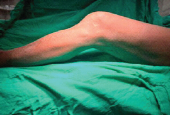
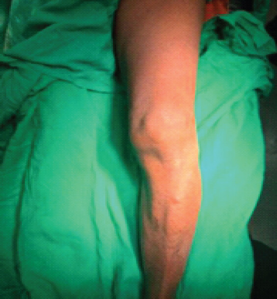
He presented to us 3 months after the injury with a fixed flexion deformity of 30°, inability to bear weight, and persistent knee pain. On examination, there was diffuse swelling and tenderness around the knee. Extension was limited to a fixed flexion deformity of 30°, with further flexion possible up to 90°. Clinical photographs demonstrate the flexion deformity and extension deficit (Fig. 1 and 2).
Imaging
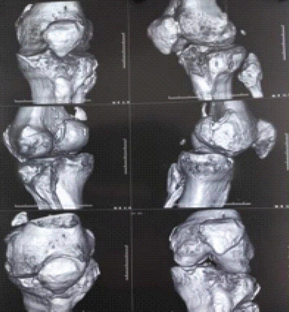
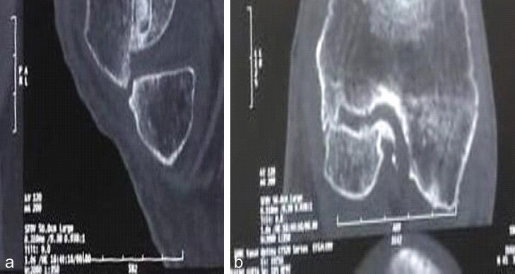
Plain radiographs demonstrated a posterior (coronal) Hoffa fracture of the femoral condyle in non-union. Computed tomography with three-dimensional reconstruction was obtained for surgical planning. Sequential sagittal bone-window reconstructions demonstrated a coronal-plane intra-articular fracture of the posterior distal femoral condyle, characteristic of a Hoffa fracture pattern (Letenneur classification). The fracture line showed classic features of chronic non-union – well-defined, smooth, sclerotic margins, a persistent radiolucent gap, and an absence of bridging callus – with displacement of the condylar fragment producing articular incongruity. Diffuse tibiofemoral joint space narrowing, subchondral sclerosis, and osteophyte formation were also present, consistent with post-traumatic osteoarthritic change (Fig. 3 and 4).
Surgical technique
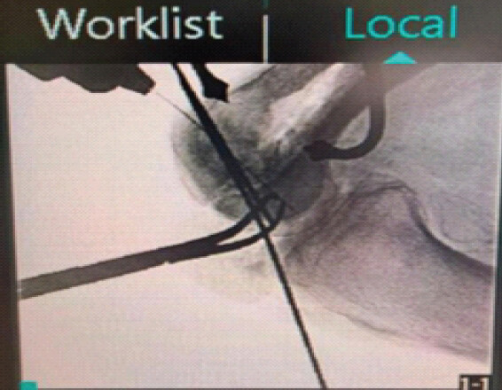
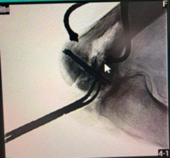
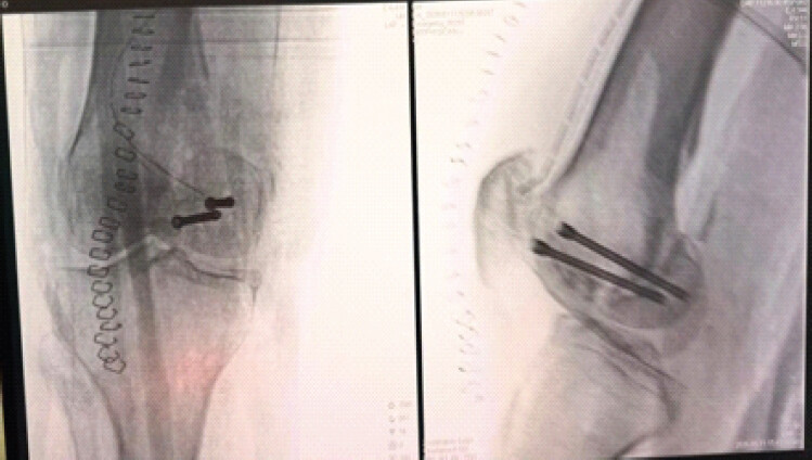
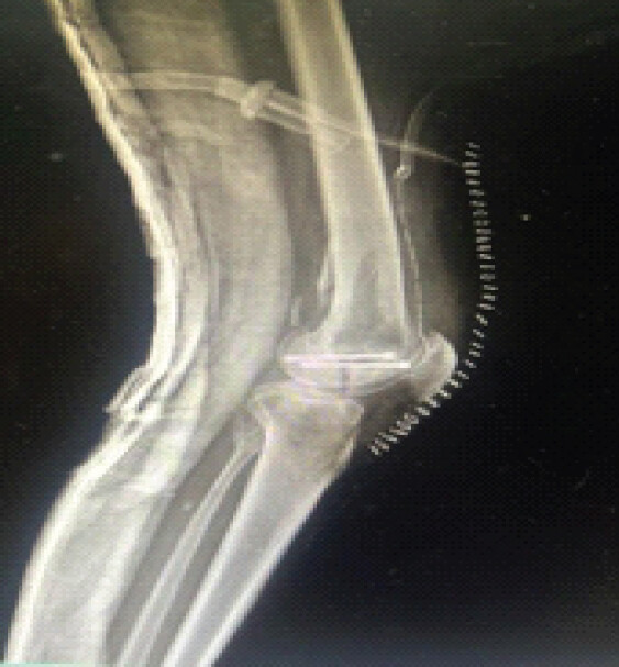
The patient underwent open reduction and internal fixation. A longitudinal skin incision was marked, and a medial parapatellar arthrotomy was performed to access the knee joint and expose the non-united fragment (Fig. 5). The fibrous non-union tissue at the fracture interface was debrided, the fracture surfaces were freshened, and the fragment was provisionally reduced and held with Kirschner wires under direct vision and fluoroscopic control (Fig. 6 and 7). Once anatomical reduction of the articular surface was confirmed on multiple fluoroscopic projections, two parallel headless compression screws were placed from anterior to posterior, perpendicular to the coronal fracture plane, over the guide wires, with the screw heads buried beneath the articular cartilage (Fig. 8 and 9). Final anteroposterior and lateral fluoroscopic images confirmed a congruent reduction and satisfactory screw position (Fig. 9). The wound was closed in layers (Fig. 5).
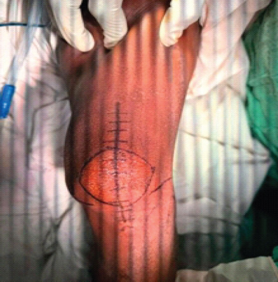
No bone graft was used at the fracture site. No drain was placed. The wound was closed in routine layers without any additional soft-tissue procedure. The limb was immobilized postoperatively in a long knee brace.
Challenges, solutions, and practical tips
Challenge 1 – Exposure and reduction through a chronic, fibrous non-union interface
At 3 months, the fracture margins were no longer clean lines but were covered by fibrous scar tissue and early sclerosis, which obliterated the normal fracture plane and made the fragment difficult to mobilize and reduce anatomically.
-
Solution: A generous medial parapatellar arthrotomy with patellar eversion/subluxation was used to bring the posterior condylar fragment into full view rather than attempting reduction through a limited anterior window. All fibrous tissue at the interface was sharply excised back to bleeding, viable bone on both fracture surfaces before any attempt at reduction.
-
Practical tip: Do not accept a fibrous “fit” as reduction. Debride until the two surfaces are congruent bone-on-bone; an interposed fibrous layer left in place is the single most common reason a non-union screw construct fails to unite even when it looks well-fixed on the table.
Challenge 2 – Fixed flexion deformity from prolonged immobilization
Three months of nil-weight-bearing immobilization in a slab had produced a 30° fixed flexion deformity. This reflected both posterior capsular contracture and secondary contracture of the hamstring muscles, which together limited exposure and the ability to bring the knee to full extension for definitive fixation and closure.
-
Solution: Circumferential release of intra-articular and periarticular adhesions was carried out as part of the same exposure, with gradual, gentle extension of the knee rather than a single forceful manipulation, to avoid an iatrogenic periarticular fracture in bone that had been disused for 3 months.
-
Practical tip: Plan the arthrolysis before you plan the fixation. A knee that cannot be brought to near-full extension on the table will not tolerate the screw trajectory you have planned on pre-operative imaging, and closure over a joint still held in flexion places the fixation under constant tensile load postoperatively.
Challenge 3 – Achieving a screw trajectory truly perpendicular to the coronal fracture plane
A coronal shear fragment is deceptive under fluoroscopy: a guide wire or screw that looks well-aimed on a standard anteroposterior or lateral knee view can still be running obliquely across the actual fracture plane or can appear to be within the condyle on one view while exiting it on another.
-
Solution: Provisional Kirschner wires were placed first and checked in more than one fluoroscopic plane before any drilling or screw insertion, adjusting the C-arm to profile the fracture line itself rather than relying on the routine knee AP/lateral series (Figs. 6 and 7).
-
Practical tip: Never advance a definitive screw over a guide wire that has been checked in only one fluoroscopic plane. Rotate the C-arm to obtain an oblique or “down-the-barrel” view of the wire relative to the fracture line before committing to the screw.
Challenge 4 – Stable interfragmentary compression in bone with early sclerotic change
Sclerotic, partially healed bone at the fracture margins offers less predictable screw purchase and compression than a fresh fracture.
-
Solution: Two parallel headless compression screws were used rather than one, both placed perpendicular to the fracture plane and countersunk beneath the articular surface, to provide rotational stability in addition to interfragmentary compression.
-
Practical tip: A single screw resists compression but not rotation. In a coronal fragment of any appreciable size, plan for at least two points of fixation, and choose a headless, cannulated, variable-pitch compression screw so the head can be buried without a countersinking step that removes additional subchondral bone.
Post-operative course
The patient was kept non-weight-bearing immediately after surgery, with the limb protected in a long knee brace. Bedside physiotherapy was started early, with active and passive straight-leg raises, static quadriceps and static hamstring strengthening, and vastus medialis obliquus strengthening, alongside range-of-motion exercises advanced in a staged manner: Knee flexion was permitted up to 30° for the first 10 post-operative days, up to 60° for the next 10 days, and up to 90° for a further 10 days, with complete knee extension maintained throughout. Partial weight-bearing was then started and continued for 1 month, after which the patient progressed to full weight-bearing. At final follow-up, the fixation was maintained on serial radiographs (Fig. 9); the patient had regained knee flexion to 100°, with the pre-operative 30° extension lag improving to 10°, and only occasional mild pain (Visual Analog Score 3/10). He returned to sedentary, desk-based activity. No post-operative complications occurred. The technical difficulties encountered were intraoperative rather than post-operative, and were attributable to the chronicity of the non-union: severe periarticular fibrosis made fracture reduction difficult, and secondary hamstring contracture had contributed to the pre-operative fixed flexion deformity (see Challenges 1 and 2, above).
Intraoperative and post-operative imaging (Figs. 5, 6, 7, 8, 9).
Discussion
Hoffa fractures represent a small fraction of femoral fractures and are frequently missed at initial presentation, particularly when a routine anteroposterior and lateral knee series is relied upon without dedicated oblique views or cross-sectional imaging [1]. In the case described here, the fracture was managed nonoperatively for 1–2 months, and the patient was then left without further surgical intervention, illustrating how a displaced coronal fragment that is not anatomically reduced and rigidly fixed can progress to non-union even when it has been recognized radiographically.
Cross-sectional imaging was central to surgical planning in this case. Sagittal bone-window CT reconstructions characterized the coronal fracture pattern in a manner consistent with the Letenneur classification and demonstrated the specific hallmarks of an established non-union – smooth, sclerotic fracture margins, a persistent radiolucent gap, and an absence of bridging callus – findings that are not always reliably appreciated on plain radiographs alone. The CT also showed diffuse tibiofemoral joint space narrowing, subchondral sclerosis, and osteophyte formation, indicating that post-traumatic osteoarthritic change had already begun to develop by the time of presentation. This is a recognized consequence of a neglected or malaligned Hoffa fragment, in which persistent articular incongruity accelerates degenerative change in the joint [7]. It underlines two practical points: first, that CT is not an optional adjunct but a planning necessity once non-union is suspected, since it defines fragment morphology and articular incongruity in a way that directly shapes the surgical approach; and second, that patients presenting this late should be counseled that anatomical union does not guarantee freedom from degenerative symptoms and that longer-term follow-up should specifically track for progression of joint space narrowing.
The published experience with Hoffa non-union is limited to a small number of reports, most describing debridement of the pseudarthrosis, bone grafting, and internal fixation, often combined with formal arthrolysis to address the associated joint stiffness [5, 6, 7]. Singh et al. treated six cases of Hoffa non-union with excision of the pseudarthrosis, bone grafting, internal fixation, and knee arthrolysis, achieving union in all patients at a mean of 16 weeks, and emphasized that careful pre-operative planning and meticulous technique – rather than any single implant choice – determined the outcome [5]. The present case supports that emphasis: The technically decisive steps were adequate exposure of the posterior fragment, complete debridement of the fibrous interface, and confirmation of a truly perpendicular screw trajectory on more than one fluoroscopic view, rather than the specific hardware used.
Headless compression screws have been used successfully in both acute and non-united Hoffa fractures [6, 8, 9]. In a comparative series of coronal plane femoral condyle fractures, Maheshwari et al. reported good functional outcomes in 87% of knees fixed with headless compression screws compared with 67% fixed with headed cancellous screws, with significantly fewer implant-related complications in the headless screw group, attributed to their ability to be buried beneath the articular surface without a prominent head [1]. This is consistent with the fixation strategy used in the present case, in which the screw heads were seated beneath the articular cartilage to minimize soft-tissue and cartilage irritation.
Approach selection is dictated by which condyle is involved: a medial parapatellar arthrotomy provides direct access to the medial condyle and, with patellar eversion, can also be extended to visualize the lateral condyle when required, whereas an isolated lateral fragment is more commonly approached laterally [2]. Regardless of the side, the exposure must be generous enough to bring the posterior fragment fully into view, since coronal fragments sit posteriorly and are easily under-visualized through a limited anterior arthrotomy – a point borne out in several published techniques that describe deliberate anterior delivery of the fragment with the knee held in flexion before reduction [8].
Rehabilitation after fixation of a non-united Hoffa fragment must balance two competing risks: stiffness from prolonged immobilization, which had already produced the patient’s 30° flexion contracture, against loss of fixation from motion introduced too early into sclerotic, previously ununited bone. In this case, that balance was managed by protecting the limb in a long knee brace and keeping it non-weight-bearing while introducing bedside range of motion in a graduated sequence – flexion to 30° for the first 10 days, 60° for the next 10, and 90° for the following 10, with full extension maintained throughout – before progressing to partial and then full weight-bearing. Muscle activation was addressed in parallel rather than deferred: active and passive straight-leg raises and static quadriceps, hamstring, and vastus medialis obliquus strengthening were started at the bedside from the early post-operative period, on the principle that periarticular muscle tone can be restored without loading the fracture, whereas restoring it only after the joint is allowed to bear weight prolongs the overall recovery. This staged protocol allowed early motion and muscle re-education to protect against recurrence of the contracture without loading the fixation before early biological consolidation had occurred.
This report has the limitations inherent to a single case: the findings and technical points described here reflect one surgeon’s experience with one fracture pattern and cannot be generalized with the certainty of a comparative series. At the reported follow-up, the patient had regained knee flexion to 100°, with the pre-operative 30° extension lag improving to 10°, only occasional mild pain, and a return to sedentary activity, but longer-term follow-up is still needed to confirm durable radiographic union, stability of fixation under full loading, further improvement in the residual extension lag, and any progression of the pre-existing post-traumatic osteoarthritic change.
Conclusion
Reconstruction of a neglected Hoffa fracture non-union is a technically demanding but reproducible procedure. The determinants of success illustrated by this case are a generous exposure that fully visualizes the posterior condylar fragment, thorough debridement of fibrous and sclerotic bone until congruent bleeding surfaces are restored, staged release of any associated flexion contracture before fixation is attempted, and confirmation of a truly perpendicular screw trajectory on more than one fluoroscopic plane before committing to definitive headless compression screw fixation. CT-based characterization of the fracture and non-union should be considered routine once non-union is suspected, both to plan the fixation and to identify early post-traumatic osteoarthritic change so that patients can be counseled accordingly. These are transferable technical lessons for any surgeon managing a delayed or neglected coronal shear fracture of the femoral condyle.
Clinical Message
In neglected Hoffa fracture non-union, CT-based planning, complete debridement to viable bone, anatomical reduction, and screw trajectories perpendicular to the fracture plane are essential; address associated contracture, and introduce protected, staged knee motion to limit stiffness.
Conflict of Interest:
Nil
Source of Support:
Nil
Consent:
The authors confirm that informed consent was obtained from the patient for publication of this article
How to Cite this Article
Chowdhary S, Dhond A, Bharti H. Hoffa Fracture Non-union Treated with Medial Parapatellar Approach and Headless Compression Screw Fixation: J Orthop Case Rep. 2026 November;16(11): 130-136. doi: 10.13107/jocr.2026.v16.i11.8414.
References
- Maheshwari V, Sharma SL, Goyal D, Qureshi P, Hussain Z.. Clinical experience with management of Hoffa fractures using headless compression screw and headed screw. J Clin Orthop Trauma 2019;10:934-40. [Google Scholar] | [PubMed]
- Orapiriyakul W, Apivatthakakul T, Buranaphatthana T.. How to determine the surgical approach in Hoffa fractures?. Injury 2018;49:2302-11. [Google Scholar] | [PubMed]
- Zhou Y, Pan Y, Wang Q, Hou Z, Chen W.. Hoffa fracture of the femoral condyle: Injury mechanism, classification, diagnosis, and treatment. Medicine (Baltimore) 2019;98:e14633. [Google Scholar] | [PubMed]
- Patel PB, Tejwani NC.. The Hoffa fracture: Coronal fracture of the femoral condyle a review of literature. J Orthop 2018;15:726-31. [Google Scholar] | [PubMed]
- Singh AP, Dhammi IK, Vaishya R, Jain AK, Singh AP, Modi P.. Nonunion of coronal shear fracture of femoral condyle. Chin J Traumatol 2011;14:143-6. [Google Scholar] | [PubMed]
- Somford MP, Van Ooij B, Schafroth MU, Kloen P.. Hoffa nonunion, two cases treated with headless compression screws. J Knee Surg 2013;26 Suppl 1:S89-93. [Google Scholar] | [PubMed]
- Zhang P, Zhang XZ, Tao FL, Li QH, Zhou DS, Liu FX.. Surgical treatment and rehabilitation for Hoffa fracture nonunion: Two case reports and a literature review. Orthop Surg 2020;12:1327-31. [Google Scholar] | [PubMed]
- Lee SY, Niikura T, Iwakura T, Sakai Y, Kuroda R, Kurosaka M.. Bicondylar Hoffa fracture successfully treated with headless compression screws. Case Rep Orthop 2014;2014:139897. [Google Scholar] | [PubMed]
- Nandy K, Raman R, Vijay RK, Maini L.. Non-union coronal fracture femoral condyle, sandwich technique : A case report. J Clin Orthop Trauma 2015;6:46-50. [Google Scholar] | [PubMed]
- Jiang YR, Wang ZY, Zhang DB, Gu GS.. Twenty-seven-year nonunion of a Hoffa fracture in a 46-year-old patient. Chin J Traumatol 2015;18:54-8. [Google Scholar] | [PubMed]
© 2026 Journal of Orthopaedic Case Reports - Published by Indian Orthopaedic Research Group
About the Authors
-

Sahil Chowdhary
-

Amit Dhond
-

Himanshu Bharti
Related Articles in Journal of Orthopaedic Case Reports
 May 1, 2026 Medial Hoffa’s Fracture Treated with Subvastus Approach – Case Series
 October 1, 2026 Single-Stage Fixation of Bilateral Open Medial Hoffa Fractures Associated with Bilateral Patella Fracture and Bilateral Distal Radius Fractures: A Case Report
 October 1, 2026 Retrograde Intramedullary Nailing Versus Locking Plate Fixation in Extra-Articular Distal Femur Fractures: A Prospective Study
 October 1, 2026 Humeral Fractures in Metabolically Compromised Bone: Outcomes of Structural Strut Onlay Allograft with Open Reduction and Internal Fixation – A Case Series of Three Patients