


Introduction
Periprosthetic joint infection (PJI) remains one of the critical post-operative complications of total knee arthroplasty (TKA) and total hip arthroplasty (THA). Reported PJI incidence for TKA and THA ranges from approximately 0.5% in the first post-operative year to 2% in 10–15 years, demonstrating its relevance as arthroplasty procedures increase in prevalence worldwide [1]. PJIs are commonly associated with prolonged hospital stays, corrective surgeries, and increased need for healthcare maintenance, making accurate diagnosis and evidence-based management essential components of modern post-arthroplasty care.
A two-stage revision approach has become the prevailing strategy for management of chronic PJI. During the first stage, all foreign material is removed, the joint is completely irrigated and debrided, and an antibiotic-loaded bone cement (ALBC) spacer is placed to maintain joint space, allowing for delivery of high local concentrations of antibiotics. After systemic antibiotic therapy and resolution of infection markers, the second stage involves removal of the spacer, repeat debridement, and reimplantation of a definitive prosthesis, frequently using antibiotic-impregnated cement [2,3]. This approach provides extensive infection-control but faces complexity in patients with concurrent fracture fixation or retained orthopedic hardware.
In combination, the presence of hardware presents a management dilemma. Available studies have primarily focused on total hip arthroplasty cases, where hardware retrieval has been associated with increased perioperative complications and long-term mortality in select patient populations [4]. Hardware removal (HWR) before conversion arthroplasty has also been explored as a preventive measure to reduce post-operative infection risk [5]. In knee arthroplasty, Fisher et al. demonstrated higher wound-related complication rates in patients undergoing TKA after tibial plateau fracture fixation, while Klatte et al. reported an elevated risk of infection in arthroplasty performed with preexisting osteosynthetic material in situ [6,7]. Altogether, these findings emphasize the increased susceptibility to infection when prosthetic implantation is pursued in the presence of residual fixation hardware.
Despite these findings, there are limited studies addressing whether partial preservation of fixation hardware can demonstrate infection clearance while maintaining fracture stability. Conventional methods favor complete HWR, which may result in loss of structural support, requiring downstream secondary fixation. A study performed by Hao et al. described a two-stage revision involving full removal of the fixation hardware, and replacement with an ALBC plate and hand-made cement spacer, eradicating the infection with delayed fracture consolidation [2]. This explores a potential opportunity for selective hardware retention – removing the infected or intra-articular portion while maintaining the stable extra-articular segments – to balance infection control with mechanical stability.
Further exploration of this theoretical approach may inform future surgical decision-making, specifically in complex femoral fracture cases with PJI complications. Investigating outcomes following staged revision with partial hardware preservation could support revisions to present management approaches and expand treatment options for patients requiring infection resolution in coordination with structural integrity maintenance.
Informed consent was obtained from the patient for the collection and publication of their clinical data and images.
Case Report
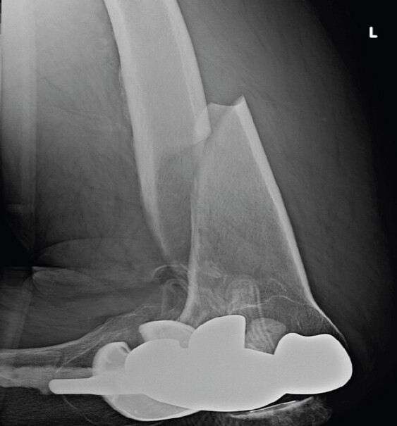
A 67-year-old male with a prior left TKA fell while walking to the bathroom in his home. He presented to the emergency department with an obvious deformity of the left lower extremity. The patient was evaluated in the ED and found to be neurovascularly intact. X-rays revealed a displaced and comminuted diaphyseal femur fracture involving the middle and distal thirds (Fig. 1). This fracture was subsequently fixed using a 14-hole distal femur locking plate with 4.5 mm non-locking cortical screws. The TKA hardware remained intact and unchanged following the surgery. The patient was evaluated at 12 days, 5 weeks, and 10 weeks after surgery with no complaints. X-rays at these visits demonstrated good alignment and progressive fracture healing. At 5 months post-operative, the patient developed loosening of the hardware and underwent debridement with revision of bone graft and placement of new screws. At this time, wound and tissue cultures grew Corynebacterium striatum, and he was subsequently placed on 6 weeks of vancomycin, which cleared the infection.
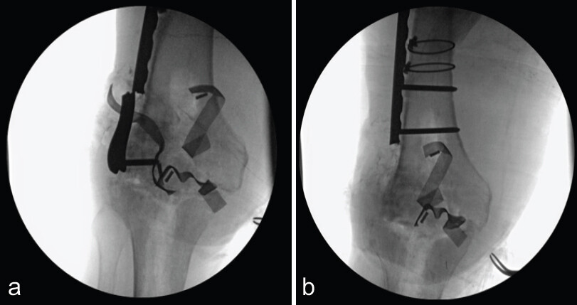
Ten months after the distal femur fracture, the patient complained of pain and swelling in the left knee. Arthrocentesis of the synovial fluid grew Staphylococcus epidermidis in the joint, and vancomycin was started. The knee was aspirated, and computed tomography (CT) scans were obtained for surgical planning. The CT scan demonstrated insufficient healing of the distal femur fracture, so along with removing the TKA hardware and placing an antibiotic spacer, the intra-articular aspect of the plate was cut off (Fig. 2a and b). The proximal part of the plate and screws remained attached to the femur to stabilize the fracture and allow for continued healing. At 4 weeks after the stage 1 revision, the patient was doing very well with no signs of persistent infection or complaints. Stage 2 revision was delayed due to poor control of diabetes (Hemoglobin A1c of 9.4%) and a urinary tract infection. The patient was able to get his A1c down to 6.5% over the course of the next few months, allowing the 2nd stage of the revision to be completed. Before this surgery, knee aspiration cultures were negative, and the patient had finished antibiotic therapy: Ciprofloxacin for his UTI.
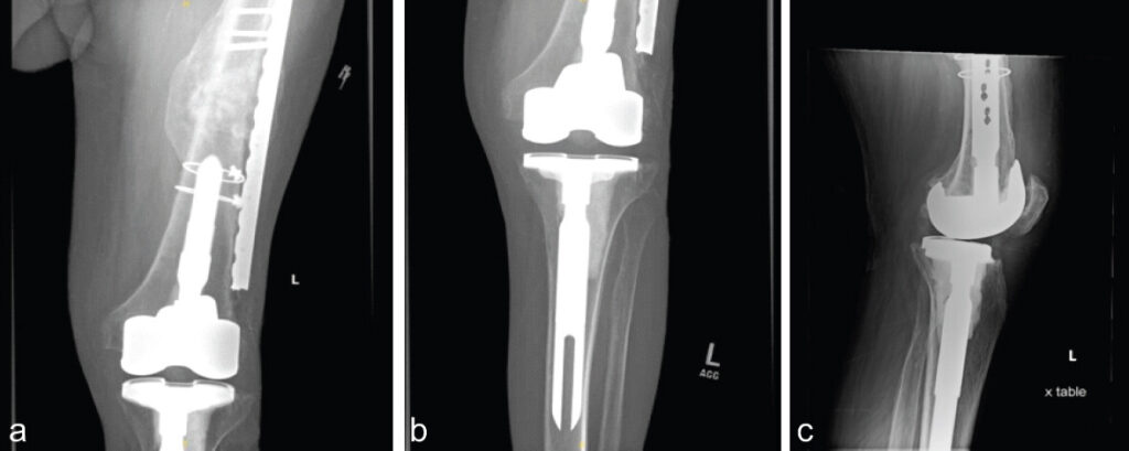
The revision surgery went as planned, using cemented hardware. Following surgery, the patient was placed on 100 mg oral doxycycline BID. Six weeks after the revision, cultures remained negative, and the patient was instructed to continue weight-bearing as tolerated. The images at 2 months showed an intact cemented TKA and lateral femoral plate (Fig. 3a, Fig. 3b, c). Most recent follow-up at 29 months demonstrated stable alignment of the knee with no further complications.
Discussion
PJIs are complications of joint arthroplasty surgeries which involve the introduction of foreign material into the human body. These infections arise from a sinus tract with the outside environment that directly communicates with the prosthesis [3]. Diagnosis involves arthrocentesis with evaluation of the fluid, specifically looking for the presence of elevated serum C-reactive protein, elevated erythrocyte sedimentation rate, neutrophils in the synovial fluid, and/or an isolated pathogen [8,9]. Common pathogens include Staphylococcus aureus in the acute setting (<3 months) and S. epidermidis in the delayed setting (3–12 months). After identifying a PJI in the case of a TKA, a staged revision is planned to remove the infected hardware and then restabilize the joint.
Standard staged management of PJIs involves the removal of all foreign material to optimize the eradication of the infection. Hardware retained during the revision process are at increased risk of causing a persistent infection due to biofilm production [10]. However, femoral fractures, critical to the stability of the lower extremity, add complexity to the management of PJIs. Partial HWR has been utilized as a method of treating infections involving deep brain stimulation, but has yet to be adopted as a standard of practice for orthopedic infections. Studies by Ginalis et al.; Fenoy and Simpson demonstrated successful clearance of atraumatic device-related infection when only pieces of the hardware were removed in patients with deep brain stimulation [11,12]. These pathogens are often similar to the ones that affect orthopedic hardware. Since the infection in this case was found in the synovial fluid, all the TKA hardware was removed. The distal end of the femoral plate closest to the joint space was cut out and removed from the femur. This procedure successfully cleared the infection without sacrificing femoral support.
Diabetes mellitus is a well-documented cause of delayed healing and increased post-operative infections. Patients with diabetes and persistent hyperglycemia remain in a pro-inflammatory state with the production of tumor necrosis factor-alpha, interleukin (IL)-1β, and IL-6 leading to increased osteoclast formation and bone resorption [13]. Fracture healing involves formation of a hematoma, granulation tissue, bony callus, and bone remodeling. Microvascular disease from a high-glucose state leads to a disruption in this process, delayed fracture union, and an increased risk of infection [14]. Therefore, careful consideration should be given to diabetic patients with femur fractures near the prosthesis. The patient in this case presented 10 months after open reduction and internal fixation (ORIF) of a femoral fracture with a PJI, aiding the decision to keep the fracture stable during the revision process.
Early identification and intervention of PJI is essential for maximizing chances of successful treatment. Additional hardware around the joint would typically be removed to decrease the chance of sustained infection. However, this case presents an alternative option for maintaining the structural integrity of the lower extremity while simultaneously eradicating the infection. Keeping the plate attached provided earlier weight bearing for the patient, which reduced the chance of developing deep vein thrombosis. Partial plate removal was an individualized treatment method that led to successful outcomes for 29 months in this patient and should be considered by surgeons who are presented with a similar problem.
Conclusion
This case report describes the successful management of a PJI following TKA in a patient with periarticular distal femur fracture fixation, using selective retention of the extra-articular hardware to maintain structural stability and minimize soft tissue insult. This 67-year-old male developed a chronic PJI with S. epidermidis after prior ORIF. Standard two-stage revision with complete HWR would have compromised fracture stability, so only the intra-articular portion of the plate was removed, while the stable proximal, extra-articular portion was left in place. Following targeted antibiotic therapy and delayed reimplantation, the infection was eradicated without any loss of mechanical integrity. At the 29-month follow-up, the patient demonstrated infection-free recovery and stable length, alignment, and rotation of the previous fracture fixation. This report highlights that partial hardware preservation is a viable alternative in complex PJI cases where both infection control and structural stability must be maintained.
Clinical Message
Partial HWR is a valid option for PJIs with additional periarticular hardware. Adequate antibiotics and debridement with a staged revision is still required, however, only partial removal of the intra-articular hardware can preserve extra-articular fracture stability. Individualized treatment plans are essential for optimizing stability and improving patient outcomes.
Conflict of Interest:
Nil
Source of Support:
Nil
Consent:
The authors confirm that informed consent was obtained from the patient for publication of this article
How to Cite this Article
Boesel J, Ifetiha I, Farmer NC, Yatsonsky D, Hanna M. Management of Periprosthetic Total Knee Infection with Partial Hardware Removal: A Case Report. J Orthop Case Rep. 2026 November;16(11): 142-146. doi: 10.13107/jocr.2026.v16.i11.8418.
References
- Tabaja H, Abu Saleh OM, Osmon DR.. Periprosthetic joint infection: What's new?. Infect Dis Clin North Am 2024;38:731-56. [Google Scholar] | [PubMed]
- Hao LJ, Wen PF, Zhang YM, Song W, Chen J, Ma T.. Treatment of periprosthetic knee infection and coexistent periprosthetic fracture: A case report and literature review. World J Clin Cases 2023;11:2321-8. [Google Scholar] | [PubMed]
- Mian HM, Lyons JG, Perrin J, Froehle AW, Krishnamurthy AB.. A review of current practices in periprosthetic joint infection debridement and revision arthroplasty. Arthroplasty 2022;4:31. [Google Scholar] | [PubMed]
- Madariaga S, Vargas-Reverón C, Tornero E, Alías A, Capurro B, Combalia A. Outcomes of hip arthroplasty with concomitant hardware removal: Influence of the type of implant retrieved and impact of positive intraoperative cultures. Arch Orthop Trauma Surg 2021;141:333-9. [Google Scholar] | [PubMed]
- Sequeira SB, Hasenauer MD, McKinstry R, Ebert F, Boucher HR.. Decreased risk of periprosthetic joint infection with concurrent hardware removal during conversion total hip arthroplasty compared to staged removal: A matched cohort analysis. J Am Acad Orthop Surg Glob Res Rev 2023;7:e2300261. [Google Scholar] | [PubMed]
- Fisher ND, Egol KA, Schwarzkopf R.. Risk factors for wound complications following conversion tka after tibial plateau fracture. Indian J Orthop 2022;56:1751-8. [Google Scholar] | [PubMed]
- Klatte TO, Schneider MM, Citak M, Oloughlin P, Gebauer M, Rueger M. Infection rates in patients undergoing primary knee arthroplasty with pre-existing orthopaedic fixation-devices. Knee 2013;20:177-80. [Google Scholar] | [PubMed]
- Parvizi J, Gehrke T.. Definition of Periprosthetic Joint Infection 2014. Available from: https://jdc.jefferson.edu/rothman_institute/49. [Last accessed on 2022 Jan 11]. [Google Scholar] | [PubMed]
- Parvizi J, Zmistowski B, Berbari EF, Bauer TW, Springer BD, Della Valle CJ. New definition for periprosthetic joint infection: From the workgroup of the musculoskeletal infection society. Clin Orthop Relat Res 2011;469:2992-4. [Google Scholar] | [PubMed]
- Tande AJ, Patel R.. Prosthetic joint infection. Clin Microbiol Rev 2014;27:302-45. [Google Scholar] | [PubMed]
- Ginalis EE, Hargreaves EL, Caputo DL, Danish SF.. Is it possible to save the deep brain stimulation hardware when presenting with wound dehiscence or hardware infection?. Stereotact Funct Neurosurg 2021;99:496-505. [Google Scholar] | [PubMed]
- Fenoy AJ, Simpson RKJr. Management of device-related wound complications in deep brain stimulation surgery. J Neurosurg 2012;116:1324-32. [Google Scholar] | [PubMed]
- Jiao H, Xiao E, Graves DT.. Diabetes and its effect on bone and fracture healing. Curr Osteoporos Rep 2015;13:327-35. [Google Scholar] | [PubMed]
- Retzepi M, Donos N.. The effect of diabetes mellitus on osseous healing. Clin Oral Implants Res 2010;21:673-81. [Google Scholar] | [PubMed]
© 2026 Journal of Orthopaedic Case Reports - Published by Indian Orthopaedic Research Group
About the Authors
-

Joseph Boesel
-

Ibrahim Ifetiha
-

Neal Chase Farmer
-

David Yatsonsky
-

Maged Hanna
Related Articles in Journal of Orthopaedic Case Reports
 June 1, 2026 Antiphospholipid Syndrome–Associated Livedoid Vasculopathy Mimicking Recurrent Periprosthetic Joint Infection after Total Knee Arthroplasty: A Case Report
 June 1, 2026 One-Stage Revision for Low-Grade Periprosthetic Infection with Staphylococcus hominis Following Cruciate-Retaining Total Knee Arthroplasty: Case Report and Literature Review
 January 1, 2026 A Step Beyond Convention: Three-Stage Revision with Limb Reconstruction System Fixator for Chronic Infected Total Knee Arthroplasty – A Case Report
 January 1, 2026 Retrograde Intramedullary Nailing Versus Locked Plating for Periprosthetic Distal Femur Fractures: A Propensity-matched Cohort from TriNetX