


Introduction
Vertebral hemangiomas (AVH) are common benign vascular lesions (10–12%) [1] accounting for 2–3% of all spinal tumors [2], and are often asymptomatic and incidentally detected. However, a small subset, termed aggressive AVH, exhibits locally invasive behavior characterized by cortical destruction, epidural extension, and neurological compromise [1,3]. These lesions account for approximately 1% of all AVH and may present with progressive myelopathy or radiculopathy [4].
Management remains controversial, and treatment options include radiotherapy, embolization, vertebroplasty, and surgical decompression and resection, often combined [5,6,7]. Pediatric cases are rare, and optimal strategies have not been well established. We present a case of aggressive vertebral hemangioma in a 12-year-old girl with progressive neurological deterioration, successfully managed with a multimodal approach and long-term follow-up.
Case Report
A 12-year-old girl presented with right-side dominant lower limb sensory loss. One month later, she developed progressive bilateral lower limb weakness, numbness, and gait imbalance. She also reported night pains; there were no bowel or bladder complaints.
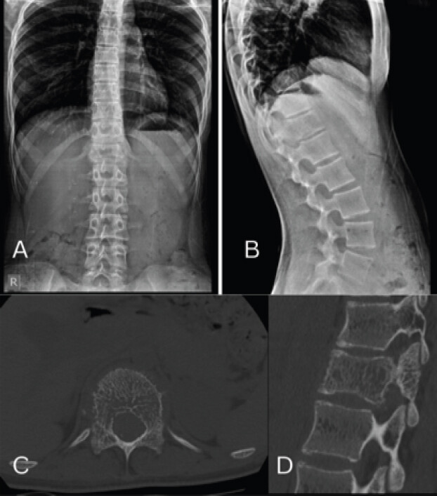
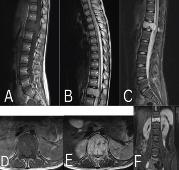
Neurological examination revealed a high-stepping, ataxic gait. Motor power was 3/5 at the hips and knees and 2/5 at the ankles (bilateral). Sensory examination revealed reduced sensation in the bilateral L4, L5, and S1 dermatomes. Deep tendon reflexes were brisk, with bilateral ankle and patellar clonus and extensor plantar responses (Nurick grade-4). Based on the biopsy findings and clinical progression, the lesion was characterized as an aggressive vertebral hemangioma (Enneking stage 3) with evolving conus compression. The patient underwent radiological investigations. (Fig. 1a and b) Anteroposterior and lateral thoracolumbar radiographs demonstrated a D12 vertebral body lesion with coarse trabeculations, (Fig. 1c and d) Pre-operative computed tomography showed Corduroy and Polka dot appearances in sagittal and axial sections. (Fig. 2a, b, c, d,e,f) Pre-operative magnetic resonance imaging demonstrated an aggressive D12 vertebral hemangioma with vertebral body involvement and epidural extension, causing significant spinal cord compression.
Pre-operative management
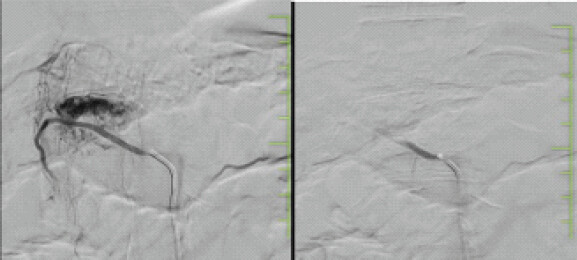
The patient underwent a pre-operative angiographic evaluation. Imaging revealed a hypervascular lesion involving the D12 vertebral body supplied by bilateral posterior intercostal arteries at T12, with an additional collateral supply from the upper thoracic arteries. The anterior spinal artery was identified at the T10 and T11 levels. Superselective embolization was performed using a microcatheter. Post-embolization angiography demonstrated stasis within the feeding vessels and the absence of tumor blush, indicating successful devascularization. The anterior spinal artery remained patent. (Fig. 3 a and b) depict the pre- and post-embolization of the D12 hemangiomatous lesion.
Surgical procedure
The patient subsequently underwent definitive surgical management consisting of D12 bi-pedicular vertebroplasty with D12 laminectomy, bilateral transpedicular decompression, and posterior instrumentation from D10 to L2 with pedicle screw fixation. Post-vertebroplasty, most of the compressive vascular granulation tissue was found to be shrunken, and the remaining vascular granulation tissue was subsequently excised, achieving adequate neural decompression. Intraoperative blood loss was approximately 600 mL. No intraoperative or immediate post-operative neurological deterioration was noted.
Histopathological findings
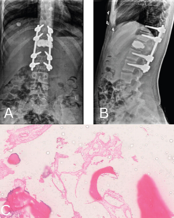
Histopathological examination revealed a vascular neoplasm composed of compact, thin-walled vascular channels of varying sizes separated by stroma. Endothelial cells showed mild nuclear enlargement and atypia without mitotic activity or necrosis (Fig 4a–c). Immunohistochemistry revealed CD34 and CD31 positivity, strongly suggesting a vascular neoplasm, as these markers are expressed in the endothelial cells. The low MIB-1 labeling index of ~1% indicated slow proliferative activity, consistent with benign biological behavior despite aggressive clinical presentation. These findings are consistent with vertebral hemangiomas.
Post-operative course and adjuvant therapy
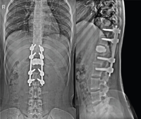
The post-operative course was uneventful. By post-operative day 2, the patient showed early neurological improvement in the left ankle power (3/5) and right ankle (2/5). She was Nurick Grade 4. Post-operative radiography was performed and found to be satisfactory (Fig. 4 a and b). The patient was discharged on post-operative day 7. At the week’s post-operative period, improvement in gait was noted with persistent right foot drop (2/5) and left ankle power (3/5). At 2 weeks, she improved to Nurick grade III. At 1 month post-operative follow-up, the right ankle power improved (3/5), while the left ankle power remained the same (3/5). The patient underwent external beam radiotherapy using the Rapid-Arc technique, receiving 4000 cGy in 22 fractions over 31 days, as per the opinion of the radiation oncologist.
Follow-up visits
-
Two months post-operatively: Gait balance was attained, and left dorsiflexion power improved (4/5), with right ankle power (3/5). Nurick Grade-2
-
Three months post-operatively: Complete recovery of right and left ankle dorsiflexion (5/5) was achieved, and the patient attained a normal neurological status and Nurick Grade 1. X-rays showed satisfactory implant positioning
-
One year post-operatively: Asymptomatic, normal activities, and X-rays showed appropriate implant positioning and spinal alignment. Nurick grade 0
-
Three years post-operatively: No recurrence; normal neurology; X-rays showed construct placement and spine alignment. Nurick grade 0
-
Five years post-operatively: No recurrence; normal neurology with well-positioned implants and spinal alignment. Nurick grade 0 (Fig. 5 a and b).
Discussion
AVH are rare entities with the potential for significant neurological compromise. Management of these tumors requires careful planning because of their high vascularity and risk of intraoperative bleeding [2,5]. Pre-operative embolization plays a critical role in reducing intraoperative blood loss and facilitating safe resection [7,8]. Surgical decompression remains the mainstay treatment for patients with neurological deficits, with or without stabilization, depending on structural involvement [4]. Vertebroplasty provides the additional benefits of mechanical stabilization and tumor devascularization [9] and also significantly reduces the need for radical surgical resection by achieving local tumor control [10]. Main indication for surgical stabilization in our case is to maintain spinal stability and to prevent post-operative deformity in patient with extensive pan vertebral involvement, requiring extensive decompression, rather than solely the lesion being a stable aggressive vertebral hemangioma [2,6]. Adjuvant radiotherapy has been shown to reduce recurrence rates and improve local control, particularly in subtotal resections [6,10]. Post-operative radiotherapy (40 Gy) with in the therapeutic range, was mainly indicated here because of locally aggressive nature of the lesion and anticipated residual intraosseous remnants even following extensive decompression to achieve complete disease free environment [3,9]. A multimodal approach combining embolization, surgery, and radiotherapy is increasingly advocated in the literature and is associated with favorable outcomes [10]. Pediatric aggressive vertebral hemangioma is extremely rare and this case adds to the limited available literature demonstrating successful long-term outcomes and that a carefully planned multimodal approach may achieve satisfactory disease control while avoiding the morbidity associated with radical surgical excision in aggressive vertebral hemangioma. Recurrence should not be considered inevitable but residual disease foci and incomplete treatment strategy can result in late recurrence [2]. Our Patient with 5 year asymptomatic and disease free follow-up is the result of implementing appropriate treatment strategy in such exceptionally rare presentation of Pediatric aggressive vertebral hemangioma.
Conclusion
AVH in pediatric patients can cause rapid neurological deterioration. Early diagnosis and a multimodal treatment strategy involving pre-operative embolization, surgical decompression, vertebral stabilization, and adjuvant radiotherapy can result in excellent neurological recovery and long-term disease control without the need for morbidly aggressive resection.
Clinical Message
Pediatric aggressive vertebral hemangiomas are exceptionally uncommon, and the literature has only a few cases reported. They are prone for causing rapid neurological deterioration, necessitates need for a multidisciplinary multimodal strategy incorporating embolization, vertebroplasty, decompression, stabilization, and post-operative adjuvant radiotherapy, avoiding the morbidity associated with en bloc vertebral resection and can achieve excellent neurological recovery and durable long-term disease control without radical resection.
Conflict of Interest:
Nil
Source of Support:
Nil
Consent:
The authors confirm that informed consent was obtained from the patient for publication of this article
How to Cite this Article
Guganesh, Ghorpade A, Swapnil K, Saikumar W, Ashutosh S. Multimodal Management of an Aggressive Vertebral Hemangioma in a Child with Neurological Deficit: A Case Report with 5-Year Follow-Up. J Orthop Case Rep. 2026 November;16(11): 175-179. doi: 10.13107/jocr.2026.v16.i11.8430.
References
- Nakahara M, Nishida K, Kumamoto S, Hijikata Y, Harada K.. A case report of spondylectomy with circumference reconstruction for aggressive vertebral hemangioma covering the whole cervical spine (C4) with progressive spinal disorder. Eur Spine J 2017;26:69-74. [Google Scholar] | [PubMed]
- Diarra MD, Zhang Z, Wang Z, Yinwang E, Li H, Wang S. Surgical options for aggressive vertebral hemangiomas:A case series, literature review and treatment recommendations. J Bone Oncol 2023;43:100515. [Google Scholar] | [PubMed]
- Fox MW, Onofrio BM.. The natural history and management of symptomatic and asymptomatic vertebral hemangiomas. J Neurosurg 1993;78:36-45. [Google Scholar] | [PubMed]
- Acosta FL, Dowd CF, Chin C, Tihan T, Ames CP, Weinstein PR.. Current treatment strategies and outcomes in the management of symptomatic vertebral hemangiomas. Neurosurgery 2006;58:287-95. [Google Scholar] | [PubMed]
- Pastushyn AI, Slin'ko EI, Mirzoyeva GM.. Vertebral hemangiomas: Diagnosis, management, natural history and clinicopathological correlates in 86 patients. Surg Neurol 1998;50:535-47. [Google Scholar] | [PubMed]
- Jiang L, Liu XG, Yuan HS, Yang SM, Li J, Wei F. Diagnosis and treatment of vertebral hemangiomas with neurologic deficit: A report of 29 cases and literature review. Spine J 2014;14:944-54. [Google Scholar] | [PubMed]
- Heyd R, Seegenschmiedt MH, Rades D, Winkler C, Eich HT, Bruns F. Radiotherapy for symptomatic vertebral hemangiomas: Results of a multicenter study and literature review. Int J Radiat Oncol Biol Phys 2010;77:217-25. [Google Scholar] | [PubMed]
- Chen HI, Heuer GG, Zaghloul K, Simon SL, Weigele JB, Grady MS.. Lumbar vertebral hemangioma presenting with the acute onset of neurological symptoms. Case report. J Neurosurg Spine 2007;7:80-5. [Google Scholar] | [PubMed]
- Kato K, Teferi N, Challa M, Eschbacher K, Yamaguchi S.. Vertebral hemangiomas: A review on diagnosis and management. J Orthop Surg Res 2024;19:310. [Google Scholar] | [PubMed]
- Cotten A, Deramond H, Cortet B, Lejeune JP, Leclerc X, Chastanet P. Preoperative percutaneous injection of methyl methacrylate and N-butyl cyanoacrylate in vertebral hemangiomas. AJNR Am J Neuroradiol 1996;17:137-42. [Google Scholar] | [PubMed]
© 2026 Journal of Orthopaedic Case Reports - Published by Indian Orthopaedic Research Group
About the Authors
-

Guganesh Palanisamy
-

Abhay Ghorpade
-

Swapnil Kumar
-

Saikumar Waghmore
-

Ashutosh Sabnis Balkrishna
Related Articles in Journal of Orthopaedic Case Reports
 September 1, 2026 Combined Ultrasound- and Fluoroscopy-guided Percutaneous Cementoplasty and Ablation for a C2 Pathologic Fracture: A Case Report
 July 1, 2025 Failure of Stentoplasty Due to Non-performance of Balloon
 May 1, 2025 To Study the Functional Outcome of Vertebroplasty versus Conservative Management in Osteoporotic Fractures
 November 1, 2026 Incidentally Detected Osseous Hemangioma of the Femoral Head: Conservative Management of a Rare Case with Review of the Literature