


Introduction
Brodie’s abscess is a localized form of subacute osteomyelitis that often presents with persistent localized pain and minimal systemic symptoms, making the diagnosis challenging. Radiographs typically demonstrate a well-defined lytic lesion with surrounding sclerosis, while magnetic resonance imaging (MRI) may reveal the characteristic penumbra sign, a feature considered highly suggestive of subacute osteomyelitis. However, this sign is not specific, and several benign and malignant bone lesions can closely mimic these imaging findings, posing a significant diagnostic challenge [1,2,3,4,5].
Distinguishing infection from malignancy is essential, as both conditions may present with similar clinical and radiological features, including bone pain, marrow edema, cortical destruction, and elevated inflammatory markers. Although MRI plays an important role in the evaluation of these lesions, imaging findings should always be interpreted in conjunction with the clinical presentation. In atypical cases, histopathological examination remains the definitive method for establishing the diagnosis [1].
Esophageal squamous cell carcinoma (ESCC) is one of the most common histological subtypes of esophageal cancer worldwide and is strongly associated with chronic tobacco and alcohol use. Bone metastasis is uncommon and most frequently involves the axial skeleton, particularly the vertebrae, ribs, and pelvis. Metastatic involvement of the femoral head is exceptionally rare and may present with non-specific symptoms, often mimicking more common orthopedic conditions [6, 7].
We report a rare case of metastatic ESCC involving the femoral head that clinically and radiologically mimicked Brodie’s abscess. The diagnosis was further complicated by concurrent methicillin-resistant Staphylococcus aureus (MRSA) infection, with positive intraoperative cultures initially supporting an infective etiology. The unexpected histopathological diagnosis of metastatic squamous cell carcinoma emphasizes the importance of obtaining tissue diagnosis in atypical bone lesions, particularly in patients with significant risk factors for malignancy.
Case Report
A 53-year-old man presented to our orthopedic outpatient department with pain in the right groin and upper thigh for 2 months. The pain had an insidious onset and gradually worsened over time. Initially intermittent, it became persistent and eventually interfered with walking and weight-bearing. There was no history of trauma, fever, night sweats, significant weight loss, or previous musculoskeletal disease. He was a chronic smoker with a smoking history of more than 30 pack-years and had no known history of malignancy or tuberculosis.
On examination, he walked with an antalgic gait and had tenderness over the right groin. Hip movements, particularly flexion and internal rotation, were painful. No local swelling, erythema, or increased local temperature was noted, and the distal neurovascular examination was normal.
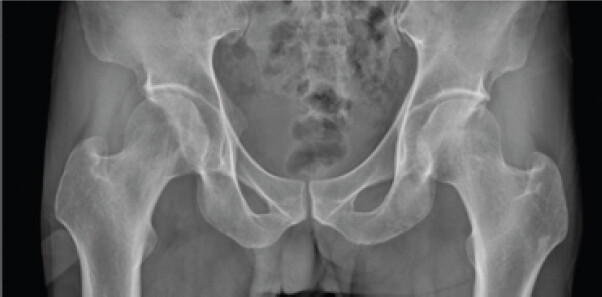
Laboratory investigations showed mild leukocytosis with elevated erythrocyte sedimentation rate (45 mm/h) and C-reactive protein levels. Plain radiographs of the pelvis revealed a well-defined lytic lesion with surrounding sclerosis involving the right femoral head (Fig. 1).
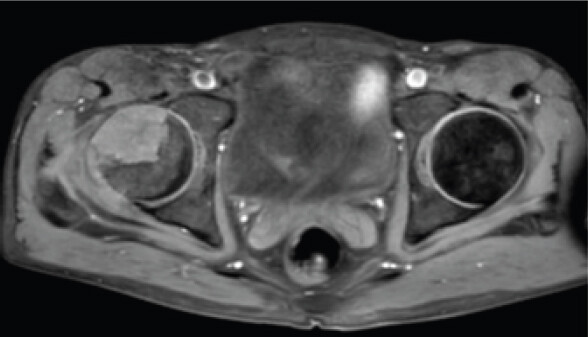
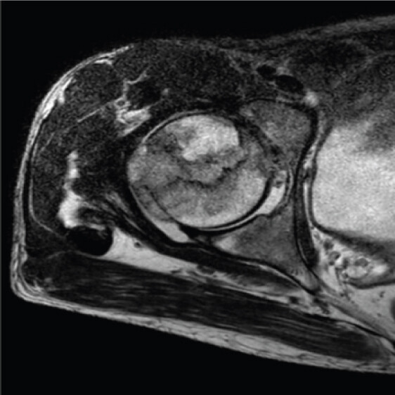
MRI of the pelvis and right hip demonstrated a well-circumscribed lesion that was hypointense on T1-weighted images and hyperintense on T2-weighted images, with peripheral rim enhancement and surrounding marrow edema after contrast administration (Fig. 2 and 3). In view of the clinical findings and characteristic MRI appearance, a provisional diagnosis of Brodie’s abscess was made.
The patient subsequently underwent core decompression of the femoral head. Intraoperatively, no pus was encountered. Instead, the lesion contained friable grey-white tissue with areas of necrosis, raising suspicion of an alternative pathology. Tissue samples were sent for microbiological culture and histopathological examination, followed by curettage and decompression of the lesion.
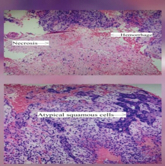
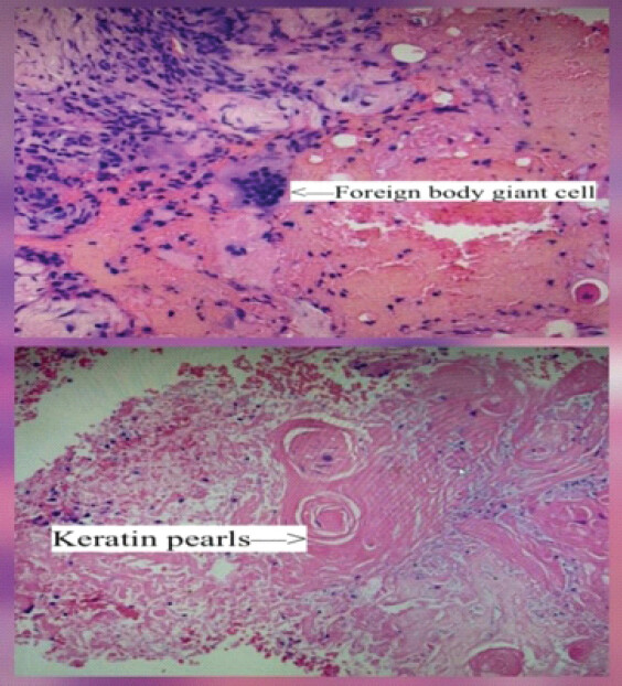
Microbiological culture grew MRSA, initially supporting the diagnosis of subacute osteomyelitis, and culture-directed antibiotic therapy was started. Histopathological examination, however, revealed metastatic squamous cell carcinoma characterized by infiltrating nests of atypical squamous cells with keratinization and intercellular bridges (Fig. 4 and 5).
A subsequent metastatic workup with contrast-enhanced computed tomography and upper gastrointestinal endoscopy demonstrated an ulcero-proliferative lesion in the mid-esophagus. Biopsy from the lesion confirmed a moderately differentiated squamous cell carcinoma, establishing the diagnosis of metastatic ESCC involving the femoral head with concurrent MRSA infection. The patient was referred to the multidisciplinary oncology team for further management. After staging, he was treated with palliative chemoradiotherapy for metastatic ESCC and remained under oncological follow-up. The clinical timeline of the patient has been summarized in Table 1.
Clinical timeline of presentation, diagnostic evaluation, and management
| Timeline | Event |
|---|---|
| 2 months | Groin pain |
| X-ray | Lytic lesion |
| MRI | Brodie’s abscess suspected |
| Surgery | Core decompression |
| Culture | MRSA |
| Histopathology | Metastatic SCC |
| Endoscopy | Mid-esophageal SCC |
| Oncology | Chemoradiotherapy |
MRI: Magnetic resonance imaging, MRSA: Methicillin-resistant Staphylococcus aureus, SCC: Squamous cell carcinoma
Discussion
Brodie’s abscess is an important differential diagnosis in patients presenting with persistent bone pain and a well-defined lytic lesion, particularly when MRI demonstrates the characteristic penumbra sign. Although this imaging feature is considered highly suggestive of subacute osteomyelitis, it is not specific and has also been described in several benign and malignant bone lesions [1,2,3,4,5]. In our patient, the clinical presentation, raised inflammatory markers, and MRI findings were all in keeping with Brodie’s abscess, making the initial diagnosis appear convincing.
The diagnostic challenge became evident only during surgery. Instead of the expected purulent collection, the lesion contained friable grey-white tissue, prompting tissue sampling for microbiological and histopathological examination. While intraoperative cultures grew MRSA and initially reinforced the diagnosis of infection, histopathology revealed metastatic squamous cell carcinoma. This case demonstrates that the presence of a positive bacterial culture does not necessarily exclude an underlying malignancy. Infection and metastatic disease may coexist within the same lesion, and histopathological examination remains essential whenever the operative findings are atypical or do not correlate with the pre-operative diagnosis [8].
Bone metastasis from ESCC is uncommon and usually occurs in advanced disease. When present, the vertebral column, ribs, and pelvis are the most frequently involved sites, whereas metastasis to the femoral head is exceptionally rare [6,7,9]. Furthermore, skeletal pain may be the first manifestation of an otherwise occult primary tumor, delaying the diagnosis of esophageal carcinoma. Lyngdoh et al. described a patient whose initial presentation was hip pain secondary to femoral metastasis from esophageal carcinoma, while Lyngdoh et al. reported occult ESCC presenting with multiple osteolytic lesions that initially mimicked multiple myeloma [10]. Unlike these reports, our patient had a solitary femoral head lesion with MRI features highly suggestive of Brodie’s abscess, and the diagnosis was further confounded by concurrent MRSA infection.
To our knowledge, reports describing metastatic ESCC of the femoral head presenting as Brodie’s abscess with concurrent MRSA infection are exceedingly rare. The combination of characteristic MRI findings, positive microbiological culture, and underlying metastatic disease created a unique diagnostic challenge. This case reinforces the importance of obtaining representative tissue for histopathological examination whenever the clinical course, imaging findings, or intraoperative appearance is atypical.
For the orthopedic surgeon, the most important lesson is that imaging and microbiological findings should not be interpreted in isolation. When clinical, radiological, and intraoperative findings are discordant, early biopsy with histopathological examination is essential to establish the correct diagnosis and ensure timely oncological management.
Conclusion
Metastatic ESCC involving the femoral head is an exceptionally rare presentation and can closely resemble Brodie’s abscess on clinical and radiological evaluation. As demonstrated in this case, even positive microbiological cultures may not represent the primary pathology. Histop
athological examination remains indispensable when evaluating atypical bone lesions, particularly in patients with risk factors for malignancy. A timely biopsy can establish the correct diagnosis and facilitate appropriate oncological management.
Clinical Message
Not every lesion that appears to be Brodie’s abscess is infectious. When clinical or intraoperative findings are unusual, biopsy should be performed to rule out an underlying malignancy.
Conflict of Interest:
Nil
Source of Support:
Nil
Consent
The authors confirm that informed consent was obtained from the patient for publication of this article
How to Cite this Article
Sharma C, Modi N, Borgohain B, Khonglah T, Mandal A. Metastatic Esophageal Squamous Cell Carcinoma of the Femoral Head Masquerading as Brodie’s Abscess with Concurrent Methicillin-Resistant Staphylococcus aureus Infection: A Diagnostic Dilemma. J Orthop Case Rep. 2026 November;16(11):293-297. doi: 10.13107/jocr.2026.v16.i11.8476.
References
- Lew DP, Waldvogel FA. Osteomyelitis. Lancet 2004;364:369-79. [Google Scholar] | [PubMed]
- Miller WB Jr., Murphy WA, Gilula LA. Brodie abscess: Reappraisal. Radiology 1979;132:15-23. [Google Scholar] | [PubMed]
- Marti-Bonmati L, Aparisi F, Poyatos C, Vilar J. Brodie abscess: MR imaging appearance in 10 patients. J Magn Reson Imaging 1993;3:543-6. [Google Scholar] | [PubMed]
- Grey AC, Davies AM, Mangham DC, Grimer RJ, Ritchie DA. The 'penumbra sign' on T1-weighted MR imaging in subacute osteomyelitis: Frequency, cause and significance. Clin Radiol 1998;53:587-92. [Google Scholar] | [PubMed]
- McGuinness B, Wilson N, Doyle AJ. The “penumbra sign” on T1-weighted MRI for differentiating musculoskeletal infection from tumour. Skeletal Radiol 2007;36:417-21. [Google Scholar] | [PubMed]
- Sung H, Ferlay J, Siegel RL, Laversanne M, Soerjomataram I, Jemal A. Global cancer statistics 2020: GLOBOCAN estimates of incidence and mortality worldwide for 36 cancers in 185 countries. CA Cancer J Clin 2021;71:209-49. [Google Scholar] | [PubMed]
- Imura Y, Yamamoto S, Wakamatsu T, Tanaka T, Tamiya H, Sugimura K. Clinical features and prognostic factors in patients with esophageal cancer with bone metastasis. Oncol Lett 2020;19:717-24. [Google Scholar] | [PubMed]
- Haruta Y, Kataoka Y, Ito H, Mori T, Matsuo M. Metastatic squamous cell carcinoma of the lung mimicking multiple bone abscesses. Respirol Case Rep 2017;5:e00256. [Google Scholar] | [PubMed]
- Shaheen O, Ghibour A, Alsaid B. Esophageal cancer metastases to unexpected sites: A systematic review. Gastroenterol Res Pract 2017;2017:1657310. [Google Scholar] | [PubMed]
- Lyngdoh BS, Dey B, Raphael V, Khonglah Y, Marak RS. Occult esophageal squamous cell carcinoma presenting with multiple bony osteolytic lesions clinically mimicking multiple myeloma: A diagnostic dilemma. J Lab Physicians 2020;12:236-9. [Google Scholar] | [PubMed]
© 2026 Journal of Orthopaedic Case Reports - Published by Indian Orthopaedic Research Group
About the Authors
-

Chirag Sharma
-

Naman Modi
-

Bhaskar Borgohain
-

Tashi G Khonglah
-

Alokananda Mandal
Related Articles in Journal of Orthopaedic Case Reports
 September 1, 2026 Askin Tumor Presents with Ewing Sarcoma of the Femur in Young Female Patient: Case Report and Literature Review
 July 1, 2026 Diagnostic Dilemma in a Distal Femoral Intramedullary Lesion: Enchondroma Mimicking Low-Grade Chondrosarcoma with Discordant Biopsy Findings: A Case Report
 June 1, 2026 Cortical Lytic Lesion of the Proximal Tibia in an 8-Year-Old Child: A Pediatric Diagnostic Conundrum
 June 1, 2026 Atypical Bilateral Cystic Foot Swellings with Osteomyelitis-Like Features: A Diagnostic Dilemma