


Introduction
Scapulothoracic dissociation is a rare but devastating injury characterized by complete disruption of the shoulder girdle’s musculoskeletal attachments from the thoracic wall, often associated with neurovascular compromise and typically resulting from high-energy trauma. Despite the profound internal disruption, the overlying skin is usually intact, hence the description of this entity as an internal or “closed” forequarter amputation.
Clinically, the condition presents with significant swelling in the shoulder region, preserved skin integrity, and varying degrees of neurological and vascular compromise – most notably, weakness, sensory disturbances, and absent pulses in the affected upper limb.
Diagnosis relies heavily on imaging, particularly anteroposterior chest radiographs, where measurement of the scapular index (ratio of distances from the midline to each scapula on an AP chest radiograph) can indicate lateral displacement of the scapula, which must be supplemented with higher imaging.
The Zelle et al.’s modification of Damschen et al.’s classification (Table 1) is a system for categorizing the severity of scapulothoracic dissociation injuries and has some predictive utility in determining the functional recovery of the affected limb.
Zelle et al.’s modification of Damschen et al. classification for scapulothoracic dissociation.
| TYPE | CLINICAL FINDINGS |
|---|---|
| 1 | ISOLATED MUSCULOSKELETAL INJURY |
| 2A | MUSCULOSKELETAL INJURY WITH VASCULAR DISRUPTION |
| 2B | MUSCULOSKELETAL INJURY WITH INCOMPLETE NEUROLOGICAL IMPAIRMENT OF UPPER EXTERMITY |
| 3 | MUSCULOSKELETAL INJURY WITH INCOMPLETE NEUROLOGICAL IMPAIRMENT OF UPPER EXTERMITY + VASCULAR DISRUPTION |
| 4 | MUSCULOSKELETAL INJURY WITH COMPLETE BRACHIAL PLEXUS AVULSION |
In this report, we describe a case of scapulothoracic dissociation managed successfully with operative skeletal stabilization of an acromioclavicular joint injury and a scapular spine fracture with innovative, custom-fabricated implants.
Case Report
A 35-year-old male was involved in a high-energy motor vehicle collision involving a truck. He was brought to the emergency department, where initial assessment revealed that he was unconscious and regained consciousness after half an hour. Diffuse swelling was observed around the right shoulder girdle, with no evidence of neurovascular deficit from C5 to T1. Clinical examination further demonstrated paradoxical breathing along with localized traumatic signs. Radiographs (Fig. 1) of the chest and right shoulder, along with computed tomography of the right shoulder (Fig. 2), revealed a laterally displaced right scapula with a fracture-dislocation of the acromion process and associated disruption of the acromioclavicular joint. Additional findings included a flail chest – multiple fractures of the right 2nd to 4th ribs. The scapular index on the right side was measured at 1.30 (Fig. 1). Further assessment demonstrated transverse process fractures from D2 to D9 along with associated muscle avulsion and presence of a hemopneumothorax for which an ICD tube insertion was done. Magnetic resonance imaging (Fig. 1) of the shoulder girdle with brachial plexus assessment showed severe contusion and edema involving the soft tissues, periscapular muscles, and fascial planes, in addition to extensive subcutaneous crepitus. The medial scapulothoracic muscles were not involved. Diffuse edema was seen around the trunks, divisions, and cords of the right brachial plexus with no obvious discontinuity along the nerve fibers involving the cervical and brachial plexus. Computed tomography (CT) arteriogram revealed no filling defects. A CT brain did not reveal any intracranial bleeds.
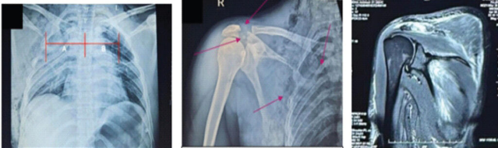
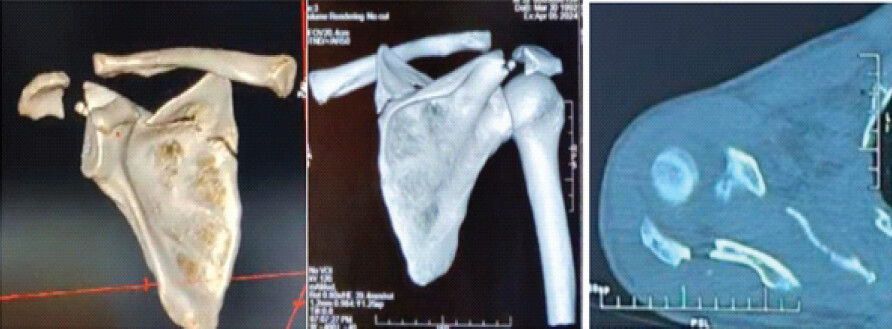
This injury was thus classified as Zelle et al. [1] Type I Right Scapulothoracic Dissociation (Table 1). Although there is no absolute indication for orthopedic intervention in this patient, we felt that there is an increased susceptibility for the patient to have an unstable shoulder girdle in the near future if left untreated.
Surgical technique
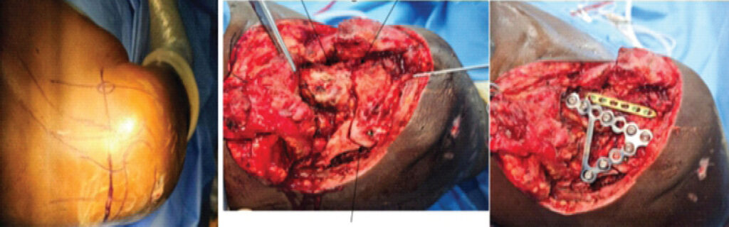
The patient was initially monitored and observed under intensive care and was taken up for the procedure 2 weeks following trauma once anesthesia deemed him fit for surgery. The patient was positioned on the operating table in a low beach-chair position with the right upper limb supported by an armboard and the arm was draped free. Using the Saber-cut approach, a skin incision was made over the posterior aspect of the angle of the acromion and extended anteriorly to the acromioclavicular joint. The incision was further extended medially along the spine of the scapula. Soft tissues were carefully dissected. The glenohumeral joint was not disturbed. The lateral insertion of the trapezius was incised and retracted medially, whereas the deltoid was partially incised and retracted laterally. The supraspinatus muscle was not incised.
The fracture site over the spine of the scapula (Fig. 3) was identified, and disruption of the acromioclavicular joint was visualized. The scapular spine fracture was reduced and stabilized using a lag screw. The acromioclavicular joint was reduced and temporarily held with Kirschner wires. The spine of the scapula, acromion process, and clavicle were then reduced and fixed with a customized 15-hole Stoppa’s (infrapectineal) plate, contoured to match the morphology of the fracture pattern. For additional stability, a 40-mm cortical screw was inserted through the plate from the clavicle to the coracoid. Furthermore, a 6-hole locking compression plate was applied to the superior surface of the clavicle and fixed with appropriately sized screws. Thorough wound lavage was performed, a drain was secured in place, and the distal pulse was confirmed to be palpable.
Outcome
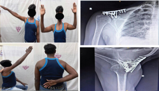
Postoperatively, the patient demonstrated progressive functional recovery. Early mobilization with active and passive range of motion was initiated to avoid complications such as muscle atony, complex regional pain syndrome, and post-traumatic periarthritis of the shoulder joint. Aggressive physiotherapy was feasible owing to the inherent stability conferred by the plate anatomy. At 2 weeks, shoulder abduction and forward flexion reached 30°. Follow-up radiographs at 5 and 12 months confirmed satisfactory fracture union without implant failure. At 5 months, abduction improved to 45° and forward flexion to 90°. By 12 months, the patient achieved abduction up to 120°, forward flexion to 150°, and external and internal rotation to 80° (Fig. 4), with no evidence of scapulothoracic instability and scapular winging.
Discussion
Advances in imaging at tertiary trauma centers have increased recognition of scapulothoracic dissociation [2], a condition with potentially fatal outcomes that warrants standardized diagnostic and management protocols. Orthopedic management of scapulothoracic dissociation begins with adherence to Advanced Trauma Life Support protocols, ensuring airway, breathing, and circulation are secured. Priority is then given to vascular injuries, which may necessitate urgent surgical repair. Subsequent steps include stabilization of associated fractures and assessment of brachial plexus integrity. A detailed diagnostic workup (Table 2) for a suspected case of scapulothoracic dissociation has been outlined, encompassing clinical, radiological, and adjunctive investigations to facilitate timely recognition and management.
A diagnostic workup for a suspected case of scapulothoracic dissociation.
| 1. INITIAL EVALUATION (ATLS PROTOCOL) |
|---|
| • To assess airway, breathing, circulation |
| • To inspect for presence of any massive swelling around shoulder joint, lateral displacement of shoulder, arm held in abduction, skin intact despite severe internal disruption |
| • To palpate for any bony discontinuity in the clavicle and to check for Scapular step-offs |
| • To assess neurovascular status: Brachial plexus (motor and sensory), distal pulses and capillary refill to be documented |
| 2. PLAIN RADIOGRAPHY (INITIAL SCREENING) |
| I. Chest X rays: |
| ♦ Scapular Index (Ratio of distance from spinous process to Scapular compared to contralateral side; >1.29 is suspicious) |
| ♦ Associated clavicle, acromioclavicular. Sternoclavicular joint disruption |
| ♦ Hemothorax, pneumothorax or rib fractures |
| II. Shoulder girdle X rays: AP, Lateral and axillary views if patient is stable |
| 3. ADVANCED IMAGING |
| I. COMPUTED TOMOGRAPHY – CHEST/ SHOULDER GIRDLE |
| ♦ Diagnosis of osseous injuries: Scapula, clavicle, AC/SC joints, and ribs |
| ♦ Quantification of scapular displacement |
| ♦ Preoperative planning |
| II. CT Angiography (CTA) |
| ♦ To assess vascular injury if suspected |
| ♦ To guide vascular repair and stenting |
| III. MRI (If patient is stable) |
| ♦ To rule out brachial plexus injuries (Root avulsions/Stretch injuries) |
In cases of non-salvageable limbs with extensive neurovascular compromise, early above-elbow amputation may be indicated. Depending on the extent of neural and musculoskeletal injury, further surgical options may include brachial plexus reconstruction or shoulder arthrodesis to optimize functional outcomes.
Disruption of two or more elements of the superior shoulder suspensory complex (SSSC) generally necessitates fixation [3]. Surgical stabilization is typically indicated to safeguard vascular repairs, prevent neurological deficit, and enable rehabilitation [4]. Early fixation has been associated with improved outcomes and fewer complications [5,6,7]. Although vascular repair remains the priority when limb viability is threatened, some authors advocate initial skeletal stabilization to protect subsequent vascular reconstructions [8].
The scapular index [9], with values >1.29 considered suggestive, has been described as a diagnostic adjunct but lacks specificity due to influences such as positioning, chest wall asymmetry, or pre-existing deformities. In this case, the index normalized on follow-up imaging, indicating either muscle sprain or atony rather than a complete muscle tear. Thus, the scapular index should be regarded only as a screening tool, with a definitive diagnosis confirmed by advanced imaging such as computed tomography/magnetic resonance imaging.
To date, no study has directly compared the outcomes of surgical fixation of clavicular fractures, acromioclavicular joint disruptions, or associated ipsilateral extremity fractures in patients with scapulothoracic dissociation against those in patients with isolated injuries. Consequently, the necessity of surgical stabilization in this condition remains uncertain. Fixation methods vary, with choices often guided by surgeon preference and available resources; reported techniques include plates, suture anchors, tightrope constructs, and mesh plates [10]. In this case, the modified infrapectineal plate (Fig. 5) provided inherent stability due to the anatomy of the implant and by addressing both the spine of the scapula fracture and the acromioclavicular disruption through a single stable construct.
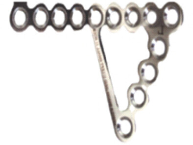
The operative technique employed in this case precludes motion at the acromioclavicular joint, which normally contributes once the arm is elevated beyond 90°. It is important to emphasize that the primary objective of this procedure is to maintain stability between the axial skeleton and the shoulder girdle, rather than to achieve conventional fracture fixation. To further augment stability, a cortical screw was inserted through the plate from the clavicle into the coracoid process. While this construct provides robust stabilization, it inevitably restricts terminal elevation of the shoulder. Consequently, implant removal will be necessary in the near future to allow restoration of the full range of shoulder motion.
Conclusion
Scapulothoracic dissociation is an uncommon, high-energy injury that necessitates a high index of suspicion for prompt diagnosis and timely intervention to optimize limb and life salvage. In the absence of standardized management protocols, thorough diagnostic evaluation and appropriate classification are essential for guiding treatment. Until evidence-based guidelines are established, management must be individualized, often requiring innovative, case-specific strategies.
Clinical Message
Scapulothoracic dissociation is a rare, potentially limb- and lifethreatening injury that may be overlooked when the associated bony injuries are not fully appreciated. This case highlights the importance of recognizing a bifocal SSSC disruption in association with a relatively subtle Type I scapulothoracic dissociation. Restoration of the integrity of the SSSC using stable fixation can help re-establish shoulder girdle alignment and provide a stable platform for functional recovery. Careful assessment of the entire shoulder girdle, including the acromion, acromioclavicular joint, clavicle, scapula, and neurovascular structures, is therefore essential in patients with suspected scapulothoracic dissociation.
Conflict of Interest:
Nil
Source of Support:
Nil
Consent
The authors confirm that informed consent was obtained from the patient for publication of this article
How to Cite this Article
Prakassh SKSR, Sameer M, Sashaank S. A Devastating Shoulder Girdle Injury and an Innovative Approach to Management – A Case Report. J Orthop Case Rep. 2026 November;16(11):287-292. doi: 10.13107/jocr.2026.v16.i11.8474
References
- Zelle BA, Pape HC, Gerich TG, Garapati R, Ceylan B, Krettek C. Functional outcome following scapulothoracic dissociation. J Bone Joint Surg Am 2004;86:2-8. [Google Scholar] | [PubMed]
- Althausen PL, Lee MA, Finkemeier CG. Scapulothoracic dissociation: Diagnosis and treatment. Clin Orthop Relat Res 2003;416:237-44. [Google Scholar] | [PubMed]
- Goss TP. Double disruptions of the superior shoulder suspensory complex. J Orthop Trauma 1993;7:99-106. [Google Scholar] | [PubMed]
- Yim J, Singleton A, Crist B, Phillips S. Open reduction internal fixation with mesh plating and suture fixation for scapulothoracic dissociation: A case report and discussion of surgical methods. JSES Rev Rep Tech 2023;3:403-9. [Google Scholar] | [PubMed]
- McCague A, Schulte A, Davis JV. Scapulothoracic dissociation: An emerging high-energy trauma in medical literature. J Emerg Trauma Shock 2012;5:363-6. [Google Scholar] | [PubMed]
- Lal H, Tanwar YS, Jaiswal A, Singh SP, Habib M. Scapulothoracic and scapholunate dissociation in the ipsilateral upper limb of a trauma victim. Chin J Traumatol 2014;17:242-5. [Google Scholar] | [PubMed]
- Sano K, Ozeki S. Treatment of combined injuries of the axillary and suprascapular nerves with scapulothoracic dissociation. J Brachial Plex Peripher Nerve Inj 2015;10:e62-5. [Google Scholar] | [PubMed]
- Lavelle WF, Uhl R. Scapulothoracic dissociation. Orthopedics 2010;33:417-21. [Google Scholar] | [PubMed]
- Choo AM, Schottel PC, Burgess AR. Scapulothoracic dissociation: Evaluation and management. J Am Acad Orthop Surg 2017;25:339-47. [Google Scholar] | [PubMed]
- Merk BR, Minihane KP, Shah NA. Scapulothoracic dissociation with acromioclavicular separation: A case report of a novel fixation method. J Orthop Trauma 2008;22:572-5. [Google Scholar] | [PubMed]
© 2026 Journal of Orthopaedic Case Reports - Published by Indian Orthopaedic Research Group
About the Authors
-

S K Srinivasa Ram Prakassh
-

Mohamed Sameer
-

Srijay Sashaank
Related Articles in Journal of Orthopaedic Case Reports
 October 1, 2026 Stress-Related Acromion Base Fracture after Clavicular Hook Plate Fixation: A Case Report
 July 1, 2026 Short-term Functional Outcomes of Shoulder Girdle Limb Salvage in Primary Bone Tumors: A 2-year Follow-up Study
 April 1, 2026 Modified Posterior Approach for Scapular Body Non-union: A Case Report
 November 1, 2024 Cuff Arthropathy, Scapular Spine Fracture Non-union, and Instability: Open Reduction, Internal Fixation & Simultaneous Reversed Shoulder Arthroplasty – A Case Report