


Introduction
Giant cell tumor (GCT) of bone is a locally aggressive, primary osteolytic neoplasm. While histologically benign, it is characterized by significant bone resorption and a recognized potential for pulmonary metastasis. GCT accounts for approximately 4–10% of all primary bone tumors and nearly 20% of benign skeletal lesions [1,2]. The tumor typically arises in the epiphyseal or epi-metaphyseal regions of long bones, with the distal femur, proximal tibia, and distal radius serving as the most frequent anatomical sites [2].
The demographic distribution of GCT is well-documented, with a peak incidence occurring in the third and fourth decades of life. Approximately 80% of cases are diagnosed in patients between the ages of 20 and 40 [2]. Conversely, the occurrence of GCT in the geriatric population – specifically those older than 70 years – is exceedingly rare, often cited as sporadic in clinical literature [3,4]. Because of this rarity, there is a paucity of data regarding how the aging skeleton influences tumor biology, surgical survivorship, and long-term functional recovery.
While standard treatment protocols are well-established for younger cohorts, elderly patients present unique challenges, including age-related comorbidities, altered bone mineral density, and different functional demands. This study aims to evaluate the clinical behavior, recurrence rates, and post-operative functional outcomes in a cohort of patients over the age of 70, providing insight into the management of this “young person’s tumor” within the geriatric demographic.
Materials and Methods
Study design and setting
This was a prospective observational study conducted at a tertiary care referral center, between January 2020 and January 2024. The study focused on the clinical management and outcomes of GCTs of bone in the geriatric population, specifically patients aged 70 years and older, with a minimum follow-up period of 24 months.
Inclusion criteria
Histologically confirmed cases of GCT of bone in patients aged 70 years and older at the time of diagnosis were included in the study. Eligible patients were those who underwent surgical treatment at our institution for either primary or recurrent lesions and had a minimum post-operative follow-up of 24 months.
Exclusion criteria
Patients younger than 70 years of age at the time of presentation were excluded from the study. Additional exclusion criteria included incomplete clinical, radiological, or follow-up data, loss to follow-up before the completion of 18 months after surgery, and unwillingness to participate in the study.
Data collection
Clinical and demographic data, including age, sex, presenting symptoms, and tumor location, were recorded. Radiographic findings and magnetic resonance imaging were reviewed pre-operatively to assess tumor aggressiveness, cortical breach, and soft tissue extension. Tumors were classified according to the Campanacci grading system. Surgical details, including the use of chemical and mechanical adjuvants and the type of reconstruction, were documented.
Surgical technique
All surgical procedures were performed by experienced orthopedic oncologic surgeons, with the choice of procedure individualized according to the tumor grade, anatomical location, extent of bone involvement, and patient comorbidities. Extended intralesional curettage was performed whenever feasible and involved the use of high-speed diamond burrs along with local adjuvants, such as phenol or hydrogen peroxide, to reduce the risk of local recurrence. Following curettage, the resulting osseous defects were reconstructed using biological methods, including fibular strut grafts, iliac crest autografts, or allografts, bone cement, or a combination of these techniques, depending on the size and location of the defect.
Internal fixation using appropriate implants, such as locking plates or cannulated cancellous screws, was performed whenever necessary to provide structural stability and facilitate early mobilization. In patients with extensive bone destruction or lesions not amenable to joint-preserving surgery, wide resection was undertaken, followed by reconstruction with a distal femoral or proximal tibial endoprosthesis (megaprosthesis) to restore limb function.
Outcome assessment
The primary outcomes evaluated in this study were disease-free survivorship, local recurrence rates, and functional recovery following surgical treatment. Functional outcomes were assessed at the final follow-up using the musculoskeletal tumor society (MSTS) scoring system [5]. Patients underwent regular oncological surveillance with serial clinical examinations and radiographic evaluations at scheduled intervals, including 6 weeks, 3 months, 6 months, and annually thereafter, to detect local recurrence or pulmonary metastasis. In addition, all post-operative complications, particularly those requiring readmission or reoperation, were systematically recorded and analyzed.
Ethical considerations
The study was approved by the Institutional Ethics Committee. Informed consent was obtained from all patients for participation in the study and for the use of their clinical and radiological data for research and publication purposes.
Results
A total of eight patients aged ≥70 years with histologically confirmed GCT of bone were included. The mean age was 75.0 ± 3.9 years, with a median of 76 years (range, 70–81 years). There were four males and four females. All tumors were Campanacci grade III. The proximal tibia was involved in six patients (75.0%) and the distal femur in two (25.0%) (Fig. 1).
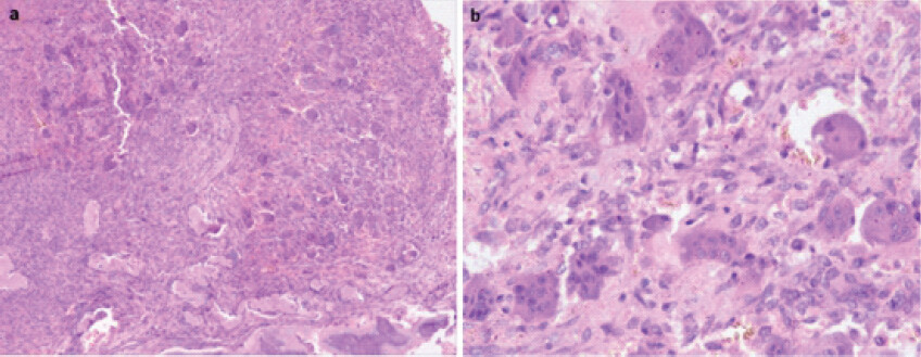
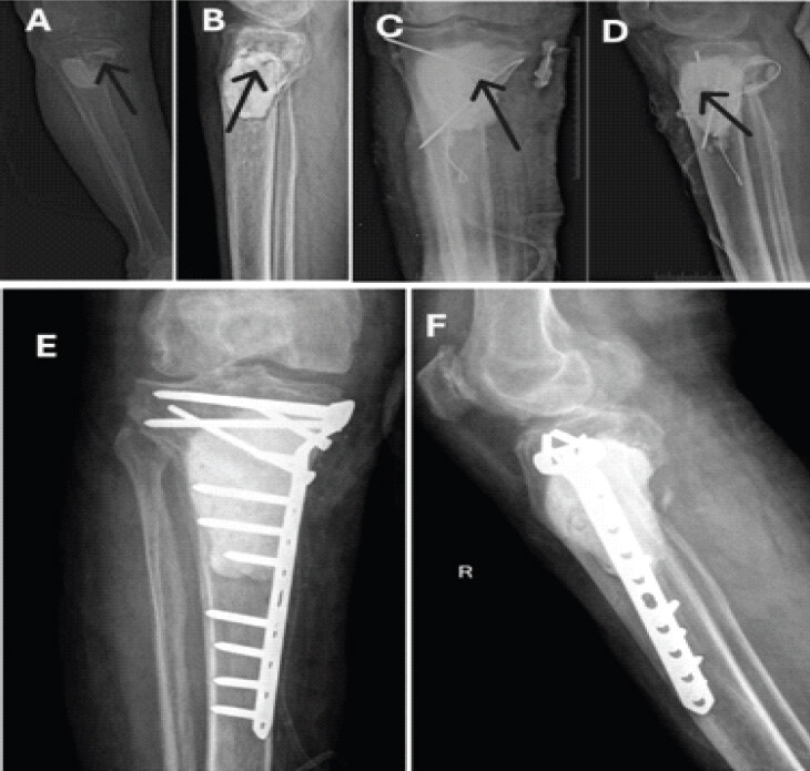
Four patients (50.0%) presented with recurrent disease; three had recurrent GCT following previous treatment elsewhere, and one had infected recurrent GCT. The infected case had previously undergone extended curettage and bone cementation and was subsequently managed with staged debridement, sinus tract excision, extended curettage, antibiotic-loaded cement and temporary K-wire fixation, followed 6 weeks later by repeat curettage, cementation and definitive plate fixation (Fig. 2).
One of the four recurrent cases, a 70-year-old woman with proximal tibial GCT, demonstrated secondary sarcomatous transformation on biopsy with concomitant pulmonary metastasis. She underwent wide resection, endoprosthetic reconstruction and medial gastrocnemius perforator flap coverage, followed by oncological treatment. She subsequently died from metastatic disease-related complications.
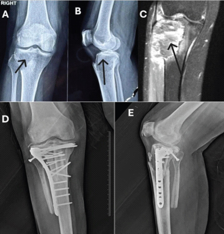
Extended curettage with reconstruction was performed in selected patients, using fibular strut and iliac crest grafts, allografts, bone cement and internal fixation as appropriate (Fig. 3). Wide resection with endoprosthetic reconstruction was performed for extensive or recurrent disease, including two distal femoral and one proximal tibial reconstruction (Fig. 4).
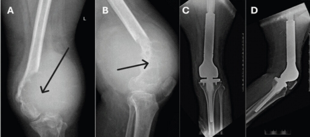
The mean follow-up was 26.4 ± 3.1 months, with a median of 25.5 months (range, 24–32 months). The mean MSTS score at final follow-up was 18.1 ± 6.4, with a median of 18.5 (range, 8–25). Higher scores were generally observed following joint-preserving procedures, while lower scores were seen after wide resection; the lowest score (8) occurred in the patient with sarcomatous transformation and pulmonary metastasis.
No new local recurrence was documented following treatment at our institution during the available follow-up. Thus, the 50.0% recurrence figure represents recurrent disease at presentation rather than post-operative recurrence. One patient (12.5%) died during follow-up from metastatic disease-related complications, while the remaining seven patients (87.5%) were alive at final follow-up. No other post-operative complication requiring hospital admission was recorded.
The details of cases are summarized in Table 1.
Demographic, Clinical and Follow-Up Details of patients with GCT above 70 years.
| No | Age/Sex | Location | grade | Treatment | Follow up months | MSTS Score | Post op complications requiring admission |
|---|---|---|---|---|---|---|---|
| 1 | 72/7 | Proximal tibia | 3 | Extended curettage + fibular strut graft + iliac crest grafting and fixation with anatomical locking plate | 28 | 24 | Nil |
| 2 | 78/M | Proximal tibia (Recurrent Case) | 3 | Two stage treatment (Infected and Recurrent case – Previously underwent Extended curettage + bone cement elsewhere) | 24 | 12 | Nil |
| Stage 1- debridement, sinus tract excision, extended curettage, and cavity filling with antibiotic bone cement, and K-wires for support. | |||||||
| 2nd stage – (After 6 weeks) cement removal, repeat extended curettage, refilling with bone cement and fixation using a proximal tibial locking plate | |||||||
| 3 | 76/F | Distal femur | 3 | Wide excision and reconstruction with distal femur Endomegaprosthesis. | 28 | 15 | Nil |
| 4 | 70/F | Proximal tibia (Recurrent case) | 3 | Patient had Giant cell tumour with secondary sarcomatous changes in biopsy with lung metastasis also. Wide resection and Endo prosthetic reconstruction and medial gastrocnemius perforator flap coverage was done and further treatment was undergone under supervision of Oncology department. | 26 | 8 | Patient expired due to metastasis related complications after 26 months of surgery |
| 5 | 77/M | Proximal tibia | 3 | Extended curettage + allografting, bone cement application, and internal fixation using a proximal tibial T-plate | 32 | 24 | Nil |
| 6 | 81/F | Proximal tibia (Recurrent case) | 3 | Extended curettage, bonne cement and internal fixation with anatomical locking plate. | 25 | 22 | Nil |
| 7 | 76/M | Distal femur (Recurrent case) | 3 | Recurrence in elsewhere operated case (Earlier extended curettage and bone cement application and fixation with anatomical locking plate was done). Wide resection and reconstruction with distal femur Endomegaprosthesis | 26 | 15 | Nil |
| 8 | 70/M | Proximal tibia | 3 | Extended Curettage, Allografting, Bone cement application and Fixation with anterolateral raft plate | 24 | 25 | Nil |
Discussion
GCT of bone is typically a disease of young and middle-aged adults, making its appearance in patients aged 70 and older genuinely unusual. Because of this rarity, what we know about how GCT behaves in the elderly comes mostly from scattered case reports and small series rather than any systematic body of evidence. Our cohort adds a meaningful data point to this gap, and several findings stand out. Every patient in the series presented with Campanacci grade III disease, half arrived with disease that had already recurred elsewhere, and one patient went on to develop secondary sarcomatous transformation with pulmonary metastasis and died as a result. Taken together, these observations point toward a more aggressive disease course in older patients than is typically assumed for GCT, and argue for a more cautious approach to treatment planning in this group [1,2].
Diagnosing GCT in an elderly patient carries its own challenges, since the differential for an osteolytic bone lesion shifts considerably with age. Past 70, metastatic disease, myeloma, lymphoma, and other primary malignant bone tumors become far more likely explanations for a destructive epiphyseal or metaphyseal lesion than a benign GCT. Malignant transformation of GCT itself, though rare, adds a further layer of complexity. For these reasons, imaging alone is not sufficient in this population – diagnosis needs to draw on clinical history, radiology, and histopathology together, with tissue diagnosis and staging completed before any definitive reconstruction is planned. This point was underscored in our series by one recurrent proximal tibial lesion that showed sarcomatous change on biopsy alongside pulmonary metastasis, a reminder of how carefully these lesions need to be worked up in older patients [3,4].
Perhaps the most notable finding was simply how advanced these tumors were at presentation. All eight cases were Campanacci grade III, reflecting substantial bone destruction and, in some cases, soft-tissue extension. Four of the eight patients (50%) had recurrent rather than primary disease, three following prior surgery at other centers, and one with an infected recurrence after earlier extended curettage and cement reconstruction.
This 50% recurrence rate stands in sharp contrast to the lower rates typically reported for conventional GCT treated with modern extended curettage techniques. We don’t have a matched younger cohort to compare against, so we can’t claim a statistically demonstrated age effect on recurrence, but the combination of uniformly high-grade presentation and substantial recurrence still suggests GCT in older patients may behave less indolently than is often assumed. Several explanations are plausible and worth investigating further: Delayed diagnosis, age-related changes in bone quality, differences in underlying tumor biology, or perhaps less definitive initial surgery [2,4,6].
One detail worth emphasizing is that all four recurrent cases had already been treated with extended curettage and biological reconstruction elsewhere – meaning recurrence occurred despite an initial strategy aimed at joint preservation. This has real implications for how we think about managing these patients. Curettage remains a reasonable option for well-selected GCTs, but when disease recurs in an elderly patient – especially with extensive bone destruction or other risk factors – it may be time to consider a more definitive oncological procedure. We’re not suggesting curettage is inherently inferior in older patients, but our data hint that the bar for moving to wide resection may need to sit lower in this group [7,8].
The most dramatic illustration of this concern was a 70-year-old woman with recurrent proximal tibial GCT that had undergone secondary sarcomatous transformation, confirmed on biopsy, alongside pulmonary metastasis at presentation. She was treated with wide resection, endoprosthetic reconstruction, and flap coverage, followed by oncological therapy, but ultimately died from complications of metastatic disease. Malignant transformation is a known but uncommon complication of GCT, and its appearance here in an already-recurrent tumor is a useful reminder to stay alert to this possibility whenever recurrent disease is being evaluated in an older patient. It also makes clear that recurrence in this age group shouldn’t automatically be treated as a low-stakes local event; it may call for full restaging and a fresh look at tumor biology [9,10].
Despite this aggressive clinical picture, the anatomical pattern of disease in our series matched what’s typically seen in younger patients. Six of eight tumors (75%) were in the proximal tibia, with the remaining two in the distal femur, meaning every lesion in the cohort was located around the knee, the classic site for GCT [2,4].
This is a useful point in its own right: Even though the clinical behavior of GCT may differ with age, its characteristic skeletal distribution appears to hold. Age or anatomical site alone shouldn’t be used to rule GCT out of the differential. A lytic lesion around the knee in an elderly patient – provided imaging and histology fit, still deserves consideration as GCT, even while remaining mindful that metastatic and other malignant lesions are statistically more likely in this age group.
These findings also carry weight for surgical decision-making. In younger patients, extended curettage with adjuvants and biological or cement reconstruction is usually favored when it can achieve good local control while preserving the native joint. Given the recurrence rate seen here, though, it’s fair to ask whether that same default should apply once a patient is past 70 [6,11].
All four recurrences in our series followed prior extended curettage and biological reconstruction performed elsewhere. By contrast, none of the patients we treated with wide resection and endoprosthetic reconstruction – for either extensive or recurrent disease – developed documented local recurrence over the available follow-up. The recurrent distal femoral case was managed with wide resection and distal femoral endoprosthetic reconstruction, and the recurrent proximal tibial case with sarcomatous change was managed the same way [2,12].
This comparison needs to be read with appropriate caution, though. The study isn’t randomized or directly comparative, and the patients who underwent wide resection were, almost by definition, those with more extensive or already-recurrent disease, so this isn’t proof that resection is categorically better than curettage. What it does support is the idea that, in elderly patients with recurrent, extensive, or structurally compromised GCT, definitive wide resection with endoprosthetic reconstruction is a reasonable option that may carry a lower risk of further local recurrence than repeating intralesional surgery. Given that functional demands are typically lower in older patients and durable oncological control matters more, the balance may tip toward a more definitive procedure in carefully chosen cases [9,13].
Notably, over roughly 2 years of follow-up, no patient treated with wide resection and megaprosthetic reconstruction developed a documented local recurrence, the one exception being the patient who already had pulmonary metastasis and sarcomatous change at presentation, and whose death was attributable to metastatic disease rather than local recurrence. The numbers here are small, but this pattern is still worth further study as a possible argument for lowering the threshold for definitive resection in elderly patients with recurrent or extensive GCT [2,9,13].
Even with advanced age and locally aggressive disease, most patients retained useful limb function after limb-salvage surgery. The mean MSTS score at final follow-up was 18.1 ± 6.4 (median 18.5, range 8–25). Lower scores clustered among patients who required wide resection and endoprosthetic reconstruction, most notably the patient with metastatic sarcomatous transformation, who scored 8, while those managed with joint-preserving procedures tended to score better, though the small sample size makes formal statistical comparison inappropriate.
These results highlight a genuine trade-off between oncological control and functional outcome. When a recurrent tumor in an elderly patient has already failed curettage, the functional gain from attempting another joint-preserving procedure has to be weighed against the risk of yet another recurrence and further surgery. In well-selected patients, durable control of the disease may reasonably take priority over preserving native joint anatomy [1,5,10,11].
The oncological outcomes here also reflect how varied geriatric GCT can be. No new local recurrence was recorded post-operatively among patients treated at our institution, with the single exception of the patient whose tumor had already undergone malignant transformation with pulmonary spread before treatment began, and who ultimately died from complications of metastatic disease.
Hence, while the rest of the cohort showed no post-operative recurrence, that shouldn’t be read as evidence that geriatric GCT is generally indolent. If anything, our findings suggest a real spectrum, from locally aggressive but manageable disease at one end to highly aggressive, recurrent disease with malignant transformation and metastatic spread at the other. That distinction matters most when assessing an elderly patient who presents with recurrence after earlier intralesional treatment.
Despite the patients’ age, definitive limb-salvage surgery proved feasible throughout this cohort, and no post-operative complication required admission apart from the disease course of the patient with sarcomatous transformation. The lack of significant perioperative morbidity here suggests that age by itself shouldn’t be treated as a barrier to appropriate oncological surgery. Treatment decisions should instead be guided by physiological reserve, comorbidities, bone quality, disease extent, recurrence status, and the functional benefit realistically on offer [4,7].
Strengths and limitations
The strength of this study lies in its focus on a rare and underreported clinical entity, GCT of bone in patients aged 70 years and older, with detailed evaluation of clinical presentation, surgical management, oncological outcomes, and functional recovery. The inclusion of histologically confirmed cases with a minimum follow-up of 24 months provides useful clinical insights.
The main limitations are the small sample size, single-center design, and relatively short follow-up, which limit the generalizability of the findings and the assessment of long-term recurrence and implant survival. The absence of a comparative control group and the heterogeneity of surgical procedures also preclude definitive conclusions regarding the superiority of any particular treatment strategy.
Conclusion
GCT of bone, although uncommon in patients aged 70 years and older, should remain an important diagnostic consideration for destructive periarticular osteolytic lesions in the elderly. In this series, tumors demonstrated the characteristic anatomical distribution around the knee, but all patients presented with Campanacci grade III disease and 50% presented with recurrent disease, suggesting a potentially more aggressive clinical presentation than that generally observed in younger patients. Notably, all recurrent cases had previously undergone extended curettage with biological reconstruction, raising concern regarding the durability of repeated intralesional treatment in this population. One recurrent tumor demonstrated secondary sarcomatous transformation with pulmonary metastasis and resulted in metastatic disease-related death. In contrast, patients undergoing wide resection and endoprosthetic reconstruction for recurrent or extensive disease had no documented local recurrence during the available follow-up. These findings suggest that, while joint-preserving surgery remains appropriate for selected elderly patients, a lower threshold for wide resection and endoprosthetic reconstruction may be considered when disease is recurrent, extensive, or structurally destructive. Larger multicentre comparative studies with longer follow-up are required to determine whether these observations represent an age-related difference in tumour behaviour and to define the optimal surgical strategy for geriatric GCT.
Clinical Message
Giant cell tumor of bone should remain a diagnostic consideration in elderly patients with destructive periarticular lesions. Histopathological confirmation and staging are essential, while recurrent or extensive disease may warrant wide resection and endoprosthetic reconstruction.
Conflict of Interest:
Nil
Source of Support:
Nil
Consent
The authors confirm that informed consent was obtained from the patient for publication of this article
How to Cite this Article
Kantiwal P, Prakash V, Suhail A, Soni BK, Meer F, Nikhil VL. Giant Cell Tumor of Bone in the Geriatric Population: A Comprehensive Study of Survivorship, Recurrence, and Functional Outcomes. J Orthop Case Rep. 2026 November;16(11):397-404. doi: 10.13107/jocr.2026.v16.i11.8508.
References
- Campanacci M. Giant cell tumor. In: Bone and Soft Tissue Tumors. Vienna. Springer Vienna 1999:99-142. Available from: https://link.springer.com/10.1007/978-3-7091-3846-5_4. [Last accessed on 2023 Jun 17]. [Google Scholar] | [PubMed] | [CrossRef]
- Eckardt JJ, Grogan TJ. Giant cell tumor of bone. Clin Orthop Relat Res 1986;204:45-58. [Google Scholar] | [PubMed]
- Mahajan NP, Sadar A, Kumar GS, Marfatia A, Sangma SM, Kondewar P. Giant cell tumor (GCT) of the third metatarsal in an elderly patient: A rare case report. J Orthop CASE Rep 2021;11:29-32. [Google Scholar] | [PubMed]
- Broehm CJ, Inwards CY, Al-Ibraheemi A, Wenger DE, Jenkins SM, Jin L. Giant cell tumor of bone in patients 55 years and older: A study of 34 patients. Am J Clin Pathol 2018;149:222-33. [Google Scholar] | [PubMed]
- Enneking WF, Dunham W, Gebhardt MC, Malawar M, Pritchard DJ. A system for the functional evaluation of reconstructive procedures after surgical treatment of tumors of the musculoskeletal system. Clin Orthop Relat Res 1993;286:241-6. [Google Scholar] | [PubMed]
- Arismawati, Raharjo P, Setiawati R. Diagnosis of bone giant cell tumor in elderly patient: A case report of an unusual case. Ann Med Surg (Lond) 2022;79:104111. [Google Scholar] | [PubMed]
- Kundu ZS, Gogna P, Singla R, Sangwan SS, Kamboj P, Goyal S. Joint salvage using sandwich technique for giant cell tumors around knee. J Knee Surg 2015;28:157-64. [Google Scholar] | [PubMed]
- Yadav SK, Rajnish RK, Prakash V, Nalwa A. Malignant giant cell tumour of distal ulna. BMJ Case Rep 2024;17:e258047. [Google Scholar] | [PubMed]
- Augustus M, Ali A, Beharry A, Armorer D, Lousaing D. Management of a giant cell tumors with megaprosthesis in a resource-limited setting. Indian J Orthop Surg 2024;10:189-94. [Google Scholar] | [PubMed]
- Gupta G, Singh A, Kala S, Saini A, Singh S, Shende S. Giant cell tumor of the proximal femur with pathological fracture of femoral neck. J Orthop Case Rep 2025;15:113-8. [Google Scholar] | [PubMed]
- Pujani M, Bahadur S, Jairajpuri ZS, Jetley S, Jameel J. Giant cell tumor bone in an elderly male- an unusual case misdiagnosed on MRI as a malignant sarcoma. Indian J Surg Oncol 2015;6:285-7. [Google Scholar] | [PubMed]
- Garg N, Tanveer N, More S, Chauhan S. Giant cell tumor of clavicle in an elderly: Rare presentation of a common tumor with review of literature; 1980-2020. J Microsc Ultrastruct 2021;10:93-5. [Google Scholar] | [PubMed]
- Ben Bouzid Y, Lamrani MO. Knee megaprosthesis: Analysis of clinical outcomes, complications, and implant survival across complex indications. J Orthop Rep 2025;4:100632. [Google Scholar] | [PubMed]
© 2026 Journal of Orthopaedic Case Reports - Published by Indian Orthopaedic Research Group
About the Authors
-

Prabodh Kantiwal
-

Vimal Prakash
-

Amir Suhail
-

Bharat Kumar Soni
-

Furqan Meer
-

V L Nikhil
Related Articles in Journal of Orthopaedic Case Reports
 May 1, 2026 Reawakening of a Giant Cell Bone Tumor after 40 Years of Silence: A Rare Case Report
 October 1, 2026 Delayed Metachronous Multicentric Giant Cell Tumor of Bone after a 16-Year Disease-Free Interval: A Case Report
 July 1, 2026 Short-term Functional Outcomes of Shoulder Girdle Limb Salvage in Primary Bone Tumors: A 2-year Follow-up Study
 January 1, 2026 Combined Approach (Surgery + Systemic Adjuvant Therapy) in a Giant Cell Tumor of Proximal Femur: A Rare Case Report