
The iatrogenic gel foam-induced early onset postoperative neurological deterioration is very debilitating, the surgeons should be more vigilant in preventing and managing it accordingly. The occurrence of postoperative neurological deterioration in patients with OPLL-induced cervical myelopathy who underwent posterior decompression is well established. However, the rare cause of early onset cord compression due to adsorbed gelatin sponge has to be considered when other common causes are ruled out. Although it is difficult to diagnose, the early suspicion and the management of such a condition are crucial to regaining the neurological function of the patient.
Dr. Arulkumar Nallakumarasamy Senior Resident, Department of Orthopaedics, All India Institute of Medical Sciences, Bhubaneshwar, Odisha, India – 751019. E-mail: arulmmcian@gmail.com
Abstract
Introduction: Gelatin foam has been regularly used the complex neuro and spinal surgeries for a long time. Apart from their hemostatic properties, these are inert and provide form an inert membrane that prevents scar adhesions o vital structures such as the brain or spinal cord.
Case Presentation: We present a case of cervical myelopathy due to an ossified posterior longitudinal ligament that underwent the instrumented posterior decompression and had neurological worsening 48 h after the index surgery. An magnetic resonance imaging showed a hematoma compressing the spinal cord which on exploration was confirmed to be a gelatin sponge. It represents the rare phenomenon of mass effect due to their osmotic properties, especially in a closed space causing neurologic deterioration.
Conclusion: We emphasize the rare cause of early onset quadriparesis after the posterior decompression due to the swollen gelatin sponge over the neural elements. The patient recovered with timely intervention.
Keywords: Cervical myelopathy, ossification of posterior longitudinal ligament, gelatin sponge, early onset post-operative neurological deterioration, case report.
Ossification of the posterior longitudinal ligament (OPLL) is routinely treated with indirect decompression by posterior laminectomy ± fusion. The procedure is comparatively effective and safer than the anterior approach when the spinal canal occupancy ratio is <50–60% [1]. However, there can be considerable blood loss from the epidural vein and bleeding raw bones, and surgeons have been using absorbable gelatins as a hemostat and effective interposing membrane over the naked spinal cord to prevent scar adhesions. These gelatins can be counterproductive, and there are few reports of complications due to their usage in the literature [2,3,4,5,6]. Here, we report a patient with cervical OPLL who underwent a routine post-instrumented decompression but deteriorated in the early post-operative period. A magnetic resonance imaging (MRI) confirmed compression by tamponade effect of gelatin sponge which was subsequently revised. The patient showed improvement.
OPLL is a common cause of cervical spinal cord compression in Asian countries. Continuous A 48-year-old male presented with difficulty in walking and clumsiness of both hands for a 1-year duration. The disease was insidious and progressive, making him unemployed through ambulatory. There were upper motor neuron signs such as increased muscle tone, exaggerated reflexes (upper and lower limb but jaw jerk was absent), a positive Hoffman sign, plantar extensors, and a motor power of 4/5 grade. On a functional score, he was 4/6 on the modified Nurick scale and 11/18 on the modified Japanese Orthopaedic Association (major) scale. An X-ray showed increased bony densities along the sub-axial region’s posterior border and degrative changes (Fig. 1a). The MRI reported a discontinuous band-like low signal intensity structure along the posterior border from C2 till C6 in the T1 sequence and hyperintensity signal in the T2 sequence abutting the ventral cord surface, suggesting OPLL causing cord compression (Fig. 1b). A computed tomography revealed a double line sign with a cortical density over the posterior longitudinal ligament, confirming the diagnosis of the long segment, mixed type of OPLL (Fig. 1c). The patient was operated on with posterior decompression C3-C6 undercutting the C2 lamina. Instrumentation was done with lateral mass screws C3-C6 and rods were carefully secured to the screws, and autologous bone grafts were placed laterally. A gelatin sponge (abgel) corresponding to the laminectomy defect’s size was placed in the midline, and the wound was closed with a drain. Postoperatively, he felt some “lightness” in his limbs and regained his pre-operative power. The immediate X-ray showed satisfactory screw placement and adequate laminectomy (Fig. 1d).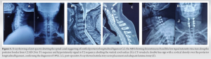
However, by 48 h of surgery, there was gradual and progressive deterioration in the muscle power of all extremities (1-2/5). There were no associated fever, headache, and nuchal rigidity episodes. An urgent repeat MRI was done which showed an en-mass heterogeneous signal at the laminectomy site compressing the spinal cord (Fig. 2a and b).
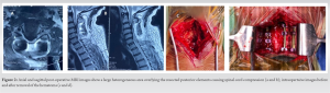
The patient was wheeled into the operating theater for re-exploration. The gel foam was found swollen and removed piece-meal, and the hematoma was evacuated (Fig. 2c and d). After achieving proper hemostasis and irrigating the surgical site with copious saline, the wound was closed with a drain. The patient started to show improvement immediately postoperatively, and by 3 weeks was able to walk with support. At 3 months, follow-up was complete ambulatory unaided, with an mJOA score improved to 14.
Neurological deterioration in a patient following is a medical emergency that needs immediate radiological assessment. Although it can be attributed to various causes, malposition of screws, loss of fixation, retained osseous fragments, or fluid collections are the common causes that need to be ruled out if the surgical procedure was uneventful. On rare occasions, gelatin foams have been implicated in causing “mass effect” and compression. Gelatin foams have been commonly used in complex neurosurgical and spine surgeries for more than 75 years [7]. These hemostatic agents control the bleeding and, more importantly, prevent adhesion between the neural elements and the other soft tissues [8]. It is also inert and biodegradable (by the 5th week) [8,9]. The mechanism of action is by forming an artificial blood clot when it comes in contact with the bleeding surfaces. This passive mode of action builds a mechanical matrix that can provide the tamponade effect within the confined space. The response can be seen as late as by post-operative day 13 [2]. Dubin et al. showed that in patients with early-onset clinical deterioration after the index spine surgery and the MRI evidence of mass on the laminectomy site due to the hematoma organized with the absorbable gelatin sponge, thus causing the cord compression [6]. Friedman et al. reported that the unusual occurrence of post-operative cauda equina syndrome (CES) after 13 days of decompression surgery for lumbar can stenosis due to a large gel foam at the laminectomy site [2]. Bessette et al. have also reported CES development postoperatively, secondary to retained gel foam pieces in a microdiscectomy patient [4]. Interestingly, Kitahara et al. reported a case of recurrent myelopathy following 6 months of cervical laminectomy [10]. The authors noticed a membrane-like structure at the laminectomy site around the exogenous surgical materials which was used during the index surgery. Herndon and colleagues described a case of gel foam-induced Brown Sequard syndrome after a week of trans-thoracic surgery for congenital kyphosis due to wedged D5 vertebra [3]. Librianto et al. reported a regained neurological outcome after the revision surgery secondary to gel foam-induced early onset cord compression following the deformity correction index surgery for thoracolumbar scoliosis [5]. Buchowski et al. had an unusual case of epidural spinal cord compression following the usage of gelatin sponges in the cannulated pedicles. The authors observed unabsorbed gelatin sponges in the canal despite the lack of any medial pedicle breach during revision surgery [11]. Friedmann et al. recommend that using only small pieces should it be necessary to use and remove them once hemostasis is achieved [2]. They advocate against using a single large piece, especially in a closed space. The sponges are squeezed to expel any trapped air bubbles before use. Nowadays, a thrombin-gelatin hemostatic matrix is commercially available and used to control bleeding in complex spine surgery [12]. Recently, in-vivo and in-vitro studies showed that the free fat transplantation at the laminectomy decreases the adhesion and the scar tissue formation to exogenous hemostatic agents [13].
When used as a large piece in an enclosed space, Gelatin sponges can cause mass effects and be disastrous for a post-operative patient. While the hardware-related causes are easily diagnosed, this rare etiology is preventable if proper precautions are undertaken.
A better understanding of the different etiologies of the early-onset post-operative neurological deterioration (specifically after posterior decompression) helps the surgeon for framing a plan of immediate interventions. The gel foam is the most commonly used hemostatic agent in spine surgeries, they are particularly essential in OPLL cases to control the bleeding. The immediate evacuation of the swollen gel foam which lodged and compresses the cord has to be done to regain the deteriorated neurological functions.
References
- 1.Lee SE, Chung CK, Jahng TA, Kim HJ. Long-term outcome of laminectomy for cervical ossification of the posterior longitudinal ligament. J Neurosurg Spine 2013;18:465-71. [Google Scholar | PubMed]
- 2.Friedman J, Whitecloud TS 3rd. Lumbar cauda equina syndrome associated with the use of gelfoam: Case report. Spine (Phila Pa 1976) 2001;26:2000-2. [Google Scholar | PubMed]
- 3.Herndon JH, Grillo HC, Riseborough EJ, Rich JC. Compression of the brain and spinal cord following use of gelfoam. Arch Surg 1972;104:107. [Google Scholar | PubMed]
- 4.Bessette MC, Mesfin A. Cauda equina syndrome caused by retained hemostatic agents. J Clin Neurosci 2015;22:1518-20. [Google Scholar | PubMed]
- 5.Librianto D, Fachrisal, Saleh I. Gelatin sponge as a rare and forgotten cause of early-onset neurological deficit post osteotomy of thoracolumbar kyphosis a case report and review of literature. Int J Surg Case Rep 2020;75:497-503. [Google Scholar | PubMed]
- 6.Dubin LM, Quencer RM, Green BA. A mimicker of a postoperative spinal mass: Gelfoam in a laminectomy site. AJNR Am J Neuroradiol 1988;9:217-8. [Google Scholar | PubMed]
- 7.Abbott WD, Coleman FC. The use of gelatin sponge in neurosurgery. J Am Med Assoc 1946;132:329-30. [Google Scholar | PubMed]
- 8.Sabel M, Stummer W. The use of local agents: Surgicel and surgifoam. Eur Spine J 2004;13:2-3. [Google Scholar | PubMed]
- 9.Özer A, Köstü B. Use of gelatin sponge affects postoperative morbidity in cesarean section patients. Med Sci Monit 2017;23:1141-5. [Google Scholar | PubMed]
- 10.Kitahara T, Hanakita J, Takahashi T. Postlaminectomy membrane with dynamic spinal cord compression disclosed with computed tomographic myelography: A case report and literature review. Spinal Cord Ser Cases 2017;3:2-3. [Google Scholar | PubMed]
- 11.Buchowski JM, Bridwell KH, Lenke LG, Good CR. Epidural spinal cord compression with neurologic deficit associated with intrapedicular application of hemostatic gelatin matrix during pedicle screw insertion. Spine (Phila Pa 1976) 2009;34:E473-7. [Google Scholar | PubMed]
- 12.Gazzeri R, De Bonis C, Galarza M. Use of a thrombin-gelatin hemostatic matrix (Surgiflo) in spinal surgery. Surg Technol Int 2014;25:280-5. [Google Scholar | PubMed]
- 13.Kiviluoto O. Use of free fat transplants to prevent epidural scar formation: An experimental study. Acta Orthop Scand 1976;47:3-75. [Google Scholar | PubMed]


Related Articles in Journal of Orthopaedic Case Reports
 April 1, 2026 Anterior Cervical Discectomy and Fusion with Polyetheretherketone Cage or Anterior Cervical Plate: A Comparative Evaluation of Short-term Outcomes
 February 1, 2026 Evaluation of the Outcome of Zero-profile Implant in the Treatment of Single-level Cervical Spondylotic Myelopathy in the Indian Scenario – A Prospective Study
 August 1, 2025 Posterior Decompression and Arthrodesis in Congenital Atlantoaxial Subluxation and Associated Complex Vertebral Artery Anomaly: A Case Report
 September 1, 2024 White Cord Syndrome Following Long Posterior Decompression