
Thumb adductor sesamoid ossification may be absent despite distal phalangeal physeal closure in patients with growth hormone deficiency, underscoring the need for cautious interpretation of simplified bone age assessment methods.
Shevaun Doyle, Hospital for Special Surgery, 535 E 70th Street, New York, NY 10021, United States. Phone: 646-797-8816. E-mail: doyles@hss.edu
Abstract
Introduction: Assessment of skeletal maturity is fundamental to paediatric orthopaedic practice, as remaining growth potential influences prognosis and treatment decisions. Ossification of the thumb adductor sesamoid is widely used as a pubertal landmark and is expected to precede distal phalangeal physeal closure in established bone age assessment methods. Deviation from this sequence is rarely described. We report a case of distal phalangeal physeal closure occurring in the absence of thumb adductor sesamoid ossification in a patient with growth hormone deficiency.
Case Report: A 15-year-old female with hypopituitarism and growth hormone deficiency was followed for adolescent idiopathic scoliosis. Despite long-term recombinant growth hormone therapy, she demonstrated delayed pubertal progression. A posteroanterior radiograph of the left hand and wrist obtained to assess skeletal maturity revealed complete distal phalangeal physeal closure across all digits without ossification of the thumb adductor sesamoid. The contralateral hand demonstrated early sesamoid ossification. At a chronological age of 15 years and 4 months, bone age was estimated at 13 years and 6 months, consistent with delayed skeletal maturation. This represents a discordant and atypical sequence of ossification.
Conclusion: This case demonstrates that thumb adductor sesamoid ossification may be absent despite distal phalangeal physeal closure in patients with endocrine dysfunction. Simplified skeletal maturity assessment methods that rely on sesamoid appearance may therefore be misleading in this population. Careful evaluation of the overall pattern of physeal development is essential when managing growth-dependent orthopaedic conditions. This report highlights clinically relevant variability in skeletal maturation in the setting of growth hormone deficiency.
Keywords: Skeletal maturity, thumb adductor sesamoid, distal phalangeal physis, growth hormone deficiency, bone age assessment.
Assessment of skeletal maturity is fundamental to paediatric orthopaedic practice, as a patient’s remaining growth significantly influences prognosis and management across a wide range of conditions, particularly when planning surgeries about the knee [1]. Hand and wrist radiographs are commonly used to evaluate skeletal age, as the appearance and fusion of secondary ossification centres and the closure of physes follow a predictable sequence during childhood and adolescence in the hand [2]. The most commonly used assessment of bone age is the Greulich and Pyle Method (GPM), introduced in the Radiographic Atlas of the Hand and Wrist, which remains the gold standard among orthopaedic surgeons. More recent methods, such as the Shorthand Bone Age (SBA) assessment, are derived from the GPM but offer a simplified approach [3]. In normal skeletal development, the final ossification centre to appear in the hand is the adductor sesamoid of the thumb. In the SBA, the thumb sesamoid typically ossifies and becomes visible on radiographs at approximately 11 years in females and 13 years in males, preceding physeal closure of the thumb and index finger distal phalanges. Closure of the thumb distal phalanx occurs at approximately 13 years in females and 15 years in males, followed by closure of the index finger distal phalanx at approximately 13.5 and 15.5 years, respectively. This sequence is also consistent with skeletal maturation patterns described by the GPM [4,5]. Few cases in the literature describe deviation from this expected temporal sequence of ossification and physeal closure. We report the case of a 15-year-old female patient with a history of hypopituitarism and delayed puberty on growth hormone (GH) therapy, who demonstrated closure of the thumb distal phalangeal physis in the absence of thumb sesamoid ossification.
A 10-year-old female was referred to paediatric endocrinology for evaluation of pathologic short stature when her paediatrician noticed a declining growth trajectory. Her medical history was significant for isolated premature thelarche diagnosed at 10 months of age, characterised by bilateral breast development with a Tanner stage 3 appearance in the absence of other pre-pubertal features. There was no known family history of early epiphyseal closure or abnormalities of skeletal maturation. Further laboratory evaluation demonstrated decreased insulin-like growth factor (IGF-1), GH, thyroid hormone, T4, and T3 levels. Magnetic resonance imaging revealed a small-volume pituitary gland, consistent with pituitary hypoplasia. These findings supported a diagnosis of hypopituitarism with associated growth hormone deficiency. Given her short stature and the presence of open physes with evidence of remaining growth potential, confirmed on-hand radiography, treatment with recombinant human growth hormone (somatotropin) was initiated. At 11 years of age, the patient presented to the orthopaedic surgery clinic for evaluation of scoliosis following observation of shoulder asymmetry on the Adams forward bend test at a routine paediatric visit. Plain radiographic evaluation demonstrated a left T3–T12 curve measuring 12° and a right T12–L4 curve measuring 14°, consistent with a diagnosis of adolescent idiopathic scoliosis (IS). Serial standing pelvic radiographs showed persistent Risser stage 0, with absent iliac apophyseal ossification findings, which indicate substantial remaining growth potential. Throughout orthopaedic follow-up, the patient exhibited persistent delayed pubertal progression, while physical examinations remained unremarkable, with no dysmorphic features or neurologic abnormalities. As part of routine scoliosis management to assess skeletal maturity and remaining growth potential, particularly in the context of persistent Risser stage 0 and premenarchal status, a hand and wrist radiograph was obtained at 15 years of age. A posteroanterior radiograph of the left hand and wrist demonstrated complete fusion of the distal phalangeal physes across all digits, along with absence of ossification of the thumb adductor sesamoid (Fig. 1a). In contrast, the right hand showed early ossification of the thumb sesamoid (Fig. 2). At a chronological age of 15 years and 4 months, bone age was estimated at 13 years and 6 months according to the Greulich and Pyle atlas, consistent with delayed skeletal maturation. The absence of sesamoid ossification despite closure of the distal phalangeal physes represents a notable deviation from the expected, consistent maturation sequence outlined in the GPM (Fig. 1b). The patient was treated with a thoracolumbosacral orthosis, a nocturnal back brace, and had correction of her scoliosis over the course of 2 years.
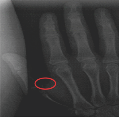
Figure 2: Anteroposterior radiograph demonstrating expected thumb adductor sesamoid ossification. Anteroposterior radiograph of the patient’s right hand obtained at 15 years and 4 months of age on the same date as the left hand radiograph was taken. An ossific nucleus of the thumb adductor sesamoid is present at the first metacarpophalangeal joint (circled). The distal phalangeal physes are fused. This radiograph demonstrates the expected appearance of sesamoid ossification at this stage of skeletal maturation.
In this case report, we describe an adolescent patient with an aberrant sequence of hand ossification, characterised by distal phalangeal physeal closure and the absence of thumb adductor sesamoid ossification. The appearance of the adductor sesamoid and physeal closure of the distal phalanx of the thumb are widely regarded as the two major landmarks in evaluating skeletal maturity during puberty and early adolescence. The sequence of hand and wrist ossification is well characterised and predictable, with the progressive appearance and fusion of multiple ossification centres that form the basis of established bone age assessment methods [7]. Accurate assessment of skeletal maturity is critical for guiding management of adolescent IS and predicting the risk of curve progression. Sanders et al. demonstrated that metacarpal and phalangeal development, specifically in the Tanner-Whiteside method, was highly correlated to curve progression in prepubescent females with IS amongst skeletal maturity parameters [8]. In maxillofacial surgery, ossification of the thumb adductor sesamoid has been extensively utilised as a marker of pubertal onset to guide the timing of growth-dependent surgical decision-making [9,10]. Previously, Hung et al. proposed the Thumb Ossification Composite Index (TOCI) as a simplified staging system for assessing skeletal maturity in patients with IS [11]. The components of the TOCI include the adductor sesamoid bone, distal phalanges of the thumb, and the lateral corner of proximal phalangeal epiphyses of the thumb to stage skeletal development. This highlights the clinical importance of individual hand ossification centres, particularly the thumb adductor sesamoid and the proximal and distal phalangeal physes, as key indicators in the assessment of skeletal maturation. The patient’s endocrinologic history of hypopituitarism provides important context for her atypical ossification pattern. Though many hormonal signals are involved in physeal maturation, including GH, IGF, thyroid hormone, oestrogen, and androgens, oestrogen primarily regulates physeal closure [12,13,14,15]. Sesamoid ossification involves Type II collagen and proteoglycans, which contribute to mineral deposition and ossification. Their growth is regulated by transforming growth factor beta and bone morphogenetic protein-2. Mechanical factors, such as shear forces and stress, can contribute to sesamoid ossification. Previous literature demonstrates that while skeletal maturity might be delayed in those with GH deficiency, bone age can be within the normal range or accelerated after patients begin GH supplementation [16,17,18,19]. Bone age progression rate is also affected by sex, GH dose, duration of GH treatment, and pubertal status. Kang et al. investigated bone age progression in 78 patients with idiopathic GH deficiency during the first 3 years of recombinant GH therapy [20]. The authors found that GH treatment was associated with accelerated skeletal maturation, with bone age advancing at a rate of approximately 1.28 years for every 1 year of chronological age. However, in the present case, the patient had been receiving GH therapy for nearly 10 years and nevertheless demonstrated absent thumb sesamoid ossification, highlighting an atypical pattern of skeletal development despite long-term treatment. Due to the complex interactions of the hypothalamic-pituitary-adrenal axis and sex hormones (oestrogens and androgens) on physeal maturation, it is difficult to determine whether GH alone contributed to the absence of sesamoid ossification in this patient. One plausible explanation suggested by Cho et al. is that GH therapy may have preferentially accelerated linear growth, while persistent deficiency of sex steroids (particularly oestrogen) selectively delayed ossification of the smaller sesamoid centre [21]. Furthermore, this patient demonstrated asymmetric development, as the right hand showed early sesamoid ossification while the left remained unossified. This case highlights the intricate relationship between growth hormone therapy and skeletal maturation, which remains important to understand when managing adolescent IS. As simplified and shorthand methods of skeletal age assessment based on hand radiographs become increasingly common, clinicians must remain attentive to potential variation in metacarpal and sesamoid development [22]. This is especially true in patients with underlying endocrine disorders. With the growing use of GH therapy, awareness of both focal and global abnormalities in skeletal maturation is essential. In summary, we report an unusual case of discordant hand ossification patterns in the setting of IS and GH deficiency.
We present the case of a patient with a medical history of hypopituitarism who was followed in the orthopaedic clinic for adolescent IS and was found to have absent ossification of the thumb adductor sesamoid despite distal phalangeal physeal closure, an uncommon and discordant finding. To the best of our knowledge, this represents the first reported case describing this atypical sequence of skeletal maturation in the setting of prolonged growth hormone therapy. In patients with a phalangeal–sesamoid discrepancy, physicians should rely more heavily on patterns of physeal closure rather than isolated ossification landmarks when assessing skeletal maturity. In paediatric growth assessment, particularly in patients monitored for orthopaedic conditions such as scoliosis, this case illustrates that the sequence of ossification events may be as clinically informative as their timing. Careful attention to deviations from expected maturation patterns may therefore provide important diagnostic insight and influence growth-dependent clinical decision-making in both orthopaedic and endocrine practice.
In patients with endocrine dysfunction, particularly those receiving growth hormone therapy, the sequence of hand ossification may not follow established norms. Distal phalangeal physeal closure can precede thumb adductor sesamoid ossification. Skeletal maturity assessment should therefore prioritise comprehensive physeal evaluation rather than isolated ossification landmarks.
References
- 1. Fabricant PD, Bram JT. Methods of assessing skeletal maturity when planning surgeries about the knee. J Am Acad Orthop Surg 2025;33:457-66. [Google Scholar] [PubMed]
- 2. Creo AL, Schwenk WF 2nd. Bone age: A handy tool for paediatric providers. Paediatrics 2017;140:e20171486. [Google Scholar] [PubMed]
- 3. Heyworth BE, Osei DA, Fabricant PD, Schneider R, Doyle SM, Green DW, et al. The shorthand bone age assessment: A simpler alternative to current methods. J Pediatr Orthop 2013;33:569-74. [Google Scholar] [PubMed]
- 4. Greulich WW, Pyle SI. Radiographic Atlas of Skeletal Development of the Hand and Wrist. 2nd Stanford: Stanford University Press; 1959. [Google Scholar] [PubMed]
- 5. Gilsanz V, Ratib O. Hand Bone Age: A Digital Atlas of Skeletal Maturity. Berlin: Springer-Verlag; 2005. [Google Scholar] [PubMed]
- 6. Understanding Growth: The Role of Hand X-Rays in Scoliosis Care. Washington University Paediatric Spinal Deformity Blog; 2024. Available from: https://sites.wustl.edu/pediatricspinaldeformity/understanding-growth-the-role-of-hand-x-rays-in-scoliosis-care [last accessed 15 Mar. 2024]. [Google Scholar] [PubMed]
- 7. Satoh M. Bone age: Assessment methods and clinical applications. Clin Pediatr Endocrinol 2015;24:143-52. [Google Scholar] [PubMed]
- 8. Sanders JO, Browne RH, McConnell SJ, Margraf SA, Cooney TE, Finegold DN. Maturity assessment and curve progression in girls with idiopathic scoliosis. J Bone Joint Surg Am 2007;89:64-73. [Google Scholar] [PubMed]
- 9. Sidlauskas A, Zilinskaite L, Svalkauskiene V. Mandibular pubertal growth spurt prediction. Part one: Method based on the hand-wrist radiographs. Stomatologija 2005;7:16-20. [Google Scholar] [PubMed]
- 10. Chapman SM. Ossification of the adductor sesamoid and the adolescent growth spurt. Angle Orthod 1972;42:236-44. [Google Scholar] [PubMed]
- 11. Hung AL, Chau WW, Shi B, Chow SK, Yu FY, Lam TP, et al. Thumb ossification composite index (TOCI) for predicting peripubertal skeletal maturity and peak height velocity in idiopathic scoliosis: A validation study of premenarchal girls with adolescent idiopathic scoliosis followed longitudinally until skeletal maturity. J Bone Joint Surg Am 2017;99:1438-46. [Google Scholar] [PubMed]
- 12. Nilsson O, Marino R, De Luca F, Phillip M, Baron J. Endocrine regulation of the growth plate. Horm Res 2005;64:157-65. [Google Scholar] [PubMed]
- 13. Stagi S, Scalini P, Farello G, Verrotti A. Possible effects of an early diagnosis and treatment in patients with growth hormone deficiency: The state of the art. Ital J Pediatr 2017;43:81. [Google Scholar] [PubMed]
- 14. Shim KS. Pubertal growth and epiphyseal fusion. Ann Pediatr Endocrinol Metab 2015;20:8-12. [Google Scholar] [PubMed]
- 15. Sarin VK, Carter DR. Mechanobiology and joint conformity regulate endochondral ossification of sesamoids. J Orthop Res 2000;18:706-12. [Google Scholar] [PubMed]
- 16. Lesage C, Walker J, Landier F, Chatelain P, Chaussain JL, Bougnères PF. Near normalisation of adolescent height with growth hormone therapy in very short children without growth hormone deficiency. J Pediatr 1991;119:29-34. [Google Scholar] [PubMed]
- 17. Frindik JP, Kemp SF, Sy JP. Effects of recombinant human growth hormone on height and skeletal maturation in growth hormone-deficient children with and without severe pretreatment bone age delay. Horm Res 1999;51:15-9. [Google Scholar] [PubMed]
- 18. Wilson DM. Regular monitoring of bone age is not useful in children treated with growth hormone. Paediatrics 1999;104:1036-9. [Google Scholar] [PubMed]
- 19. Hopwood NJ, Hintz RL, Gertner JM, Attie KM, Johanson AJ, Baptista J, et al. Growth response of children with non-growth-hormone deficiency and marked short stature during three years of growth hormone therapy. J Pediatr 1993;123:215-22. [Google Scholar] [PubMed]
- 20. Kang MJ, Kim EY, Shim YS, Jeong HR, Lee HJ, Yang S, et al. Factors affecting bone age maturation during 3 years of growth hormone treatment in patients with idiopathic growth hormone deficiency and idiopathic short stature: Analysis of data from the LG growth study. Medicine (Baltimore) 2019;98:e14962. [Google Scholar] [PubMed]
- 21. Cho JH, Jung HW, Shim KS. Growth plate closure and therapeutic interventions. Clin Exp Pediatr 2024;67:553-9. [Google Scholar] [PubMed]
- 22. Zverev S, Tenner ZM, Coladonato C, Lazar-Antman M. The rising popularity of growth hormone therapy and ensuing orthopaedic complications in the paediatric population: A review. Children (Basel) 2024;11:1354. [Google Scholar] [PubMed]


Related Articles in Journal of Orthopaedic Case Reports
 October 10, 2023 Preoperative Mohs Paste Treatment for a Subcutaneous Sarcoma and a Skin Ulcer to Prevent Intraoperative Bleeding
 June 10, 2024 Minimally Invasive Osteotomy for Correction of Post-Traumatic Tibia Internal Rotation Deformity: A Case Report
 August 1, 2025 Coxa Vara of Unknown Etiology: A Unique Case Report
 January 1, 2026 Bridge Plating of Second and Third Carpometacarpal Fracture Dislocations: A Case Report of Rare Injury