
Turret exostosis of the proximal phalanx, though rare, should be considered in patients with post-traumatic finger swellings, and complete surgical excision guided by histopathology ensures excellent functional recovery with minimal recurrence.
Dr Samyabrata Das, Department of Orthopaedic Surgery, North Bengal Medical College and Hospital, Siliguri, West Bengal, India. E-mail: sam.orthopaedics04@gmail.com
Abstract
Introduction: Turret exostosis is an uncommon benign osteocartilaginous lesion that originates from the cortical surface of bone, most commonly affecting the phalanges of the hand. It is considered a reactive subperiosteal bone proliferation that typically develops following minor trauma. Because its clinical and radiological appearance may resemble other surface bone lesions such as osteochondroma and bizarre parosteal osteochondromatous proliferation (Nora’s lesion), accurate diagnosis is important. Involvement of the proximal phalanx is particularly uncommon.
Case Report: A 32-year-old male reported with a progressively growing swelling on the dorsal surface of his right middle finger for 1 year following trauma, which was accompanied by pain and discomfort for 8 months. Radiographs demonstrated a surface-based ossified lesion arising from the proximal phalanx without continuity with the medullary canal. Magnetic resonance imaging revealed an osteocartilaginous lesion without intramedullary extension. The lesion was treated with complete surgical excision. Histopathological examination confirmed the diagnosis of turret exostosis. The patient showed no signs of recurrence at 1-year follow-up and continued to be asymptomatic with a full range of motion.
Conclusion: Turret exostosis of the proximal phalanx is an uncommon lesion that can mimic other periosteal tumours. Careful clinicoradiological evaluation with histopathological confirmation is essential for accurate diagnosis. Complete surgical excision results in excellent functional recovery with minimal recurrence.
Keywords: Turret exostosis, osteocartilaginous lesion, proximal phalanx, Nora’s lesion, periosteal tumor.
Turret exostosis is an uncommon benign osteocartilaginous lesion arising from the cortical surface of bone and most frequently involving the phalanges of the hand. It is believed to represent a reactive subperiosteal ossification that occurs following minor or repetitive trauma to the digits [1,2]. Patients typically present with a gradually growing firm swelling over the finger, which may be accompanied by stiffness, pain, or limitation of joint mobility, depending upon the extent and location of the lesion. Radiographically, turret exostosis typically appears as a well-defined ossified mass arising from the cortical surface of bone without continuity with the medullary canal. This feature helps differentiate it from conventional osteochondroma [3]. However, the clinical and radiological findings may overlap with other periosteal lesions such as periosteal chondroma and bizarre parosteal osteochondromatous proliferation (BPOP), commonly referred to as Nora’s lesion [4,5]. Therefore, histopathological examination is essential for establishing the definitive diagnosis. Most reported cases involve the distal or middle phalanges of the hand, whereas involvement of the proximal phalanx is relatively rare [1,6]. Due to its rarity and potential for misdiagnosis, awareness of this entity is important for appropriate management. Complete surgical excision is the preferred treatment, with excellent functional results and a low chance of recurrence [1,6]. We present a rare case of turret exostosis arising from the proximal phalanx of the middle finger and discuss its clinical features, radiological findings, surgical management, and histopathological characteristics.
Patient information:
A 32-year-old male patient who predominantly used his right hand presented with a progressively growing swelling on the dorsal part of his right middle finger’s proximal phalanx (Fig. 1). There were no noteworthy comorbidities or prior medical history for the patient.
History of present illness:
The swelling had been present for approximately 1 year and was initially painless. Pain developed about 4 months after the onset of swelling and gradually increased in intensity. The pain was aggravated by finger movements. The patient reported a history of trivial trauma to the affected finger approximately 1 year before presentation. Mild restriction of flexion at the proximal interphalangeal (PIP) joint was noted.
Clinical examination:
On examination, a firm, non-mobile swelling measuring approximately 2 × 1 cm was present over the dorsal surface of the proximal phalanx of the right middle finger. The lesion was tender on palpation. There were no visible signs of inflammation, and the skin above seemed normal. Mild restriction of PIP joint flexion was observed, whereas extension was preserved. Neurovascular examination of the finger was normal.
Investigations:
Plain radiographs of the hand (anteroposterior and oblique views) demonstrated a well-defined ossified lesion arising from the cortical surface of the proximal phalanx without continuity with the medullary canal (Fig. 2). Magnetic resonance imaging (MRI) showed a surface-based osteocartilaginous lesion with a thin cartilage cap and no intramedullary extension (Fig. 3). Based on these findings, the differential diagnoses included turret exostosis, osteochondroma [3], periosteal chondroma, and BPOP [4,5].
Treatment:
Surgical excision of the lesion was performed under regional anaesthesia. Intraoperatively, a well-circumscribed bony mass that measured approximately 2 × 1 cm was identified arising from the dorsal cortex of the proximal phalanx. The lesion was completely excised along with its base (Fig. 4). There was no involvement of the extensor tendon or surrounding soft tissues. Post-operative radiographs confirmed complete removal of the lesion (Fig. 5).
Histopathological findings:
The diagnosis of turret exostosis was confirmed by histopathological analysis, which demonstrated a cartilage cap made of mature hyaline cartilage with fibrous perichondrium and underlying mature trabecular bone [2].
Follow-up and outcome:
The post-operative period was uneventful. At 3-month follow-up, the patient demonstrated good finger mobility. The patient showed no signs of recurrence at the 1-year follow-up, and both the proximal and distal interphalangeal joints had a full range of motion (Fig. 6 and 7).
Turret exostosis is a rare benign osteocartilaginous lesion believed to arise from ossification of a subperiosteal haematoma following trauma [1]. Most frequently, the hand’s phalanges are affected, particularly the distal and middle phalanges, whereas involvement of the proximal phalanx is relatively uncommon [1,6]. The literature has also included a number of case reports detailing turret exostosis affecting various digits and anatomical locations [7,8,9,10]. The presence of preceding trauma in our patient further supports the reactive nature of this lesion. A distinct ossified mass that emerges from the cortical surface of the bones, lacking corticomedullary continuity, is the radiographic hallmark of turret exostosis, which helps distinguish it from conventional osteochondroma [3]. MRI is helpful in evaluating the cartilage cap and ruling out intramedullary extension. An important differential diagnosis is BPOP, also known as Nora’s lesion, which typically demonstrates more aggressive growth and has a higher recurrence rate following excision [4,5]. Distinguishing between these entities may be challenging on imaging alone, making histopathological examination crucial for definitive diagnosis. Turret exostosis is still best treated with complete surgical excision. Adequate removal of the lesion along with its base reduces the risk of recurrence and restores normal finger function [1,6]. In the present case, complete excision resulted in excellent functional recovery with no recurrence during 1 year of follow-up.
Turret exostosis is an uncommon benign osteocartilaginous lesion that may develop following minor trauma and can present as a dorsal finger swelling. Because its clinical and radiological features may mimic other periosteal lesions, accurate diagnosis requires careful clinicoradiological assessment and histopathological confirmation. Complete surgical excision results in excellent functional recovery with minimal recurrence.
A slowly enlarging dorsal finger swelling following trauma should prompt consideration of turret exostosis in the differential diagnosis. Awareness of this entity and timely surgical excision after appropriate imaging can prevent diagnostic confusion and ensure good functional recovery.
References
- 1. Cañueto J, Santos-Briz A, Yuste-Chaves M, Nieto G, Unamuno P. Turret exostosis or acquired osteochondroma. Actas Dermosifiliogr 2011;102:474-5. [Google Scholar] [PubMed]
- 2. Mruthyunjaya M, Nekkanti S, Venkateshaiah S. Turret exostosis with swan neck deformity: an unusual presentation. Arch Hand Microsurg 2018;23:277-80. [Google Scholar] [PubMed]
- 3. Murphey MD, Choi JJ, Kransdorf MJ, Flemming DJ, Gannon FH. Imaging of osteochondroma: Variants and complications with radiologic-pathologic correlation. Radiographics 2000;20:1407-34. [Google Scholar] [PubMed]
- 4. Nora FE, Dahlin DC, Beabout JW. Bizarre parosteal osteochondromatous proliferations of the hands and feet. Am J Surg Pathol 1983;7:245-50. [Google Scholar] [PubMed]
- 5. Meneses MF, Unni KK, Swee RG. Bizarre parosteal osteochondromatous proliferation of bone (Nora’s lesion). Am J Surg Pathol 1993;17:691-7. [Google Scholar] [PubMed]
- 6. Ababneh FA, Essa MM, Qasaimeh MA, Khub RA, Beidas MJ. Case report of a rare thumb exostosis. Int J Res Med Sci 2022;10:728-30. [Google Scholar] [PubMed]
- 7. Kang ST, Kim TH, Kim HW. Huge turret exostosis of metacarpus: A case report. J Korean Bone Joint Tumour Soc 2012;18:109-12. [Google Scholar] [PubMed]
- 8. Baidriss Y, Moudoud Y, Serraji A, Aguenaou O, Fekhouai MR, Bassir RA, et al. Solitary osteochondroma of the metacarpal: A case report. MedPeer 2025; 2(3). [Google Scholar] [PubMed]
- 9. Mohanna PN, Moiemen NS, Frame JD. Turret exostosis of the thumb. Br J Plast Surg 2000;53:629-31. [Google Scholar] [PubMed]
- 10. Kontogeorgakos VA, Lykissas MG, Mavrodontidis AN, Sioros V, Papachristou D, Batistatou AK, et al. Turret exostosis of the hallux. J Foot Ankle Surg 2007;46:130-2. [Google Scholar] [PubMed]


Related Articles in Journal of Orthopaedic Case Reports
 July 1, 2026 Bizarre Parosteal Osteochondromatous Proliferation: A Case Series and Literature Review
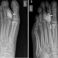 February 10, 2024 Bizarre Parosteal Osteochondromatous Proliferation (Nora’s Lesion) Of the Second Proximal Phalanx Encasing the Flexor Tendon of the Foot: A Case Report
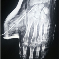 June 10, 2023 Giant Cell Tumor of Thumb Proximal Phalanx-A Case Report
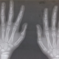 September 10, 2021 A Rare Presentation with Segmental Gap Non-union in Proximal Phalanx Fracture of Thumb in Child: A Case Report