
Adjunctive use of BMP-2 in staged revision for periprosthetic joint infection can enhance biologic acetabular bone restoration, facilitating successful reconstruction of severe defects.
Dr Vaibhav Bagaria, Department of Orthopaedics, Sir H.N. Reliance Foundation Hospital and Research Centre, Mumbai, Maharashtra, India. E-mail: bagariavaibhav@gmail.com
Abstract
Introduction: Severe acetabular bone loss in the setting of periprosthetic joint infection (PJI) presents a significant reconstructive challenge, particularly in biologically compromised environments where conventional graft incorporation may be unreliable. Bone Morphogenetic Proteins (BMPs) are potent osteoinductive agents, but their role in post-infective acetabular reconstruction remains limited.
Case Report: We report the case of a 34-year-old male with sickle cell disease who presented with chronic infection and loosening of a total hip arthroplasty, associated with severe acetabular bone loss (Paprosky IIIA). A two-stage revision strategy was employed. During the first stage, following implant removal and extensive debridement, the acetabular defect was reconstructed using a composite graft consisting of morselised allograft, antibiotic-loaded calcium sulphate, bone marrow aspirate, and recombinant human BMP-2. Radiological evaluation at 3.5 months demonstrated robust graft consolidation and restoration of the medial acetabular wall. After confirmation of infection eradication, second-stage reimplantation was successfully performed using a porous acetabular component with stable fixation.
Conclusion: This case demonstrates that adjunctive use of BMP can facilitate biologic restoration of acetabular bone stock in complex PJI scenarios, enabling effective reconstruction during staged revision arthroplasty. While promising, the use of BMP in this setting remains off-label and warrants careful patient selection, with further studies needed to establish its safety and efficacy.
Keywords: Bone morphogenetic protein, periprosthetic joint infection, acetabular bone defect, revision total hip arthroplasty, two-stage revision, osteoinduction.
Periprosthetic joint infection (PJI) remains one of the most devastating complications following arthroplasty procedures, often leading to implant loosening, progressive bone loss, compromised soft tissues, and substantial patient morbidity. Despite advances in diagnostic techniques and antimicrobial management, PJI continues to represent a major cause of revision arthroplasty worldwide [1,2,3]. Furthermore, outcomes following revision procedures remain challenging, with reinfection remaining a significant concern even after staged reconstruction [4].
Bone defect restoration is a crucial aspect of revision arthroplasty surgeries. Various management options are available depending on the magnitude, shape, anatomical position, and pathology of the bone defect. Acetabular defects resulting from chronic periprosthetic hip joint infection and repeated surgical interventions significantly limit reconstructive options and increase the risk of mechanical failure. The primary goal of acetabular reconstruction is to achieve secure fixation while restoring bone stock and maintaining long-term biological and mechanical stability.
Traditional strategies for managing acetabular bone loss include both biological and non-biological techniques. Available reconstructive options include structural allografts, impaction grafting, porous metal augments, reinforcement rings, cup-cage constructs, and custom triflange acetabular components [5,6,7,8]. While these techniques can provide mechanical support, successful graft incorporation remains dependent upon host biology, which is frequently compromised by infection, previous surgeries, and poor bone quality.
Bone morphogenetic proteins (BMPs) are potent osteoinductive growth factors that stimulate mesenchymal stem cell differentiation into osteoblasts while enhancing angiogenesis and endochondral bone formation [9,10]. BMP-2 and BMP-7 are the most extensively studied members of the transforming growth factor-β superfamily. Experimental and clinical studies have demonstrated the ability of BMPs to promote bone regeneration and improve healing in complex orthopaedic scenarios [2,10,11,12]. In parallel, advances in bone graft substitutes and biologic augmentation strategies have expanded opportunities for enhancing graft incorporation in biologically compromised environments [13]. While BMPs have been widely investigated in spinal fusion surgery, fracture healing, trauma reconstruction, and treatment of non-unions, their role in revision arthroplasty, particularly in post-infective acetabular defects, remains poorly defined [9,11,12]. The available literature describing BMP use in chronic PJI-associated acetabular bone loss is sparse, and concerns regarding cost, heterotopic ossification, and off-label application continue to limit widespread use. This case report describes the successful use of recombinant human BMP-2 as an osteoinductive adjunct in the management of severe acetabular bone loss following chronic PJI and implant failure and discusses its potential role in facilitating biological restoration of bone stock before second-stage reconstruction.
A 34-year-old male with sickle cell disease (diagnosed in 2012) and secondary bilateral hip arthritis underwent left total hip arthroplasty (THA) elsewhere in January 2022. He presented it to our institution in May 2022 for right hip pain. Examination revealed right leg shortening (3.5 cm) and limited motion. Laboratory values were within normal limits, including vitamin D (50 ng/mL). Imaging confirmed right hip arthritis with protrusion. An uncomplicated right THA was performed in June 2022. (Fig. 1). In September 2024, he presented with left hip pain, antalgic gait, and functional disability. The right hip was asymptomatic. Radiographs and a computed tomography (CT) scan revealed gross loosening of the left acetabular component with severe medial wall deficiency, central/superior migration of the prosthesis, and a peri-acetabular fluid collection (Fig. 2). CT-guided aspiration fluid was positive for Enterococcus faecium on the Biofire® joint infection panel (Figs. 3).
A two-stage revision strategy was initiated:
Stage I (Excision arthroplasty and debridement):
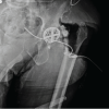
The left hip was approached through the previous posterior incision. A purulent collection was encountered. The loose acetabular cup was removed. An extended trochanteric osteotomy was required to extract a well-fixed femoral stem, which was later stabilised with cerclage wires (Fig. 4). After extensive debridement of necrotic tissue, the resulting massive acetabular defect (Paprosky IIIA) was filled with a composite of morselised allograft bone, antibiotic-loaded calcium sulphate beads (Stimulan®), bone marrow aspirate concentrate, and recombinant human BMP-2 (total dose 4.2 mg). Intraoperative tissue cultures confirmed Enterococcus faecium. The patient received culture-directed intravenous antibiotics for 6 weeks under infectious disease supervision. Serial monitoring showed normalisation of inflammatory markers (erythrocyte sedimentation rate 33 mm/h, C-reactive protein 0.9 mg/dL at 6 weeks).
Bone formation phase:
Follow-up radiographs and CT scans at 3.5 months demonstrated excellent consolidation of the graft, reconstitution of the medial acetabular wall, and healing of the osteotomy site (Fig. 5a,b,c).
Stage II (revision arthroplasty):
Before reimplantation, an ultrasound-guided aspiration of the hip yielded negative cultures. The patient underwent left revision THA using a porous titanium acetabular component with multiple screws for supplemental fixation, utilising the restored bone stock. A modular, tapered, fluted femoral stem was used (Fig. 6a).
Postoperative course:
The patient was mobilised with touchdown weight-bearing for 2 weeks, progressing to full weight-bearing. At the 2-month follow-up, he was pain-free with markedly improved function. Radiographs showed maintained implant position, graft incorporation, and no signs of loosening or infection recurrence (Fig. 6b).
This case highlights the challenges associated with managing severe acetabular bone loss in the setting of chronic PJI. Two-stage revision arthroplasty remains the gold standard for chronic infection because it offers reliable infection eradication while providing an opportunity for biological and mechanical reconstruction [1,3,4]. Contemporary evidence suggests that although infection control rates are generally favourable, reinfection and reconstruction failure remain significant concerns, particularly in cases involving extensive bone loss [14]. The defect encountered in the present case involved substantial medial wall and superior dome destruction (Paprosky IIIA). Contemporary management of such defects frequently relies on porous metal augments, trabecular metal components, cup-cage constructs, structural allografts, or custom implants [5,6,7,8]. While these techniques provide mechanical stability, restoration of host bone stock remains challenging in previously infected environments. The innovative aspect of this case was the use of recombinant human BMP-2 as an osteoinductive adjunct during the first stage of revision surgery. BMPs are known to stimulate osteoblastic differentiation, angiogenesis, and new bone formation, thereby enhancing the biological environment for graft incorporation [9,11,12]. Combined with morselised allograft, bone marrow aspirate concentrate, and antibiotic-loaded calcium sulphate, BMP-2 contributed to a biologically active graft composite possessing osteoconductive, osteogenic, and osteoinductive properties [13]. Radiographic evidence of rapid consolidation and restoration of the medial acetabular wall within 3.5 months supports the biological effectiveness of this strategy. Similar biological principles have been successfully applied in complex orthopaedic reconstruction and trauma settings, where BMP-mediated enhancement of bone regeneration has facilitated healing of challenging defects [10,12]. The advantages of BMP use in this setting include enhanced osteoinduction, potentially accelerated graft incorporation, and improved biological fixation potential for subsequent revision components. This may reduce reliance on complex mechanical solutions such as cages or custom implants [5,7,8]. However, important limitations remain. The use of BMP in acetabular reconstruction is off-label and associated with concerns regarding heterotopic ossification, increased cost, and limited long-term clinical evidence [2,9,11]. Furthermore, the use of biologic augmentation in infected arthroplasty remains controversial and should only be considered following meticulous debridement, effective infection control, and appropriate antibiotic therapy [3,4,15,16]. Further prospective studies are necessary to establish the safety, efficacy, and cost-effectiveness of BMP-assisted reconstruction in revision arthroplasty.
This case illustrates that BMP can act as a powerful osteoinductive adjunct in staged revision for PJI, transforming a severely compromised acetabular defect into a reconstructable bed through enhanced biologic restoration. While its use remains selective and off-label, it expands the reconstructive armamentarium in challenging scenarios. Rigorous clinical evaluation is essential to establish its long-term safety, indications, and economic viability.
BMP-2 can serve as a valuable osteoinductive adjunct in selected cases of PJI with major acetabular defects, promoting biologic bone restoration and enabling successful revision arthroplasty.
References
- 1. Tande AJ, Patel R. Prosthetic joint infection. Clin Microbiol Rev 2014;27:302-45. [Google Scholar] [PubMed]
- 2. Bagaria V. Bone morphogenic protein: Current state of the field and the road ahead. J Orthop 2005;2:e3. [Google Scholar] [PubMed]
- 3. McNally M, Sigmund I, Hotchen A, Sousa R. Making the diagnosis in prosthetic joint infection: A European view. EFORT Open Rev 2023;8:253-63. [Google Scholar] [PubMed]
- 4. Doski S, Sebastiao A, Thayaparan P. The investigation and management of peri-prosthetic joint infection after total knee arthroplasty: An update based on the latest British Orthopaedic Association standard and speciality standard guidelines. Cureus 2024;16: [Google Scholar] [PubMed]
- 5. Sporer SM, Paprosky WG. Acetabular revision using a trabecular metal acetabular component for severe acetabular bone loss associated with a pelvic discontinuity. J Arthroplasty 2006; 21(6) Suppl 2: 87-90. [Google Scholar] [PubMed]
- 6. Beckmann NA, Bitsch RG, Gondan M, Schonhoff M, Jaeger S. Comparison of the stability of three fixation techniques between porous metal acetabular components and augments. Bone Joint Res 2018;7:282-8. [Google Scholar] [PubMed]
- 7. Sanghavi SA, Paprosky WG, Sheth NP. Evaluation and Management of Acetabular Bone Loss in Revision Total Hip Arthroplasty: A 10-Year Update. J Am Acad Orthop Surg 2024;32:e466-75. [Google Scholar] [PubMed]
- 8. Pandey AK, Zuke WA, Surace P, Kamath AF. Management of acetabular bone loss in revision total hip replacement: A narrative literature review. Ann Jt 2023;9: [Google Scholar] [PubMed]
- 9. Carreira AC, Lojudice FH, Halcsik E, Navarro RD, Sogayar MC, Granjeiro JM. Bone morphogenetic proteins: Facts, challenges, and future perspectives. J Dent Res 2014;93:335-45. [Google Scholar] [PubMed]
- 10. Tan Q, Li J, Yin Y, Shao W. The role of growth factors in the repair of motor injury. Front Pharmacol 2022;13: [Google Scholar] [PubMed]
- 11. Garrison KR, Shemilt I, Donell S, Ryder JJ, Mugford M, Harvey I, et al. Bone morphogenetic protein (BMP) for fracture healing in adults. Cochrane Database Syst Rev 2010;2010:CD006950. [Google Scholar] [PubMed]
- 12. Vaibhav B, Nilesh P, Vikram S, Anshul C. Bone morphogenic protein and its application in trauma cases: A current concept update. Injury 2007;38:1227-35. [Google Scholar] [PubMed]
- 13. Nashi N, Kagda FH. Current concepts of bone grafting in trauma surgery. J Clin Orthop Trauma 2023;43: [Google Scholar] [PubMed]
- 14. Goud AL, Harlianto NI, Ezzafzafi S, Veltman ES, Bekkers JE, Van Der Wal BC. Reinfection rates after one- and two-stage revision surgery for hip and knee arthroplasty: A systematic review and meta-analysis. Arch Orthop Trauma Surg 2023;143:829-38. [Google Scholar] [PubMed]
- 15. Qasim SN, Swann A, Ashford R. The DAIR (debridement, antibiotics and implant retention) procedure for infected total knee replacement – a literature review. SICOT J 2017;3:2. [Google Scholar] [PubMed]
- 16. Bengtson S, Knutson K. The infected knee arthroplasty. A 6-year follow-up of 357 cases. Acta Orthop Scand 1991;62:301-11. [Google Scholar] [PubMed]


Related Articles in Journal of Orthopaedic Case Reports
 June 1, 2026 Revision of a Dislocated Articulating Antibiotic Hip Spacer as an Alternative to Girdlestone Arthroplasty: A Case Report
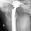 May 1, 2026 A Novel Failure Interface in Constrained Total Hip Arthroplasty: Dissociation at the Constrained Liner Insert-Bipolar Head Junction Following Periprosthetic Joint Infection
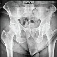 January 1, 2025 A Rare Case of Staphylococcus Caprae Periprosthetic Hip Infection with Unusual Clinical Presentation
 July 1, 2026 Custom-Made Monoflange Acetabular Component for Revision Hip Arthroplasty in Paprosky Type IIIA Defect: A Case Report