

The prone “chicken-wing” position provides optimal exposure of the ventromedial scapula, enabling safe en bloc excision of osteochondroma while minimizing risk to the serratus anterior and long thoracic nerve.
Dr. Abhishek Dussa, Department of Orthopaedics, Dr. Vasantrao Pawar Medical College Hospital and Research Center, Nashik, Maharashtra, India. Email: abhishekdussa21@gmail.com
Abstract
Introduction: Ventromedial scapular osteochondroma is an uncommon cause of scapulothoracic impingement and “pseudo-winging.” Its deep anatomical location often leads to delayed diagnosis, particularly when symptoms mimic snapping scapula syndrome.
Case Report: A 10-year-old boy presented with a 2-month history of left upper back pain and difficulty wearing a school backpack. Examination revealed terminal restriction of shoulder motion and medial scapular pseudo-winging. Magnetic resonance imaging confirmed a 3.5 × 2.2 cm osteochondral lesion arising from the ventral surface of the left scapula. The lesion was completely excised through a posterior approach with the patient in a prone “chicken-wing” position.
Conclusion: Optimal patient positioning is critical for safe surgical clearance of ventral scapular tumors. Complete excision leads to immediate relief of mechanical symptoms and excellent functional recovery.
Keywords: Osteochondroma, ventral scapula, pseudo-winging, “chicken-wing” position, pediatric orthopedics.

Osteochondroma is the most common benign primary bone tumor. It typically arises from the metaphysis of long bones and is characterized by a cartilage-capped bony projection continuous with the cortex and medullary canal [1]. Although these tumors are usually asymptomatic and found incidentally, symptoms may develop when the lesion causes mechanical irritation, compression of adjacent structures, deformity, or, in rare cases, malignant transformation. Scapular osteochondromas are uncommon, accounting for only 3–4.6% of all osteochondromas [2,3], and lesions arising from the ventral or ventromedial surface are particularly rare [4]. Because of the deep anatomical location of the scapula against the thoracic cage, ventral lesions are rarely palpable as a typical mass. Instead, patients often present with “snapping scapula syndrome,” crepitus, restricted shoulder motion, or “pseudo-winging,” a static deformity where the scapula is physically displaced from the ribs by the tumor, mimicking neuromuscular winging [3,5,6]. The deep-seated nature of these tumors poses a significant surgical challenge regarding access and visualization. To our knowledge, limited literature emphasizes the role of intraoperative positioning in facilitating safe excision of ventral scapular lesions. This case report details the clinical management and surgical excision of a rare ventral scapular osteochondroma in a pediatric patient. We specifically highlight the utility of the prone “chicken-wing” position, maximum internal rotation, and slight extension of the affected limb, which biomechanically tilts the scapular border away from the thoracic wall to “open” the subscapular space [7]. This technique facilitates safe, muscle-sparing en bloc resection, ensuring the restoration of a full range of motion (ROM) and resolution of symptomatic pseudo-winging.
A 10-year-old male presented to our orthopedic clinic with a complaint of pain in the left upper back region for a duration of 2 months. The pain was specifically associated with daily activities such as wearing a school backpack and caused significant discomfort when lying in the supine position. The patient’s mother had noticed a gradually enlarging mass during this period. There was no history of trauma, fever, or constitutional symptoms.
Clinical examination:
Inspection revealed a visible prominence over the medial border of the left scapula, suggestive of pseudo winging (Fig. 1). On palpation, a firm, non-tender mass measuring approximately 2.5 × 1.5 cm was palpable deep to the posteromedial border of the scapula. The mass moved in conjunction with scapular motion. ROM of the shoulder joint was terminally restricted, particularly during overhead activities. No dynamic winging was observed, helping differentiate from long thoracic nerve palsy, and the distal neurovascular status was intact.
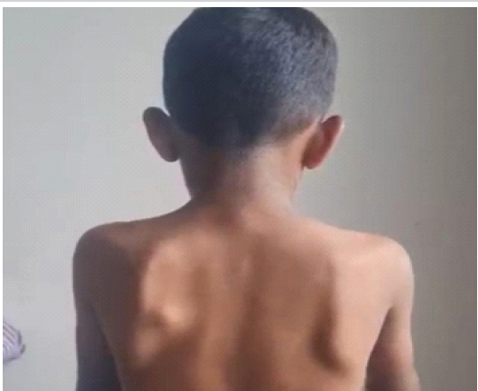
Figure 1: Pre-operative clinical photograph showing prominence of the medial border of the left scapula suggestive of pseudo-winging. The deformity is static in nature and accentuated by the underlying ventral scapular lesion.
Investigations:
Plain radiographs (anteroposterior and scapular Y views) revealed a well-circumscribed osseous outgrowth arising from the ventral surface of the left scapula, measuring approximately 3 cm (Fig. 2). The lesion demonstrated cortical and trabecular continuity with the parent bone, which was further confirmed on computed tomography with three-dimensional (3D) reconstruction (Fig. 3). Magnetic resonance imaging (MRI) provided further detail, showing a 3.5 × 2.2 cm pedunculated bony outgrowth from the medial border of the lower scapular blade. A 2.5 mm cartilage cap was noted, causing indentation of the subscapularis muscle (Fig. 4).
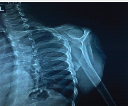
Figure 2: Scapula Y view radiograph demonstrating a well-defined osseous outgrowth arising from the ventral aspect of the scapula. The lesion shows continuity with the underlying cortex and medullary canal, suggestive of an osteochondroma.
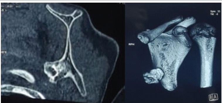
Figure 3: Axial computed tomography (CT) image and three-dimensional (3D) CT reconstruction of the left scapula demonstrating a well-defined pedunculated bony outgrowth arising from the ventral (costal) surface of the scapula, which shows continuity with the cortex and medullary canal consistent with osteochondroma.
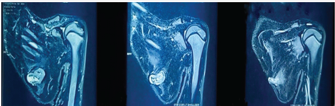
Figure 4: Magnetic resonance imaging of the left shoulder demonstrating a pedunculated lesion arising from the ventral surface of the scapula. The lesion shows a cartilage cap and causes indentation of the adjacent subscapularis muscle. No features suggestive of malignant transformation are noted.
Surgical treatment:
Under general anesthesia, the patient was placed in the prone “chicken-wing” position. The affected arm was placed in maximum internal rotation with slight extension (hand positioned over the lower back) (Fig. 5). This biomechanical maneuver tilts the medial border of the scapula away from the rib cage, effectively “opening” the subscapular space and eliminating the need for excessive muscular retraction. A longitudinal incision was made along the medial border of the scapula. The trapezius and rhomboid muscles were carefully retracted. The pedunculated lesion was identified on the costal surface (Fig. 6). Using a curved osteotome, an en bloc excision was performed at the base of the stalk (Fig. 7). Special care was taken to include the entire cartilaginous cap to minimize the risk of recurrence. The bone base was cleared with a nibbler to ensure a smooth surface. The gross specimen of the excised osteochondroma demonstrated a smooth cartilaginous cap and measured approximately 3.5 cm × 2.2 cm (Fig. 8).
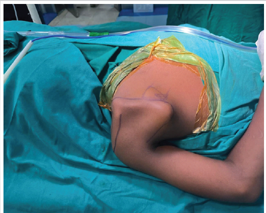
Figure 5: Intraoperative photograph demonstrating the prone “chicken-wing” position, with the affected upper limb placed in maximum internal rotation and slight extension.
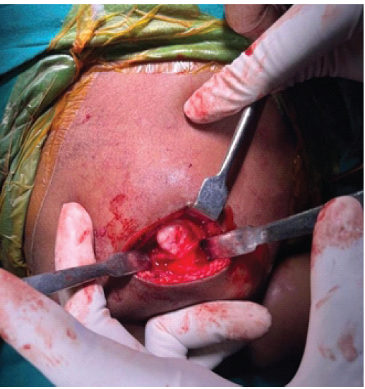
Figure 6: Intraoperative photograph showing a pedunculated osteochondroma arising from the costal (ventral) surface of the scapula. The lesion is exposed through a posterior approach with careful retraction of surrounding soft tissues, clearly demonstrating its stalk and orientation prior to excision.
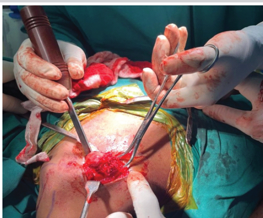
Figure 7: Intraoperative photograph demonstrating en bloc excision of the pedunculated osteochondroma using a curved osteotome at the base of the stalk. Care was taken to completely remove the lesion along with the cartilaginous cap to minimize the risk of recurrence.
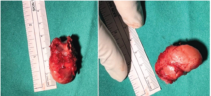
Figure 8: Gross specimen of the excised osteochondroma demonstrating a well-defined pedunculated bony mass with a smooth cartilaginous cap. The lesion measures approximately 3.5cm × 2.2 cm.
Follow-up:
The post-operative period was uneventful. Histopathological examination confirmed the diagnosis of a benign osteochondroma, showing a hyaline cartilage cap overlying organized bone trabeculae. At the 6-week follow-up, the patient reported complete relief of mechanical symptoms. Clinical evaluation showed a restored scapular contour, resolution of the pseudo-winging, and a return to full unrestricted ROM.
Osteochondromas involving the scapula are rare, and those arising from the ventral surface are particularly uncommon due to the extensive muscular coverage of the subscapularis and serratus anterior [8]. These benign developmental lesions typically grow during skeletal maturity and remain asymptomatic unless they interfere with adjacent structures. In the scapula, the mechanical interface between the ventral surface and the rib cage is narrow; therefore, even a small lesion can lead to significant symptomatic “snapping scapula syndrome” or “pseudo-winging” [8]. The clinical presentation in our 10-year-old patient, difficulty wearing a backpack and discomfort in the supine position, is characteristic of ventral lesions, which physically displace the scapula from the thoracic wall. As noted by Barapatre et al., clinical presentation often mimics neuromuscular winging, but “pseudo-winging” is distinguished by its static nature and the presence of a palpable mass that moves with the scapula [5,8]. While plain radiographs, specifically the “Scapular Y” view, can identify the lesion, 3D computed tomography and MRI are vital for defining the stalk’s orientation and the thickness of the cartilage cap, which is a key predictor for potential malignant transformation [9,10]. The “chicken-wing” positioning used in this case is a crucial technical pearl for surgeons. By placing the arm in maximum internal rotation with slight extension, the scapula is distracted and tilted away from the rib cage. This maneuver “opens” the subscapular space, providing the safest corridor for a muscle-sparing approach along the medial border while minimizing the risk of injury to the long thoracic nerve or serratus anterior [8,11]. Consistent with the existing literature, we recommend en bloc excision at the base of the stalk. Complete removal of the cartilaginous cap is mandatory to minimize the risk of local recurrence, which has been reported when remnants are left behind [12,13]. This case reinforces the need for high clinical suspicion in pediatric patients presenting with unexplained scapular prominence. Timely surgical intervention through a posterior approach in the “chicken-wing” position results in immediate symptomatic relief and excellent functional outcomes, allowing children to return to routine activities without restriction.
Ventral scapular osteochondromas should be considered in cases of unexplained scapular prominence and mechanical symptoms in the pediatric population. Due to the tumor’s deep anatomical location against the thoracic cage, symptoms such as “snapping scapula” or difficulty with daily activities, such as wearing a backpack, must be met with a high index of clinical suspicion to prevent prolonged diagnostic delays. Our report highlights that meticulous surgical planning is the cornerstone of successful management. The utilization of the prone “chicken-wing” position—specifically maximum internal rotation with slight extension—is a vital technical pearl that biomechanically tilts the scapula to “open” the subscapular space. This maneuver provides an optimal surgical corridor for safe, muscle-sparing en bloc resection while protecting the long thoracic nerve. As demonstrated in this patient, timely surgical intervention results in immediate relief of mechanical impingement, restoration of a normal scapular contour, and excellent functional recovery, allowing a rapid return to unrestricted school and play activities.
Scapular prominence with mechanical symptoms in children should prompt evaluation for structural causes such as ventral scapular osteochondroma, with careful distinction from neuromuscular winging. Early imaging aids accurate diagnosis; however, optimal surgical exposure remains the key to successful management. The prone “chicken-wing” position is a critical technical modification that is simple and reproducible, enabling safe en bloc excision while protecting the serratus anterior and long thoracic nerves, resulting in superior functional outcomes.
References
- 1. Ogawa K, Inokuchi W. Solitary osteochondroma of the ventral scapula associated with large bursa formation and pseudowinging of the scapula: A case report and literature review. Case Rep Orthop 2018;2018:5145642. [Google Scholar] [PubMed]
- 2. Jindal M. Delayed presentation of osteochondroma at superior angle of scapula-a case report. J Orthop Case Rep 2016;6:32-4. [Google Scholar] [PubMed]
- 3. Pawar E, Modi N, Yadav AK, Mhatre J, Khemkar S, Mishra S. A solitary ventral scapular osteochondroma causing pseudo-winging of scapula: A case report. J Orthop Case Rep 2021;11:82-5. [Google Scholar] [PubMed]
- 4. Donati DM, Staals EL. Osteochondroma. In: Diagnosis of Musculoskeletal Tumors and Tumor-Like Conditions: Clinical, Zadiological and Histological Correlations – the Rizzoli Case Archive. Berlin: Springer; 2024. p. 75-80. [Google Scholar] [PubMed]
- 5. Mohsen MS, Moosa NK, Kumar P. Osteochondroma of the scapula associated with winging and large bursa formation. Med Princ Pract 2006;15:387-90. [Google Scholar] [PubMed]
- 6. Seth A, Naqvi M, Yadav U, Karumuri K, Annapareddy A, Reddy AV. Unveiling scapular pseudo-winging: A case report on ventral osteochondroma of the scapula in a 21-year-old. J Orthop Case Rep 2023;13:99-104. [Google Scholar] [PubMed]
- 7. Luna‐Lizárraga CR, Berebichez‐Fastlicht E, Berebichez‐Fridman R. Bilateral elastofibroma dorsi managed with single‐stage resection in the prone position: A case report. Clin Case Rep 2025;13:e71051. [Google Scholar] [PubMed]
- 8. Barapatre J, Gayan M, Islam Z, Dhurwe A, Buragohain CR. Symptomatic ventromedial scapular osteochondroma presenting with restriction of shoulder movements: A case report. J Orthop Case Rep 2026;16:212-6. [Google Scholar] [PubMed]
- 9. Kitsoulis P, Galani V, Stefanaki K, Paraskevas G, Karatzias G, Agnantis NJ, et al. Osteochondromas: Review of the clinical, radiological and pathological features. In Vivo 2008;22:633-6. [Google Scholar] [PubMed]
- 10. Murphey MD, Choi JJ, Kransdorf MJ, Flemming DJ, Gannon FH. Imaging of osteochondroma: Variants and complications with radiologic-pathologic correlation. Radiographics 2000;20:1407-34. [Google Scholar] [PubMed]
- 11. Wiater JM, Flatow EL. Long thoracic nerve injury. Clin Orthop Relat Res 1999;368:17-27. [Google Scholar] [PubMed]
- 12. Krishnan UK. Dahlin’s Bone Tumors : General Aspects and Data on 10,165 Cases. In: Unni K, Inwards CY. Dahlin’s bone tumors: General aspects and data on 10,165 cases: Sixth edition. 2012. 1–416 p. [Google Scholar] [PubMed]
- 13. Heck RK. Benign Bone Tumors and Non Neoplastic Condition Simulation Bone Tumors. In: Terrycanale S, Beaty JH, editors. Campbell’s Operative Orthopedics. Philadelphia, PA: Mosby, Scientific Research Publishing; 2008. p. 855-7. [Google Scholar] [PubMed]




Related Articles in Journal of Orthopaedic Case Reports
 July 1, 2026 Beyond the Biopsy: Macrodystrophia Lipomatosa Unmasked After False Osteochondroma Diagnosis
 February 1, 2026 Symptomatic Ventromedial Scapular Osteochondroma Presenting with Restriction of Shoulder Movements: A Case Report
 October 10, 2023 Unveiling Scapular Pseudo-winging: A Case Report on Ventral Osteochondroma of the Scapula in a 21-year-old
 October 1, 2026 A Solitary Large Calcaneal Osteochondroma in a Skeletally Mature Patient: Case Report