

Occult foreign bodies should always be included in the differential diagnosis of chronic paediatric ankle swelling. Ultrasonography is a crucial imaging modality for detecting radiolucent foreign bodies when MRI findings are inconclusive. Timely surgical exploration following appropriate imaging leads to definitive diagnosis and complete symptom resolution.
Pujith Chandra, Department of Orthopaedics, Seth G.S. Medical College and KEM Hospital, Mumbai, Maharashtra, India. E-mail: pujith.chandra@gmail.com
Abstract
Introduction: Chronic ankle swelling in the paediatric population presents a diagnostic challenge due to a wide range of differential diagnoses and often non-specific clinical features. Occult retained foreign bodies, particularly radiolucent organic materials, such as wood or thorns, are an underrecognised cause of persistent swelling, especially in children who walk barefoot and may not recall trivial penetrating injuries. Delayed presentation and secondary inflammatory changes frequently obscure the primary pathology, leading to prolonged diagnostic delays.
Case Report: We report the case of a 10-year-old boy who presented with progressive medial ankle swelling for 18 months without a definite history of trauma, infection, or cutaneous lesions. Clinical examination revealed a firm, tender peri-ankle swelling with preserved joint motion. Plain radiographs were normal, and magnetic resonance imaging (MRI) demonstrated flexor tendon tenosynovitis without identifying a causative lesion. Subsequent ultrasonography revealed a linear echogenic structure suggestive of a retained foreign body. Surgical exploration confirmed and enabled removal of a wooden foreign body measuring 1.7 × 0.3 cm, with histopathology demonstrating a chronic inflammatory foreign body reaction. The patient achieved complete symptomatic resolution post-operatively.
Conclusion: Occult foreign bodies should be considered in the differential diagnosis of chronic paediatric ankle swelling, even in the absence of trauma history or characteristic imaging findings. Ultrasonography is a valuable diagnostic adjunct when radiographs and MRI are inconclusive. Early recognition and timely surgical intervention result in excellent outcomes and prevent unnecessary investigations and complications.
Keywords: Occult foreign body, chronic ankle swelling, paediatric foot and ankle.

Retained foreign bodies in paediatric patients are frequently occult due to trivial or forgotten trauma, the radiolucent nature of organic materials, and delayed clinical presentation [1]. Such cases often present as chronic swelling without a definite history of injury. Occult foreign bodies are a recognised cause of persistent swelling in the paediatric extremities, particularly in regions where barefoot walking is common and injuries involving wooden or thorn fragments are frequent [1,2]. The foot, especially the plantar and peri-ankle region, is the most commonly involved site [1]. Delayed presentation may result in secondary inflammatory changes, such as tenosynovitis, granuloma formation, abscess, or sinus tract formation, thereby obscuring the primary pathology and making diagnosis challenging [3]. This case is unique in that a deeply embedded, radiolucent organic foreign body presented solely as chronic peri-ankle swelling without antecedent trauma or cutaneous signs, closely mimicking inflammatory and neoplastic conditions. The prolonged diagnostic delay despite multiple clinical evaluations underscores the limitations of routine radiography and highlights the critical importance of maintaining a high index of suspicion and utilising advanced imaging modalities. This report is therefore valuable in raising awareness of occult foreign bodies as an under-recognised cause of chronic paediatric foot and ankle swelling, with important implications for timely diagnosis and avoidance of unnecessary investigations and interventions.
A 10-year-old male child presented with a history of persistent swelling over the right ankle for 18 months. The child and parents reported multiple minor falls during play over the preceding years but denied any specific penetrating injury. There was no history of fever, local wound, or persistent or intermittent discharge. The patient complained of a gradually progressive swelling associated with intermittent pain, which was exacerbated by activity. There was no clinical history suggestive of infection or inflammatory arthritis.
Clinical examination:
Right ankle:
Local examination of the right ankle revealed a diffuse, firm, tender swelling measuring approximately 8 × 4 cm over the medial aspect of the ankle, inferior to the medial malleolus (Fig. 1). The swelling was fixed to deeper planes but not adherent to the overlying skin. The skin was normal, with no evidence of a scar, sinus, or discharge. Ankle range of motion was full and painless. Neurovascular examination was normal. Gait analysis revealed restricted and painful plantar flexion.
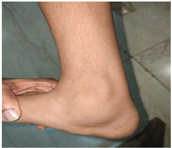
Figure 1: Medial aspect of ankle showing swelling inferior to medial malleolus with no scar or discharging sinus.
Investigations:
Plain radiographs of the ankle and foot were unremarkable. In view of the chronicity of symptoms, magnetic resonance imaging (MRI) of the ankle was performed. MRI demonstrated moderate fluid along the tendon sheaths of the tibialis posterior, flexor hallucis longus, and flexor digitorum longus tendons, suggestive of tenosynovitis, with secondary synovial inflammation (Figs. 2, 3, 4). No definite foreign body was identified, a known limitation of MRI in detecting radiolucent organic materials, such as wood or thorns [3,4]. Subsequent ultrasonography of the ankle revealed a linear echogenic structure with posterior acoustic shadowing, suggestive of a retained foreign body with a surrounding collection (Fig. 5), consistent with prior reports highlighting the superior sensitivity of ultrasonography for radiolucent foreign bodies [4,5,6].
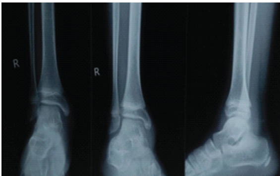
Figure 2: X-ray radiograph of right ankle of patient shows normal study.
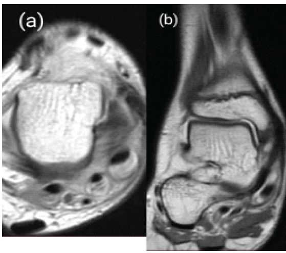
Figure 3: (a) T1 shows axial cut of ankle with tenosynovitis of tibialis posterior tendon, (b) T1 image of magnetic resonance imaging ankle in coronal view showing tenosynovitis of tibialis posterior with no obvious evidence of foreign body.
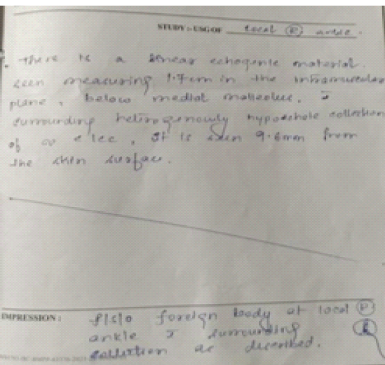
Figure 4: Sonography report of the ankle, which was the index investigation hinting toward a retained foreign body.
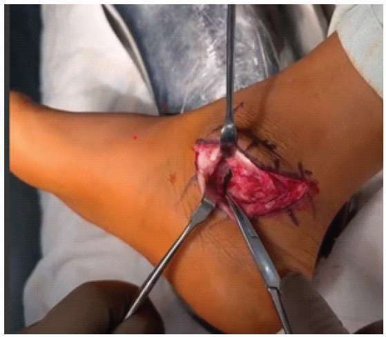
Figure 5: Intraoperative image of showing a jet-black structure seen after incising the flexor retinaculum.
Surgical procedure:
The patient underwent surgical exploration under spinal anaesthesia with tourniquet control. An L-shaped incision was made posteroinferior to the medial malleolus. Following incision of the flexor retinaculum, oedematous tibialis posterior and flexor digitorum longus tendons were exposed. Tenosynovectomy of the involved tendons was performed. Inferior to the tendons, granulation tissue was identified, and tissue samples were obtained. Following thorough irrigation, a small jet-black linear structure was identified and removed. The foreign body measured approximately 1.7 × 0.3 cm and was confirmed to be a wooden fragment (Fig. 5, 6).
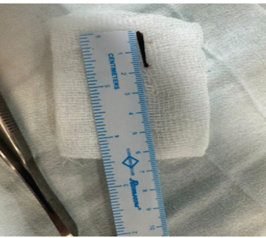
Figure 6: Gross specimen of wooden foreign body removed of size 1.7 cm × 0.3 cm.
Intraoperative findings:
Microbiological cultures of the intraoperative specimens showed no growth. Histopathological examination revealed chronic inflammatory infiltrates composed predominantly of phagocytes and plasma cells, consistent with a foreign body–induced inflammatory reaction (Fig. 7).
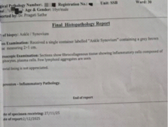
Figure 7: Final histopathological report showing findings of chronic inflammation.
Outcome:
The post-operative period was uneventful. At the 6-month follow-up, the child had complete resolution of symptoms with restoration of a painless, full range of ankle movements and normal gait (Fig. 8).
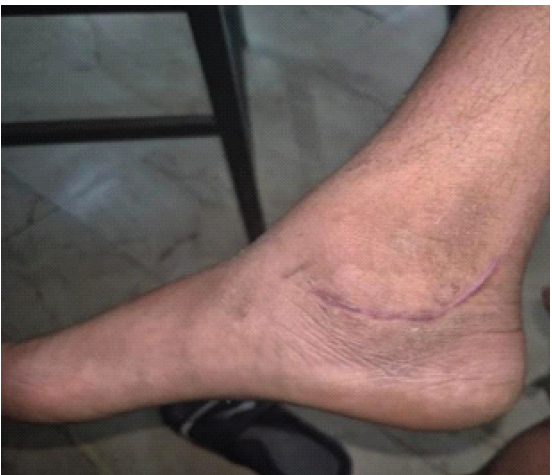
Figure 8: Healed suture site and reduction in swelling.
Chronic ankle swelling in the paediatric population poses a significant diagnostic challenge due to its broad differential diagnosis and frequently non-specific clinical features. The absence of a definite history of trauma, trivial penetrating injuries that are often forgotten, and the predominance of radiolucent organic foreign bodies, such as wood or thorns, contribute to delayed diagnosis [1,3,7]. Paediatric orthopaedic literature emphasises that occult foreign bodies must be considered in children presenting with persistent foot or ankle swelling, particularly in environments where barefoot walking is common [1,2,8]. A systematic approach to chronic paediatric ankle swelling begins with careful clinical assessment, followed by appropriate imaging. Differential diagnoses include tarsal coalition; accessory ossicles; ganglion or synovial cysts; inflammatory arthropathies, such as juvenile idiopathic arthritis; chronic osteomyelitis; benign soft-tissue tumours; and rarely malignant lesions [1,2]. Although foreign bodies represent a small proportion of cases, they are frequently overlooked due to normal radiographs and lack of corroborative history [3]. Plain radiographs are often unrevealing when the retained object is radiolucent. MRI, while sensitive for detecting secondary inflammatory changes, such as tenosynovitis and synovitis, may fail to directly identify wooden or plant-based foreign bodies [3,4]. In the present case, MRI findings reflected secondary pathology without identifying the primary aetiology, thereby contributing to diagnostic ambiguity. Ultrasonography plays a crucial complementary role in such cases. It is cost-effective, widely available, and highly sensitive for detecting radiolucent foreign bodies, particularly wood and thorns [4,5,6,9,10]. Ultrasonography can also delineate associated collections and inflammatory changes, aiding surgical planning. In this case, ultrasonography was pivotal in raising suspicion of a retained foreign body when MRI findings were non-specific. Definitive diagnosis of occult foreign bodies is often established intraoperatively. Surgical exploration in this patient revealed a wooden foreign body with an atypical presentation, accounting for chronic tenosynovitis and prolonged symptoms. Histopathological findings supported a chronic foreign body reaction, while sterile cultures were consistent with a long-standing, non-infective inflammatory process.
The definitive diagnosis in occult foreign body cases is often established intra-operatively. In the present patient, surgical exploration revealed a wooden foreign body with an atypical presentation, surrounded by inflamed and unhealthy tissue, explaining the chronic tenosynovitis and persistent symptoms. Histopathological findings of chronic inflammatory infiltrates further supported the diagnosis, while sterile cultures were consistent with a long-standing, non-infective inflammatory process. This case highlights the importance of maintaining a high index of suspicion for occult foreign bodies in children presenting with chronic ankle swelling, even in the absence of trauma history or classical imaging findings. Early utilisation of ultrasonography and timely surgical exploration can prevent prolonged morbidity, unnecessary investigations, and potential complications, such as sinus formation, secondary infection, or osteomyelitis.
Chronic ankle swelling in children may represent an occult retained foreign body despite the absence of trauma history or cutaneous signs. Radiolucent organic materials can evade detection on plain radiographs and MRI, presenting only with secondary inflammatory changes. Early consideration of this diagnosis can prevent prolonged morbidity and unnecessary investigations.
References
- 1. Tachdjian MO, editor. Tachdjian’s Pediatric Orthopaedics. 5th ed. Philadelphia, PA: Elsevier; 2014. [Google Scholar] [PubMed]
- 2. Campbell WC, Canale ST, Beaty JH. Campbell’s Operative Orthopaedics. 14th ed. Philadelphia, PA: Elsevier; 2021. [Google Scholar] [PubMed]
- 3. Peterson JJ, Bancroft LW, Kransdorf MJ. Wooden foreign bodies: Imaging appearance. AJR Am J Roentgenol 2002;178:557-62. [Google Scholar] [PubMed]
- 4. Graham DD Jr. Ultrasound in the emergency department: Detection of wooden foreign bodies in the soft tissues. J Emerg Med 2002;22:75-9. [Google Scholar] [PubMed]
- 5. Shiels WE 2nd, Babcock DS, Wilson JL, Burch RA. Localization and guided removal of soft-tissue foreign bodies with sonography. AJR Am J Roentgenol 1990;155:1277-81. [Google Scholar] [PubMed]
- 6. Boyse TD, Fessell DP, Jacobson JA, Lin J, Van Holsbeeck MT, Hayes CW, et al. US of soft-tissue foreign bodies and associated complications. Radiographics 2001;21:1251-6. [Google Scholar] [PubMed]
- 7. Yanay O, Vaughan DJ, Diab M, Brownstein D, Brogan TV. Retained wooden foreign body in a child’s thigh complicated by severe necrotizing fasciitis: A case report and discussion of imaging modalities for early diagnosis. Pediatr Emerg Care 2001;17:354-5. [Google Scholar] [PubMed]
- 8. Laor T, Barnewolt CE. Nonradiopaque penetrating foreign body: “A sticky situation”. Pediatr Radiol 1999;29:702-4. [Google Scholar] [PubMed]
- 9. Crawford R, Matheson AB. Clinical value of ultrasonography in the detection and removal of radiolucent foreign bodies. Injury 1989;20:341-3. [Google Scholar] [PubMed]
- 10. Rockett MS, Gentile SC, Gudas CJ, Brage ME, Zygmunt KH. The use of ultrasonography for the detection of retained wooden foreign bodies in the foot. J Foot Ankle Surg 1995;34:478-84; discussion 510-1. [Google Scholar] [PubMed]






Related Articles in Journal of Orthopaedic Case Reports
 May 1, 2026 Surgical Management of Painful Os Vesalianum: A Case Report and Literature Review
 July 10, 2021 Reviewers Acknowledgement & Photo-Gallery July 2021
 May 10, 2020 Non-union after Fracture of the Anterior Tibial Spine in a 6-Year-old Child: A Case Report
 August 1, 2026 Reverse Shoulder Arthroplasty with Virtual Implant Positioning System in the Management of Massive Rotator Cuff Tear with Rotator Cuff Arthropathy