

• Glomus tumors are frequently seen in subungual regions of hands • The foot and other regions are less commonly involved • The classical triad of pain, cold hypersensitivity, and point tenderness gives a clue to diagnosis • MRI is the investigation of choice • Excision of a tumour is curative
Dr Jeevan Vijay Rajasekhar, Department of Orthopaedics, All India Institute of Medical Sciences, Mangalagiri, Andhra Pradesh, India. E-mail: vijayjeevan2@gmail.com
Abstract
Introduction: Glomus tumours are benign vascular hamartomas that lie in the reticular layer as an arteriovenous anastomosis in the dermis. A glomus tumour is usually located in the subungual region of the digits. They occur more often in the hand (75%) than elsewhere and are located beneath the fingernail in 25–65% of patients. Clinical and radiological (magnetic resonance imaging [MRI]) usually clinch the diagnosis in the majority of cases. Twenty-five percent are not subungual, however, and these may pose a difficult diagnostic problem.
Case Report: A 40-year-old male, a known case of cardiac disease with a pacemaker in situ, experiencing pain on the lateral aspect of the hindfoot following trauma, was diagnosed with a soft tissue injury. The patient presented with persistence of pain and development of paraesthesia as a new symptom over 1 year. The diagnosis of neuroma of the sural nerve was made on the premise of localised pain and paraesthesia in a patient with a prior history of a blunt trauma, supported by ultrasonography. Intraoperatively, the nerve was found intact, and tumour tissue was located within the muscle, which was excised. Histopathology confirmed a glomus tumour. The patient was symptom-free on the immediate post-operative day.
Conclusion: A glomus tumour is a rare but important differential diagnosis for localised foot pain and must be evaluated with an MRI. In cases where an MRI is not feasible, the surgery must be performed, keeping the option of vigilant tissue exploration to locate any lesion. Complete excision of the tumour is curative.
Keywords: Glomus tumour, foot pain, excision.

Glomus tumours are benign hamartomas originating from the glomus body [1]. The glomus body is composed of vascular structures, nerve cells, and smooth muscle cells. It is found in the dermis throughout the body, with the highest concentrations being in the hands and feet, especially in the fingertips and under the nail bed. The glomus body is responsible for thermoregulation. Glomus tumours are typically characterised by painful, subcutaneous nodules that are usually located in the subungual region of the digits [2]. Glomus tumours commonly occur in middle age, especially in the fourth and fifth decades of life, with females affected more often than males [3]. Up to 75% of these lesions are found in the hand, and 65% are located in the fingertip. The apparatus itself consists of an afferent vessel, a Sucquet-Hoyer canal composed of a channel surrounded by large polygonal cells and multiple shunts. Histologic findings include endothelial pericytes and numerous non-myelinated nerve fibres. These findings explain the classic triad of symptoms seen: (1) cold hypersensitivity, (2) paroxysmal pain, and (3) pinpoint pain. Symptoms are remarkable in the severity of the sharp lancinating pain brought on by either cold exposure or even light touch. Physical examination is unremarkable in 50% of lesions; however, a localised bluish discolouration in the nail bed, with or without nail plate ridges, strongly suggests the diagnosis [4]. Radiological evaluation includes ultrasound and magnetic resonance imaging (MRI) [5]. Glomus tumours appear as dark, well-delineated lesions on T1-weighted and bright on T2-weighted MRI. MRI has been successful at pinpointing lesions as small as 2 mm in the fingertip [6]. Treatment of symptomatic lesions is surgical excision. The rate of recurrence may be as high as 20%. If symptoms continue longer than 3 months after excision, the surgeon should strongly consider re-exploration because the incidence of multiple lesions is high [7]. The literature on glomus tumours in the hand is extensive, while those in the foot are sparse [8]. Trehan et al., in their literature, reported glomus tumours in the subungual regions of the foot [9]. The presence of non-subungual lesions in the foot is remote, and they are found around the tips of toes. In this report, we describe the work-up, management, and recent concepts of a glomus tumour in a non-subungual location presenting as a neuroma.
A 40-year-old male patient has presented with continuous dull aching pain and tingling sensation in the lateral aspect of the left foot for 1 year. There was a history of trauma (a twisted injury of the foot). Conservative management with compression bandages, ice packs, and pain medications was prescribed. The patient developed paraesthesia over the lateral aspect of the hindfoot with persistent pain increasing in severity when the patient wore footwear. The patient was a known case of cardiac disease with a pacemaker in situ.
Clinical examination:
There was no visible swelling in the foot. A localised tenderness was found on the lateral border of the hindfoot. On tapping, there was a tingling sensation noticed by the patient, which was also painful. There was no distal motor or sensory loss. However, paraesthesia was present in the affected area.
Radiological evaluation:
Ultrasound examination of the suspected lesion showed a well-defined lesion in the subcutaneous plane, probably a neuroma of the branch of the sural nerve (Fig. 1). MRI could not be done due to a cardiac pacemaker.
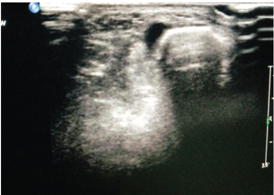
Figure 1: Ultrasonography of the lesion showing a hypointense oval shadow in the subcutaneous plane.
Treatment:
Henna was used under ultrasound guidance to localise the lesion pre-operatively (Fig. 2).
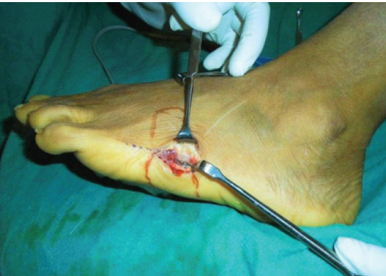
Figure 2: Henna (brown irregular line) applied to localize the lesion.
Surgical exploration showed an intact branch of the sural nerve. Further exploration of the underlying muscle revealed a pearly white, approximately 0.5 × 1 cm2 lesion which was excised and sent for biopsy (Figs. 3 and 4).
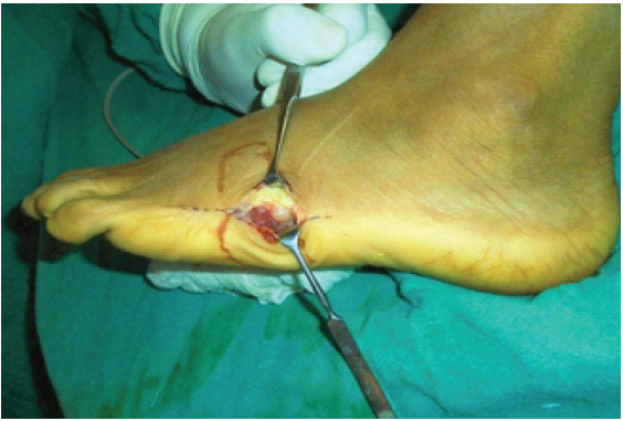
Figure 3: Intraoperative finding of an intramuscular pearly white lesion.
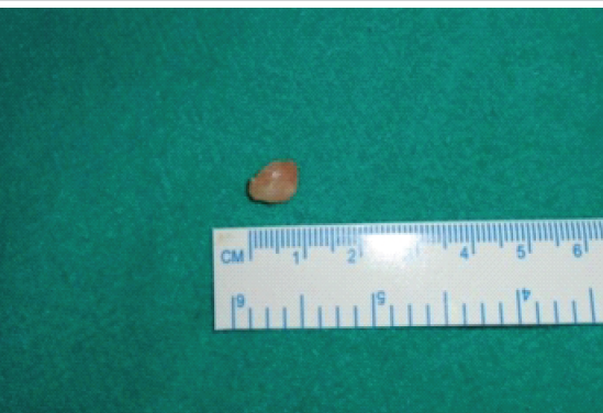
Figure 4: Completely excised lesion.
Histopathology revealed blood vessels with numerous glomus cells, which are round to oval with acidophilic cytoplasm. Immunohistochemistry showed positivity of smooth muscle actin ` in glomus cells. These features are consistent with a glomus tumour (Figs. 5 and 6). The patient was symptom-free with no signs of recurrence after 1 year.
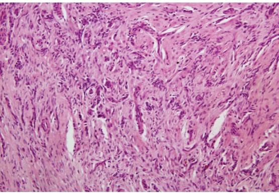
Figure 5: Blood vessels lined by endothelial cells with numerous glomus cells.
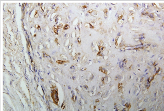
Figure 6: Immunohistochemistry showing positivity of smooth muscle actin in glomus cells.
Glomus tumours are rare benign soft tissue tumours that arise from the glomus body. Glomus tumours are most commonly found in distal extremities in the nail bed and account for 1–5% of all soft tissue tumours of the hand and foot. About 75% of glomus tumours occur in the hand, and 60% are subungual [1]. Other sites include the wrist, forearm, and foot, but the tumour can occur anywhere in the body. Glomus tumours have also been described at unusual sites, such as the wrist, forearm, foot, bone, eyelid, colon, rectum, kidney, and cervix. Intramuscular glomus tumours are very rare and present a diagnostic challenge [2]. We report a case of an intramuscular glomus tumour in the lateral aspect of the hindfoot presenting as a neuroma. Pain, cold sensitivity, and point tenderness are the characteristic triad of symptoms of a glomus tumour. Clinical examination should include the Love test – direct pressure on the tumour by a small, firm object, such as a pinhead, causes excruciating pain, whereas pressure applied slightly to one side of it elicits no pain. The cold test – immersing the involved hand or digit in ice water also causes discomfort. The Hildreth test – The induction of transient ischaemia to the affected area with use of a tourniquet. A reduction of pain should be noted upon induction of ischaemia, with a sudden return of pain to the affected area with release of the tourniquet [10]. The clinical presentation of subungual glomus tumours is classical as described above. However, when presented in other locations, the clinical presentation has variances. In our case, the triad was not found and hence posed a difficulty in diagnosis. MRI and bone scans may prove helpful in diagnosing these tumours. MRI is considered to be the imaging modality of choice for glomus tumours. MRI has been reported to detect glomus tumours as small as 2 mm in diameter. Glomus tumours have the appearance of a high-signal central dot surrounded by a zone of low-signal intensity. They have low signal intensity on T1-weighted images and marked hyperintensity on T2-weighted images [11]. In our case, MRI could not be done due to a cardiac pacemaker, and hence, the diagnosis of neuroma based on ultrasonography was clinically correlated and surgery performed. Glomus tumours can be removed with the patient under local anaesthesia and should be accurately localised by marking the lesion under ultrasonography guidance just before surgery [12]. Meticulous and complete excision of the usually well-encapsulated lesions is curative. Recurrence of symptoms should raise the suspicion of a residual tumour or multiple tumours. After surgery, our patient was relieved of symptoms. Non-operative treatments, such as sclerotherapy and laser therapy, with suboptimal to good outcomes are documented in the literature, especially for those in non-subungual locations [13]. A larger study needs to be done before this modality is advocated. Finally, histopathology is the gold standard. A biopsy of the lesion revealed a glomus tumour. Glomus cells are specialised peri-vascular muscle cells that are round or cuboidal “epithelioid” cells with perfectly round nuclei and have acidophilic cytoplasm. Non-myelinated nerve fibres, which are intermixed with thick-walled capillaries, are responsible for the lancinating pain.
A glomus tumour is an important differential diagnosis for localised foot pain and must be evaluated with an MRI. Non-subungual location and non-classical triad of symptoms may lead to misdiagnosis of the lesion. In cases where MRI is not feasible, the surgery must be performed, keeping the option of vigilant tissue exploration to locate any lesion. Complete excision of the tumour is curative.
A glomus tumour presents most frequently in the subungual regions of the hand. The foot is rather a rare location. The pathognomonic triad of pain, cold hypersensitivity, and point tenderness may be absent in locations other than subungual regions. An MRI must be included in the evaluation. Surgical exploration and complete excision are essential for symptom relief.
References
- 1. Altarifi ME, Kalas A, Alnajjar A, Yasin Dali M, Alkhowailed MS. Glomus tumor: A rare differential diagnosis for subungual lesions. Radiol Case Rep 2024;19:6034-8. [Google Scholar] [PubMed]
- 2. Stewart DR, Sloan JL, Yao L, Mannes AJ, Moshyedi A, Lee CC, et al. Diagnosis, management, and complications of glomus tumours of the digits in neurofibromatosis type 1. J Med Genet 2010;47:525-32. [Google Scholar] [PubMed]
- 3. Seo JH, Lee HS, Kim SW, Jeong JJ, Choi YR. Subungual glomus cell proliferation in the toe: A case report. J Foot Ankle Surg 2014;53:628-30. [Google Scholar] [PubMed]
- 4. El Jouari O, Gallouj S, Elloudi S, Senhaji G, Rimani M, Mernissi FZ. A painless glomus tumour: A case report. J Med Case Rep 2018;12:302. [Google Scholar] [PubMed]
- 5. Del Carpio GS, Burgos EM, Kreilinger JJ, Taboada DB, Pensado MP, Tapia Viñé M, et al. Case series of extradigital glomus tumours: imaging findings, differential diagnosis and radiologic-pathologic correlation. Egypt J Radiol Nucl Med 2024;55:10. [Google Scholar] [PubMed]
- 6. Kim SW, Jung SN. Glomus tumour within digital nerve: A case report. J Plast Reconstr Aesthet Surg 2011;64:958-60. [Google Scholar] [PubMed]
- 7. Jalan D, Elhence A, Rathore DS, Maley DK. A recurred subungual glomus tumour of the thumb. BMJ Case Rep 2016;2016:bcr2015212963. [Google Scholar] [PubMed]
- 8. Workman M, Saragas N, Ferrao P. Glomus tumours in the foot. J Foot Ankle 2020;14:183-6. [Google Scholar] [PubMed]
- 9. Trehan SK, Soukup DS, Mintz DN, Perino G, Ellis SJ. Glomus tumours in the foot: Case series. Foot Ankle Spec 2015;8:460-5. [Google Scholar] [PubMed]
- 10. Patel NA, Hulst Z, Dehner C, Blacklidge D. Symptomatic glomus tumour in the foot: A case report. Foot Ankle Surg: Tech Rep Cases 2024;4:100360. [Google Scholar] [PubMed]
- 11. Baek HJ, Lee SJ, Cho KH, Choo HJ, Lee SM, Lee YH, et al. Subungual tumors: Clinicopathologic correlation with US and MR imaging findings. Radiographics 2010;3:1621-36. [Google Scholar] [PubMed]
- 12. Chen SH, Chen YL, Cheng MH, Yeow KM, Chen HC, Wei FC. The use of ultrasonography in preoperative localization of digital glomus tumors. Plast Reconstr Surg 2003;112:115-9, discussion 120. [Google Scholar] [PubMed]
- 13. Barnes L, Estes SA. Laser treatment of hereditary multiple glomus tumours. J Dermatol Surg Oncol 1986;12:912-5. [Google Scholar] [PubMed]






Related Articles in Journal of Orthopaedic Case Reports
 October 1, 2026 Arthroscopic Piecemeal Excision of a Large Intra-articular Infrapatellar Osteochondroma of the Knee: A Case Report
 October 1, 2026 Early Recognition of Glomus Tumor of Hand Using Clinical Tests: Experience from an Indian Tertiary Center
 September 1, 2026 Glomus Tumor of the 4th Toe: A Case Report
 September 1, 2026 The Tiny Tumor with Disproportionate Pain: Subungual Glomus Tumor of the Thumb Managed with Nail-Sparing Excision: A Case Report