

A posterior acetabular wall fracture can project over the femoral head-neck junction on anteroposterior pelvis radiographs and mimic a subcapital femoral neck fracture; intraoperative fluoroscopic reassessment and willingness to abort surgery when findings are discordant with pre-operative imaging prevent unnecessary operative intervention.
Mr. Graydon Dietrich, Loma Linda University School of Medicine, Loma Linda, California, USA 92354. E-mail: gdietrich@students.llu.edu
Abstract
Introduction: Radiographic interpretation of femoral neck fractures may be complicated by projectional anatomy. A fracture of the posterior acetabular wall can project over the femoral head-neck junction on anteroposterior pelvis radiographs and mimic a subcapital femoral neck fracture. While the literature extensively addresses occult (false-negative) hip fractures, false-positive interpretations, where acetabular fracture pathology is misidentified as a femoral neck fracture, are rarely reported. To the best of our knowledge, reports describing this specific false-positive diagnostic pitfall are uncommon in the literature.
Case Report: An 85-year-old Caucasian woman presented with left hip pain and difficulty ambulating following a twist and short fall mechanism of injury. Radiographs were initially interpreted as demonstrating a subcapital femoral neck fracture, and the patient was scheduled for operative fixation with cannulated screws. Intraoperative fluoroscopic images suggested the irregularity was more consistent with a posterior acetabular wall fracture rather than a fracture of the femoral neck. Surgery was aborted. Subsequent computed tomography confirmed a non-displaced fracture of the posterior wall of the acetabulum. She was managed non-operatively with protected weight bearing and physical therapy. At 2-week follow-up, she was ambulating with a cane and walker and reported satisfactory functional recovery.
Conclusion: A posterior acetabular wall fracture may mimic a subcapital femoral neck fracture on plain radiographs. Recognition of this pitfall is important to avoid unnecessary operative intervention. When radiographic findings are inconsistent with intraoperative fluoroscopic findings, surgeons should abort the planned procedure and obtain confirmatory imaging before proceeding.
Keywords: Acetabular fracture, femoral neck fracture, radiographic mimic, hip fracture, case report, posterior acetabular wall, diagnostic pitfall, intraoperative fluoroscopy, false-positive fracture.

Femoral neck fractures are among the most common orthopedic emergencies, accounting for nearly half of femur fractures and disproportionately affecting elderly patients [1]. Plain radiography, typically anteroposterior (AP) pelvis and lateral hip, remains the initial imaging modality of choice, and current guidelines indicate that when a fracture is clearly demonstrated, additional imaging is often unnecessary before operative planning [2]. The literature extensively addresses occult hip fractures (radiographically negative but clinically present), with meta-analyses reporting that 39% of elderly patients with negative radiographs but clinical suspicion for hip fracture harbor surgical fractures on advanced imaging [3]. However, false-positive interpretations, where non-femoral neck pathology is misidentified as a femoral neck fracture, are rarely described. The hip contains multiple anatomic variants that may mimic pathologic abnormalities on imaging, which can lead to misinterpretation if not recognized [4,5]. Radiographic interpretation is further complicated by normal anatomic variants and projectional overlap that may simulate disease processes on plain films [6]. Acetabular fractures are particularly challenging on plain radiographs, with limited sensitivity for detecting articular step deformities [7], and the overlapping shadows of the acetabular columns may create apparent lucencies simulating femoral neck fracture lines [8,9,10]. We present a case of an acetabular fracture misinterpreted as a subcapital femoral neck fracture, discovered only when intraoperative fluoroscopy failed to confirm the expected fracture. This case highlights two points: First, the overlapping radiographic anatomy of the acetabulum and femoral neck on AP pelvis views, which can allow a posterior acetabular wall fracture to be misinterpreted as a subcapital femoral neck fracture; and second, the value of intraoperative vigilance and the willingness to abort surgery when fluoroscopic findings do not match preoperative expectations.
Patient demographics and history:
An 85-year-old Caucasian woman with a history of hypertension and osteoarthritis presented with left hip pain and difficulty ambulating. She was using a cane for ambulation.
Additional past medical history included hypothyroidism and a prior lumbar decompression (L3-L4) performed in January 2018. Relevant surgical history included bilateral cataract surgery and a prior breast reduction. Active medications at the time of presentation included ibandronate, L-thyroxine 75 mcg, losartan 50 mg, lovastatin 40 mg, prednisone 10 mg (tapering course), and diclofenac 0.1% eye drops. The patient denied tobacco use, illicit drug use, and excessive alcohol use. She was married, retired, and had no known drug allergies. Her family history was non-contributory. The mechanism of injury was a twisting motion, causing the patient to sit down hard, consistent with a fall, occurring on February 10, 2024, resulting in an acute onset of left hip pain. The patient reported no prior left hip pain before this event.
Clinical findings – initial orthopedic evaluation (February 29, 2024):
Physical examination of the left hip revealed no gross deformity, erythema, swelling, or ecchymosis on inspection. Passive and active range of motion was significantly limited due to severe pain, and there was tenderness to palpation in the groin. The patient had a markedly limited ability to ambulate, taking only a step or two while leaning heavily on a cane. The left lower extremity was neurovascularly intact, with motor strength 5/5 throughout all bilateral lower extremity myotomes. Vital signs: blood pressure 135/83 mmHg, heart rate 75 bpm, height 157.5 cm (5 ft 2 in), weight 57.6 kg (127 lbs), body mass index 23.2 kg/m².
Timeline:
Table 1 presents the chronological sequence of events.
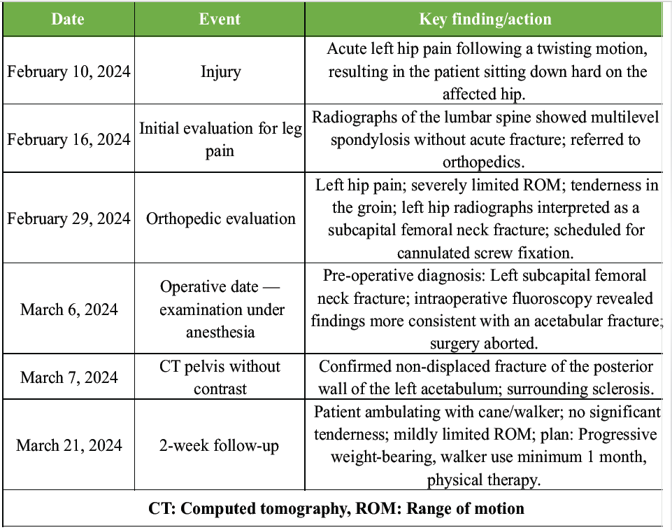
Table 1: Timeline of clinical events
Diagnostic assessment:
At the initial orthopedic visit, plain radiographs of the left hip were obtained (Fig. 1).
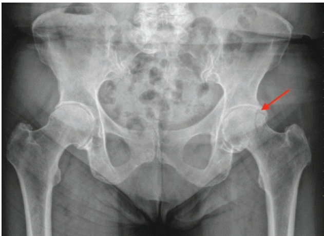
Figure 1: Anteroposterior radiograph of the left hip at initial orthopedic evaluation. Anteroposterior radiograph of the left hip at initial orthopedic evaluation (February 29, 2024), before attempted operative fixation. The linear density at the femoral head-neck junction (arrow), later identified as a posterior acetabular wall fracture projected over the proximal femur, was initially interpreted as a subcapital femoral neck fracture.
The radiographic interpretation identified an irregularity at the femoral head–neck junction, consistent with a subcapital femoral neck fracture. Based on this interpretation, the patient was scheduled for cannulated screw fixation. Intraoperatively, on March 6, 2024, with the preoperative diagnosis of a left subcapital femoral neck fracture, fluoroscopic images on the table (Fig. 2) suggested findings more consistent with a left posterior acetabular wall fracture.
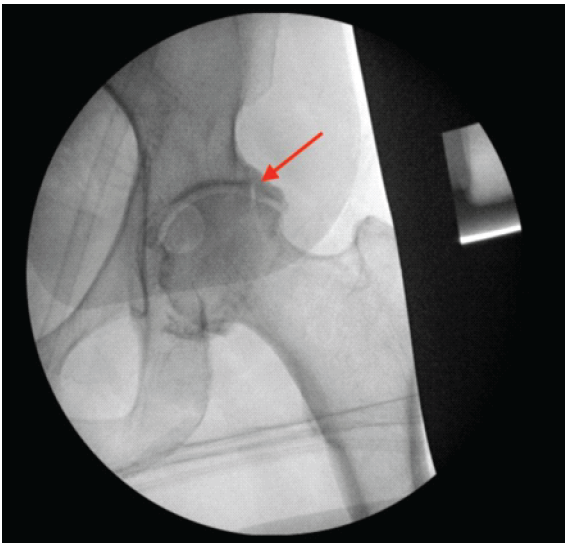
Figure 2: Intraoperative fluoroscopy of the left hip during attempted cannulated screw fixation. Intraoperative fluoroscopic images of the left hip obtained during the planned cannulated screw fixation (March 6, 2024), demonstrating the absence of the expected femoral neck fracture line and findings more consistent with posterior acetabular wall pathology.
Fluoroscopic imaging did not show the expected femoral neck fracture line. In addition, visualization of a linear density attributed to the posterior acetabular wall further suggested an alternate diagnosis. The decision was made to avoid any surgical intervention. CT imaging of the pelvis without contrast was obtained on March 7, 2024 (Figs. 3 and 4).
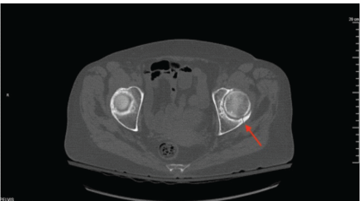
Figure 3: Axial computed tomography of the pelvis showing left posterosuperior acetabular fracture. Computed tomography of the pelvis without contrast (March 7, 2024). Axial reconstructions demonstrate a linear fracture of the posterosuperior aspect of the left acetabulum without significant displacement. Surrounding sclerosis is noted. Moderate bilateral hip osteoarthritis is visible.
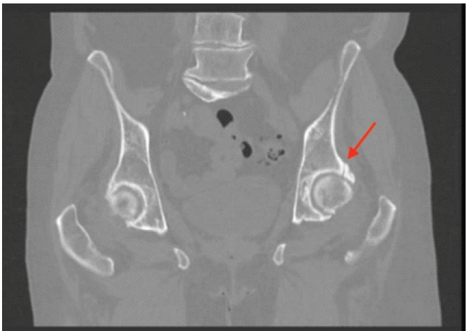
Figure 4: Coronal computed tomography of the pelvis showing left posterosuperior acetabular fracture. Computed tomography of the pelvis without contrast (March 7, 2024). Coronal reconstructions demonstrate a linear fracture of the posterosuperior aspect of the left acetabulum without significant displacement. Surrounding sclerosis is noted. Moderate bilateral hip osteoarthritis is visible.
Computed tomography (CT) demonstrated a linear fracture of the posterosuperior aspect of the left acetabulum without significant displacement. The surrounding bone was sclerotic, suggesting an acute or prior injury. No other fracture was identified. All four pubic rami were intact. Moderate osteoarthritis of both hips was noted. CT impression: Age-indeterminate fracture of the posterosuperior left acetabulum, non-displaced fracture of the posterior wall of the left acetabulum, initial encounter. The patient also demonstrated a limited range of motion of the hip secondary to ankylosis, identified during operative examination under anesthesia.
Therapeutic intervention:
Following the revised diagnosis of a non-displaced posterior wall acetabular fracture, the decision was made to manage the patient non-operatively. The patient was instructed to slowly progress to weight-bearing as tolerated on the left lower extremity. She was instructed to use a walker for ambulation for a minimum of 1 month. Physical therapy was initiated to improve the strength and range of motion of the left lower extremity. The primary change in management was the intraoperative decision to abort the planned cannulated screw fixation after fluoroscopic reassessment altered the diagnosis from a femoral neck fracture to a posterior acetabular wall fracture. No pharmacologic interventions were initiated specifically for fracture management; the patient’s existing medications, including ibandronate for bone density, were continued as previously prescribed.
Follow-up and outcomes:
At the 2-week follow-up visit on March 21, 2024, the patient reported satisfactory recovery and was ambulating independently with a cane and at times a walker. Physical examination demonstrated a mildly limited range of motion of the left hip with no significant tenderness to palpation. The left lower extremity remained neurovascularly intact.
The clinical plan included continued progressive weight-bearing as tolerated, walker use for a minimum of 1 additional month, and continuation of physical therapy. The patient was to follow up on an as-needed basis. There were no problems with adherence to restrictions, and the patient did not have any complications during treatment. The patient expressed satisfaction with the care she received and was relieved that the finding had been identified before any surgical intervention. She had questions regarding the examination performed and wanted to discuss further treatment options. She was seen again at a different follow-up appointment for an unrelated complaint of shoulder pain and has continued to do well. To date, she has not undergone hip surgery.
Diagnostic pitfall: Acetabular fracture versus femoral neck fracture:
Plain radiographs have well-documented limitations for proximal femur fracture detection, but the literature focuses almost exclusively on false negatives (missed fractures) rather than false positives (misidentified fractures) [1,3]. Arlachov and Ibrahem Adam published a review of mimics of a femoral neck fracture, noting that several conditions can simulate femoral neck fractures and emphasizing the need for systematic and careful observation [4]. This diagnostic challenge is further reflected in the acetabulum, where fractures may be radiographically occult or initially misinterpreted due to subtle displacement and complex overlapping anatomy. Even when present, acetabular fractures may not be evident on initial AP radiographs, contributing to delayed or incorrect diagnosis [11]. Acetabular fractures are notoriously difficult to characterize on plain films, and the challenge is compounded by the complex three-dimensional anatomy of the acetabulum and the superimposition of acetabular walls, columns, and obturator rings on a two-dimensional AP view [7,8,9,10]. Durkee et al. describe the radiographic landmarks of the acetabulum, including the iliopectineal line, ilioischial line, and anterior and posterior acetabular wall lines, which can be difficult to evaluate when overlapping with the proximal femur [8]. Scheinfeld et al., similarly, emphasize that although radiographs remain the initial study, CT is often essential for accurate characterization. Failure to accurately diagnose hip pathology on initial imaging can result in inappropriate management or delay in appropriate treatment [9]. This case demonstrates a false-positive diagnosis of femoral neck fracture caused by an acetabular fracture, specifically, a posterior acetabular wall fracture whose fracture line projected over the femoral head–neck junction on AP pelvis radiographs, creating a linear density that was interpreted as a subcapital femoral neck fracture. This is distinct from the well-described false-negative problem of occult femoral neck fractures. On AP pelvis radiographs, a posterior acetabular wall fracture may project over the femoral head–neck junction and create a linear density that mimics a subcapital femoral neck fracture, particularly in the setting of osteoarthritis or coxa profunda, as was present in this patient [4,9]. The presence of surrounding sclerosis on CT in this case, suggesting a possible subacute component to the injury, may have further contributed to the well-defined linear appearance of the fracture line on the plain radiograph.
Intraoperative decision-making:
The surgeon’s decision to abort surgery when fluoroscopic findings did not match preoperative expectations represents sound clinical judgment. Proceeding with cannulated screw placement without confirmed fracture visualization would have been technically inappropriate and potentially harmful. This case reinforces the principle that intraoperative findings must match pre-operative expectations, and when they do not, surgeons must reassess. Intraoperative fluoroscopy is the standard of care for cannulated screw fixation of femoral neck fractures, used primarily for surgical guidance rather than as a diagnostic tool. When the surgeon was unable to visualize the expected fracture during fluoroscopic surgical planning and found a linear density suggesting a posterior acetabular wall fracture, aborting the procedure to obtain additional imaging was the correct decision.
Strengths and limitations:
Strengths of this report include detailed documentation of the intraoperative decision-making process and multimodal imaging correlation. Limitations include the single-patient nature of this report and limited long-term follow-up data. In addition, the absence of a formal radiology report for the initial plain radiographs limits complete radiographic documentation. The American College of Radiology appropriateness criteria state that when a fracture is demonstrated, additional imaging is not routinely performed for treatment planning, and proceeding to surgery when two experienced clinicians both interpreted the radiographs as showing a subcapital femoral neck fracture is consistent with standard practice [2]. CT imaging prompted a change in surgical plan, from internal fixation to arthroplasty, in approximately 21% of cases among elderly patients with non-displaced femoral neck fractures, underscoring the limitations of plain radiographs alone in guiding treatment decisions [12]; however, current guidelines classify CT as “usually appropriate,” rather than required, in the setting of radiographically apparent hip fractures [2]. The final CT diagnosis of an acetabular fracture explains why the radiographs were misinterpreted. This case illustrates that even within an appropriate standard-of-care workup, a posterior acetabular wall fracture can create a compelling false-positive appearance of femoral neck fracture on plain radiographs.
A posterior acetabular wall fracture may mimic a femoral neck fracture on plain radiographs when the fracture line projects over the femoral head-neck junction on AP pelvis views. Recognition of this pitfall is important to avoid unnecessary operative intervention. The radiographic appearance may be compelling enough that experienced clinicians operating within the standard of care proceed to surgery, only for the true diagnosis to be revealed through intraoperative fluoroscopy. This case underscores that the diagnosis of femoral neck fracture on plain radiographs is not always definitive and that acetabular fracture pathology can create a virtually indistinguishable radiographic appearance. Accurate interpretation of imaging and use of confirmatory modalities are essential to prevent misdiagnosis and inappropriate surgical planning. Intraoperative vigilance remains essential; when fluoroscopic findings do not match preoperative expectations, surgery should be aborted and additional imaging obtained.
When pre-operative radiographs suggest a subcapital femoral neck fracture, but intraoperative fluoroscopy does not confirm the expected fracture pattern, surgeons should abort the planned procedure rather than proceed with fixation. In these cases, obtain computed tomography imaging to evaluate for an alternative diagnosis, including an acetabular fracture, before returning to the operating room. This intraoperative vigilance prevents unnecessary surgical intervention in patients whose true pathology may be better managed non-operatively.
References
- 1. Schroeder JD, Turner SP, Buck E. Hip fractures: Diagnosis and management. Am Fam Physician 2022;106:675-83. [Google Scholar] [PubMed]
- 2. Bartolotta RJ, Ha AS, Bateni CP, Chen KC, Dvorzhinskiy A, Flug J et al. ACR appropriateness criteria® acute hip pain: 2024 Update. J Am Coll Radiol 2025;22:S3-13. [Google Scholar] [PubMed]
- 3. Haj-Mirzaian A, Eng J, Khorasani R, Raja AS, Levin AS, Smith SE, et al. Use of advanced imaging for radiographically occult hip fracture in elderly patients: A systematic review and meta-analysis. Radiology 2020;296:521-31. [Google Scholar] [PubMed]
- 4. Arlachov Y, Ibrahem Adam R. Acute hip pain: Mimics of a femoral neck fracture. Clin Radiol 2018;73:773-81. [Google Scholar] [PubMed]
- 5. Chang CY, Huang AJ. MR imaging of normal hip anatomy. Magn Reson Imaging Clin N Am 2013;21:1-19. [Google Scholar] [PubMed]
- 6. Keats TE, Anderson MW. The pelvic girdle. In: Atlas of Normal Roentgen Variants that May Simulate Disease. 9th ed. Philadelphia, PA: Elsevier Saunders; 2012. p. 4-84. [Google Scholar] [PubMed]
- 7. Borrelli J Jr., Goldfarb C, Catalano L, Evanoff BA. Assessment of articular fragment displacement in acetabular fractures: A comparison of computerized tomography and plain radiographs. J Orthop Trauma 2002;16:449-56; discussion 456-7. [Google Scholar] [PubMed]
- 8. Durkee NJ, Jacobson J, Jamadar D, Karunakar MA, Morag Y, Hayes C. Classification of common acetabular fractures: Radiographic and CT appearances. AJR Am J Roentgenol 2006;187:915-25. [Google Scholar] [PubMed]
- 9. Scheinfeld MH, Dym AA, Spektor M, Avery LL, Dym RJ, Amanatullah DF. Acetabular fractures: What radiologists should know and how 3D CT can aid classification. RadioGraphics 2015;35:555-77. [Google Scholar] [PubMed]
- 10. Lawrence DA, Menn K, Baumgaertner M, Haims AH. Acetabular fractures: Anatomic and clinical considerations. AJR Am J Roentgenol 2013;201:W425-36. [Google Scholar] [PubMed]
- 11. Guerado E, Cano JR, Cruz E. Occult acetabular fracture in elderly patients. Open Orthop J 2012;6:582-6. [Google Scholar] [PubMed]
- 12. Zamora T, Klaber I, Ananias J, Bengoa F, Botello E, Amenabar P, et al. The influence of the CT scan in the evaluation and treatment of nondisplaced femoral neck fractures in the elderly. J Orthop Surg (Hong Kong) 2019;27(2):2309499019836160. [Google Scholar] [PubMed]







Related Articles in Journal of Orthopaedic Case Reports
 October 1, 2026 Primary Total Hip Arthroplasty for a Displaced Femoral Neck Fracture in an Above-the-Knee Amputee: A Case Report
 September 1, 2025 Failure of the Femoral Neck System in a Young Patient: A Case Report
 July 1, 2025 A Large-diameter Corona Mortis Artery Encountered During Surgery for Acetabular Fracture: A Case Report and Review of Anatomical Significance
 October 1, 2026 Posterior Hip Dislocation with Ipsilateral Subtrochanteric Femur Fracture: A Rare Case Report